
Article Figures & Data
Figures
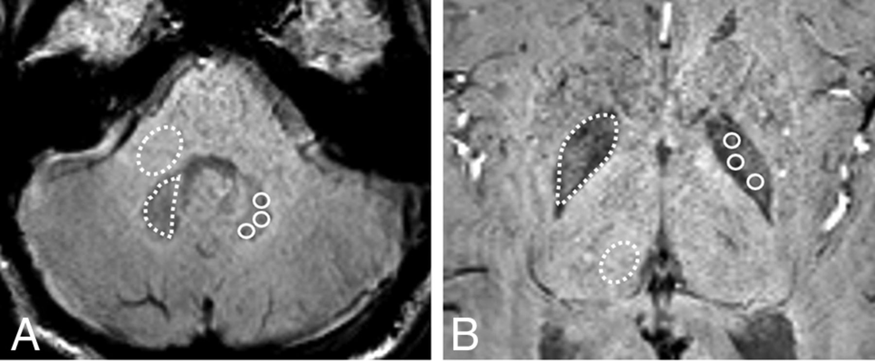
- FIG 1.
SWI at the level of the DN (A) and GP (B) used for drawing the ROIs in the DN and GP with the corresponding MCP and Th for normalization, respectively. Dotted lines illustrate the freehand ROI used to obtain SWImean from the DN, GP, MCP, and Th; solid lines illustrate the circular ROIs used to obtain SWImin from the DN and GP.
- FIG 2.
Boxplots of the mean DN-to-MCP ratios on SWI and T1WI among the linear GBCA, macrocyclic GBCA, and control groups. The error bars for boxplots represent the minimum and maximum data points within each group, and the Middle box represents the 25th, 50th, and 75th percentiles of the data within each group.
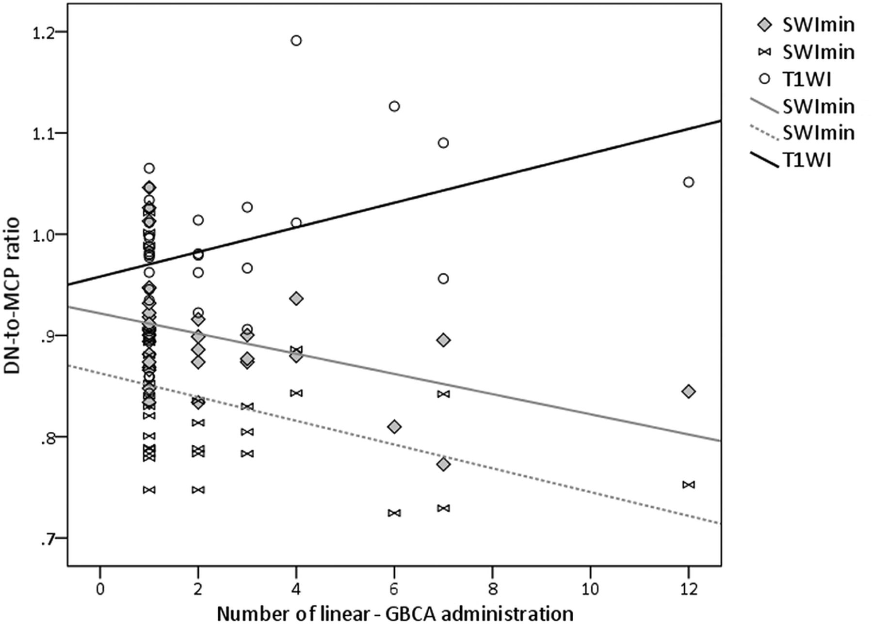
- FIG 3.
Scatterplot of the DN-to-MCP ratios for SWImean, SWImin, and T1WI versus the number of intravenous linear GBCA administrations, with linear regression lines for each group.
- FIG 4.
A 13-year-old male patient without gadolinium exposure (A and B), an age- and sex-matched patient after 5 doses of macrocyclic GBCA (C and D), and another 13-year-old adolescent boy after 7 doses of linear GBCA administration (E and F). Note the brighter signal in the DN on T1WI (E) and lower signal in the DN on SWI (F), which is not present in the other 2 children. (White arrows represent DN)
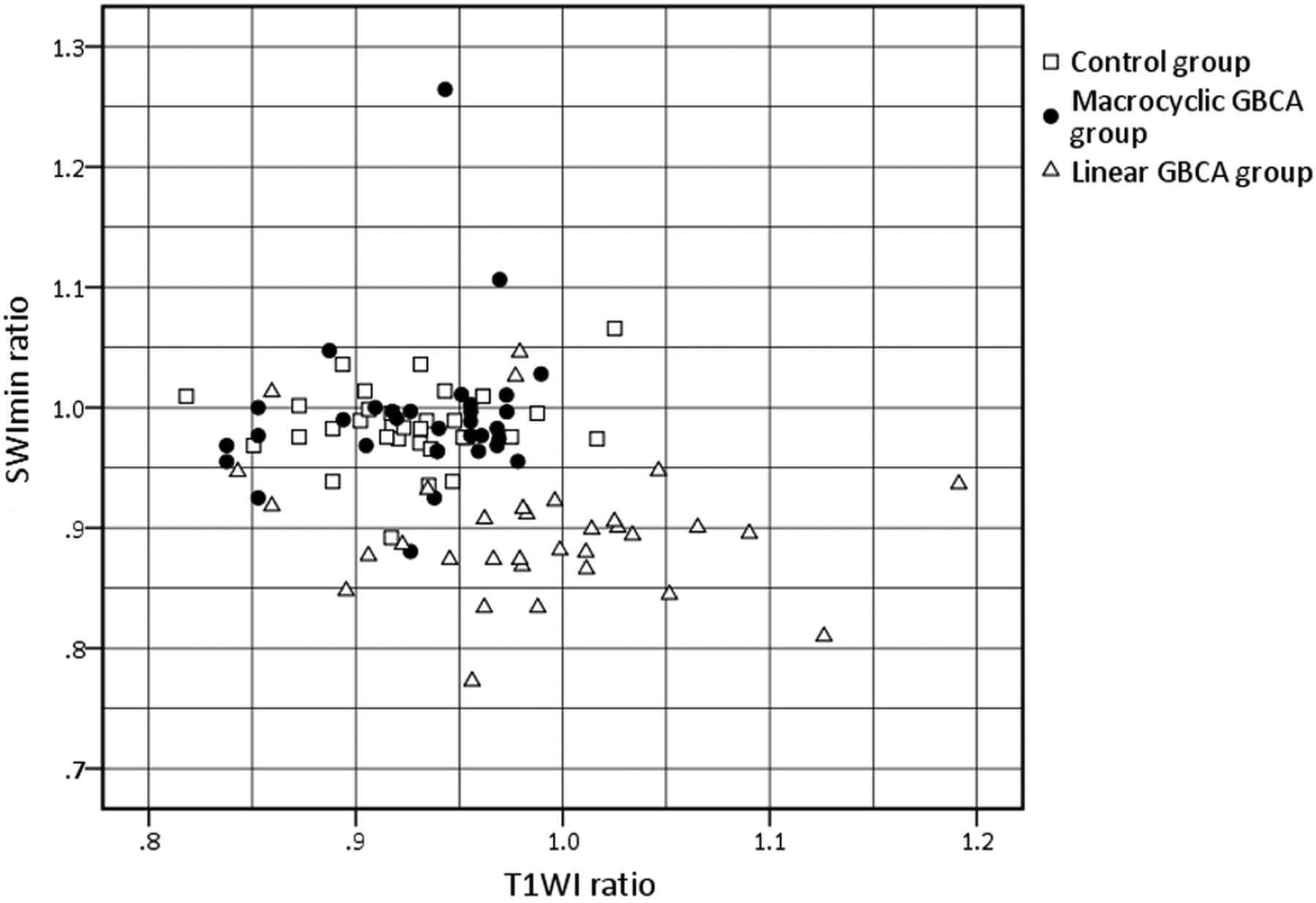
- FIG 5.
Scatterplot of the DN-to-MCP ratios on unenhanced T1WI and SWI. Increased T1WI and decreased SWImean ratios in the linear GBCA group compared with macrocyclic GBCA and control groups are seen.
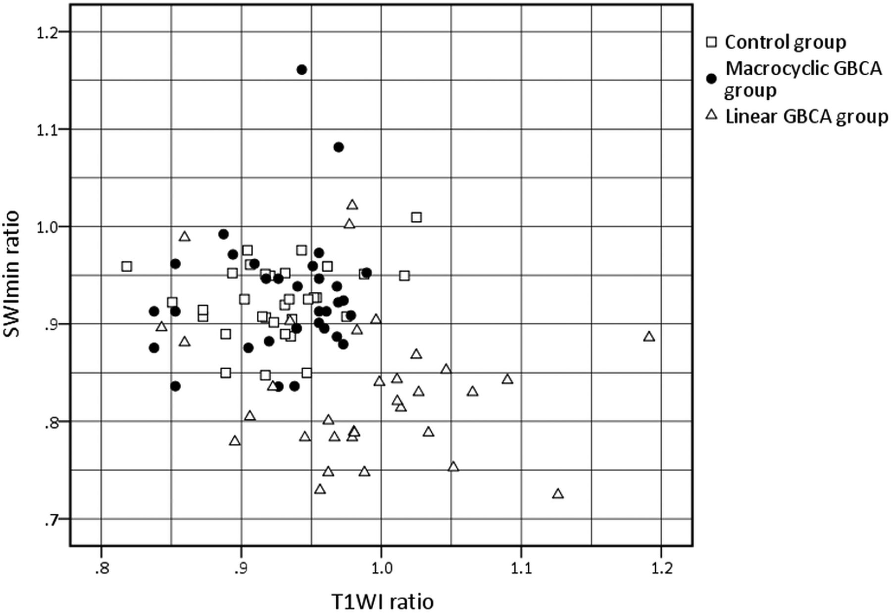
- FIG 6.
Scatterplot of the DN-to-MCP ratios on unenhanced T1WI and SWI. Note increased T1WI and decreased SWImin ratios in the linear GBCA group compared with macrocyclic GBCA and control groups.


Tables
Exclusion Parameters Control Group Macrocyclic GBCA Group Linear GBCA Group Initially selected 57 64 61 Age younger than 2 years 3 5 6 Renal dysfunction 2 0 0 Hepatic dysfunction 3 2 2 Lesion in the cerebellum, midbrain, corpus striatum, or pulvinar of the thalamus 3 2 1 Missing documentation of the prior contrast agent 8 11 7 Missing or unsatisfactory T1WI 2 2 3 Missing or unsatisfactory SWI 3 4 3 Different sequences and parameters between comparison T1WIs 0 5 6 Final No. of groups 33 33 33 Characteristic Linear GBCA–Exposed Children Macrocyclic GBCA–Exposed Children GBCA-Naive Control Subjects Age (yr) Mean (SD) 8.2 (SD, 5.4) 8.2 (SD, 5.1) 8.2 (SD, 3.6 ) Range 2–18 2–18 2–18 Sex Male 23 23 23 Female 10 10 10 Mean (SD) No. of contrast-enhanced MR imaging 2.4 (SD, 2.4) 4.03 (SD, 2.9) 0 Mean (SD) time interval between MR imaging (mo) 7.16 (SD, 7.5) 7.4 (SD, 7.3) 0 Mean (SD) accumulated gadolinium dose (mmol) 17.5 (SD, 24.1) 13.3 (SD, 11.9) 0 Diagnosis Intracranial neoplasm 7 7 10 Extracranial neoplasm 2 0 1 Pituitary abnormality 3 6 2 Orbit/optic nerve pathology 6 7 2 Adrenoleukodystrophy 5 5 10 Benign conditions 10 8 8 History of radiation treatment 3 2 3 History of chemotherapy 6 5 4 History of neurosurgery 2 2 3 ↵a Data are number of patients unless indicated otherwise and mean (SD).


Jump to section
Related Articles
Cited By...
- No citing articles found.