Advertisement

Main menu

  • Home
  • Content
    • Current Issue
    • Accepted Manuscripts
    • Article Preview
    • Past Issue Archive
    • Video Articles
    • AJNR Case Collection
    • Case of the Week Archive
    • Case of the Month Archive
    • Classic Case Archive
  • Special Collections
    • AJNR Awards
    • Low-Field MRI
    • Alzheimer Disease
    • ASNR Foundation Special Collection
    • Photon-Counting CT
    • View All
  • Multimedia
    • AJNR Podcasts
    • AJNR SCANtastic
    • Trainee Corner
    • MRI Safety Corner
    • Imaging Protocols
  • For Authors
    • Submit a Manuscript
    • Submit a Video Article
    • Submit an eLetter to the Editor/Response
    • Manuscript Submission Guidelines
    • Statistical Tips
    • Fast Publishing of Accepted Manuscripts
    • Graphical Abstract Preparation
    • Imaging Protocol Submission
    • Author Policies
  • About Us
    • About AJNR
    • Editorial Board
    • Editorial Board Alumni
  • More
    • Become a Reviewer/Academy of Reviewers
    • Subscribers
    • Permissions
    • Alerts
    • Feedback
    • Advertisers
    • ASNR Home

User menu

  • Alerts
  • Log in

Search

  • Advanced search
American Journal of Neuroradiology

American Journal of Neuroradiology

ASHNR American Society of Functional Neuroradiology ASHNR American Society of Pediatric Neuroradiology ASSR
  • Alerts
  • Log in

Advanced Search

  • Home
  • Content
    • Current Issue
    • Accepted Manuscripts
    • Article Preview
    • Past Issue Archive
    • Video Articles
    • AJNR Case Collection
    • Case of the Week Archive
    • Case of the Month Archive
    • Classic Case Archive
  • Special Collections
    • AJNR Awards
    • Low-Field MRI
    • Alzheimer Disease
    • ASNR Foundation Special Collection
    • Photon-Counting CT
    • View All
  • Multimedia
    • AJNR Podcasts
    • AJNR SCANtastic
    • Trainee Corner
    • MRI Safety Corner
    • Imaging Protocols
  • For Authors
    • Submit a Manuscript
    • Submit a Video Article
    • Submit an eLetter to the Editor/Response
    • Manuscript Submission Guidelines
    • Statistical Tips
    • Fast Publishing of Accepted Manuscripts
    • Graphical Abstract Preparation
    • Imaging Protocol Submission
    • Author Policies
  • About Us
    • About AJNR
    • Editorial Board
    • Editorial Board Alumni
  • More
    • Become a Reviewer/Academy of Reviewers
    • Subscribers
    • Permissions
    • Alerts
    • Feedback
    • Advertisers
    • ASNR Home
  • Follow AJNR on Twitter
  • Visit AJNR on Facebook
  • Follow AJNR on Instagram
  • Join AJNR on LinkedIn
  • RSS Feeds

AJNR Awards, New Junior Editors, and more. Read the latest AJNR updates

Research ArticleHead and Neck Imaging

Optimal Duration of MRI Follow-up to Safely Identify Middle Ear Residual Cholesteatoma

A.-L. Fourez, M. Akkari, G. Gascou, P.-H. Lefevre, C. Duflos, A. Kaderbay, M. Mondain and F. Venail
American Journal of Neuroradiology April 2021, DOI: https://doi.org/10.3174/ajnr.A7116
A.-L. Fourez
aFrom the Department of ENT and Head and Neck Surgery (A.-L.F., M.A., A.K., M.M., F.V.) and Neuroradiology (G.G., P.-H.L.), University Hospital Gui de Chauliac, and Department of Medical Information and Biostatistics (C.D.), University Hospital La Colombière, University of Montpellier, Montpellier, France
  • Find this author on Google Scholar
  • Find this author on PubMed
  • Search for this author on this site
  • ORCID record for A.-L. Fourez
M. Akkari
aFrom the Department of ENT and Head and Neck Surgery (A.-L.F., M.A., A.K., M.M., F.V.) and Neuroradiology (G.G., P.-H.L.), University Hospital Gui de Chauliac, and Department of Medical Information and Biostatistics (C.D.), University Hospital La Colombière, University of Montpellier, Montpellier, France
  • Find this author on Google Scholar
  • Find this author on PubMed
  • Search for this author on this site
  • ORCID record for M. Akkari
G. Gascou
aFrom the Department of ENT and Head and Neck Surgery (A.-L.F., M.A., A.K., M.M., F.V.) and Neuroradiology (G.G., P.-H.L.), University Hospital Gui de Chauliac, and Department of Medical Information and Biostatistics (C.D.), University Hospital La Colombière, University of Montpellier, Montpellier, France
  • Find this author on Google Scholar
  • Find this author on PubMed
  • Search for this author on this site
  • ORCID record for G. Gascou
P.-H. Lefevre
aFrom the Department of ENT and Head and Neck Surgery (A.-L.F., M.A., A.K., M.M., F.V.) and Neuroradiology (G.G., P.-H.L.), University Hospital Gui de Chauliac, and Department of Medical Information and Biostatistics (C.D.), University Hospital La Colombière, University of Montpellier, Montpellier, France
  • Find this author on Google Scholar
  • Find this author on PubMed
  • Search for this author on this site
  • ORCID record for P.-H. Lefevre
C. Duflos
aFrom the Department of ENT and Head and Neck Surgery (A.-L.F., M.A., A.K., M.M., F.V.) and Neuroradiology (G.G., P.-H.L.), University Hospital Gui de Chauliac, and Department of Medical Information and Biostatistics (C.D.), University Hospital La Colombière, University of Montpellier, Montpellier, France
  • Find this author on Google Scholar
  • Find this author on PubMed
  • Search for this author on this site
  • ORCID record for C. Duflos
A. Kaderbay
aFrom the Department of ENT and Head and Neck Surgery (A.-L.F., M.A., A.K., M.M., F.V.) and Neuroradiology (G.G., P.-H.L.), University Hospital Gui de Chauliac, and Department of Medical Information and Biostatistics (C.D.), University Hospital La Colombière, University of Montpellier, Montpellier, France
  • Find this author on Google Scholar
  • Find this author on PubMed
  • Search for this author on this site
  • ORCID record for A. Kaderbay
M. Mondain
aFrom the Department of ENT and Head and Neck Surgery (A.-L.F., M.A., A.K., M.M., F.V.) and Neuroradiology (G.G., P.-H.L.), University Hospital Gui de Chauliac, and Department of Medical Information and Biostatistics (C.D.), University Hospital La Colombière, University of Montpellier, Montpellier, France
  • Find this author on Google Scholar
  • Find this author on PubMed
  • Search for this author on this site
  • ORCID record for M. Mondain
F. Venail
aFrom the Department of ENT and Head and Neck Surgery (A.-L.F., M.A., A.K., M.M., F.V.) and Neuroradiology (G.G., P.-H.L.), University Hospital Gui de Chauliac, and Department of Medical Information and Biostatistics (C.D.), University Hospital La Colombière, University of Montpellier, Montpellier, France
bInstitute for Neurosciences de Montpellier (F.V.), Institut National de la Santé et de la Recherche Médicale U1051 and University of Montpellier, Montpellier, France
  • Find this author on Google Scholar
  • Find this author on PubMed
  • Search for this author on this site
  • ORCID record for F. Venail
  • Article
  • Figures & Data
  • Supplemental
  • Info & Metrics
  • Responses
  • References
  • PDF
Loading

Article Figures & Data

Figures

  • FIG 1.
    • Download figure
    • Open in new tab
    • Download powerpoint
    FIG 1.

    Flowchart of patient inclusion and follow-up imaging.

  • FIG 2.
    • Download figure
    • Open in new tab
    • Download powerpoint
    FIG 2.

    Duration of follow-up in participants with or without residual cholesteatoma Solid and dotted lines depict participants with or without residual cholesteatoma, respectively, followed up across time in months. The mean duration of follow-up is 54.3 months in the residual cholesteatoma group and 74.1 months in the no residual cholesteatoma group.
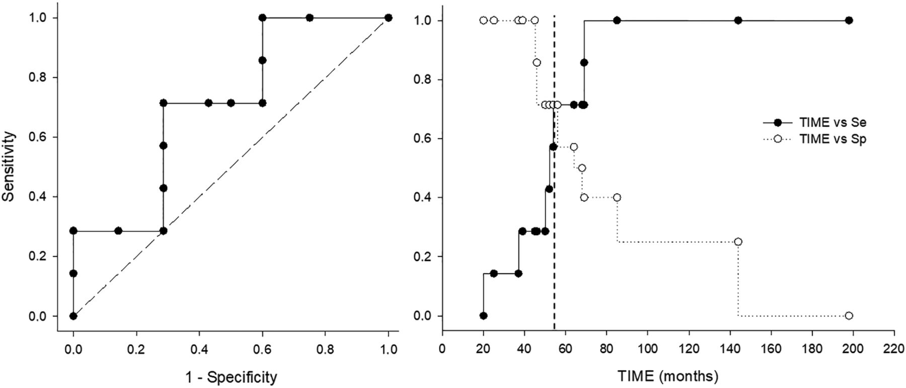
  • FIG 3.
    • Download figure
    • Open in new tab
    • Download powerpoint
    FIG 3.

    Diagnostic value of non-EPI DWI as a function of time. The left panel shows the receiver operating characteristic curve of sensitivity according to 1-specificity. The best diagnostic values (sensitivity  = 0.71, specificity = 0.71, Youden index = 0.43) are reached at 56 months postoperatively. The right panel shows the values of sensitivity and specificity as a function of time. Sensitivity increases from 0 to 1 by 85 months postoperatively. Specificity changes from 1 for MRIs performed up to 45 months to 0 for those performed from 198 months after the operation. Se indicates sensitivity; sp, specificity.

PreviousNext
Back to top
Advertisement
Print
Download PDF
Email Article

Thank you for your interest in spreading the word on American Journal of Neuroradiology.

NOTE: We only request your email address so that the person you are recommending the page to knows that you wanted them to see it, and that it is not junk mail. We do not capture any email address.

Enter multiple addresses on separate lines or separate them with commas.
Optimal Duration of MRI Follow-up to Safely Identify Middle Ear Residual Cholesteatoma
(Your Name) has sent you a message from American Journal of Neuroradiology
(Your Name) thought you would like to see the American Journal of Neuroradiology web site.
CAPTCHA
This question is for testing whether or not you are a human visitor and to prevent automated spam submissions.
Cite this article
A.-L. Fourez, M. Akkari, G. Gascou, P.-H. Lefevre, C. Duflos, A. Kaderbay, M. Mondain, F. Venail
Optimal Duration of MRI Follow-up to Safely Identify Middle Ear Residual Cholesteatoma
American Journal of Neuroradiology Apr 2021, DOI: 10.3174/ajnr.A7116

Citation Manager Formats

  • BibTeX
  • Bookends
  • EasyBib
  • EndNote (tagged)
  • EndNote 8 (xml)
  • Medlars
  • Mendeley
  • Papers
  • RefWorks Tagged
  • Ref Manager
  • RIS
  • Zotero
0 Responses
Respond to this article
Share
Bookmark this article
Optimal Duration of MRI Follow-up to Safely Identify Middle Ear Residual Cholesteatoma
A.-L. Fourez, M. Akkari, G. Gascou, P.-H. Lefevre, C. Duflos, A. Kaderbay, M. Mondain, F. Venail
American Journal of Neuroradiology Apr 2021, DOI: 10.3174/ajnr.A7116
del.icio.us logo Twitter logo Facebook logo Mendeley logo
  • Tweet Widget
  • Facebook Like
  • Google Plus One
Purchase

Jump to section

  • Article
    • Abstract
    • MATERIALS AND METHODS
    • RESULTS
    • DISCUSSION
    • CONCLUSIONS
    • Footnotes
    • References
  • Figures & Data
  • Supplemental
  • Info & Metrics
  • Responses
  • References
  • PDF

Related Articles

  • PubMed
  • Google Scholar

Cited By...

  • No citing articles found.
  • Crossref (7)
  • Google Scholar

This article has been cited by the following articles in journals that are participating in Crossref Cited-by Linking.

  • Beyond the otoscope: an imaging review of congenital cholesteatoma
    Guillaume Vangrinsven, Anja Bernaerts, Filip Deckers, Joost van Dinther, Andrzej Zarowski, Bert De Foer
    Insights into Imaging 2024 15 1
  • Cholesteatom: Nach Resektion Überwachung mit Bildgebung für mindestens 5 Jahre
    Neuroradiologie Scan 2022 12 02
  • Evaluation of Cholesteatoma
    Anne K. Maxwell, Stephen R. Hoff
    Otolaryngologic Clinics of North America 2025 58 1
  • Cholesteatom: nach Resektion Überwachung mit Bildgebung für mindestens 5 Jahre
    Onkologie up2date 2022 04 03
  • Evaluation of the effectiveness of bone obliteration in cholesteatoma surgery
    J. Chomarat, C. Fabre, S. Schmerber, Raphaële Quatre
    European Archives of Oto-Rhino-Laryngology 2025
  • Modern aspects of diagnosing purulent pathology of the ear (Review of cases of middle ear cholesteatoma using non-EPI DWI MRI)
    Asel Imangalieva, Dina Kussainova, Otabek Tajibaev, Segizbayeva Aliya
    Journal of Otology 2025 20 2
  • Recidivism and Recurrence
    Shayna Portanova Cooperman, Kevin Wong, Tiffany Peng Hwa, Jennifer Alyono
    Otolaryngologic Clinics of North America 2025 58 1

More in this TOC Section

  • ASL Sensitivity for Head and Neck Paraganglioma
  • Post SRS Peritumoral Hyperintense Signal of VSs
  • Contrast Enhanced Pituitary CISS/FIESTA
Show more Head and Neck Imaging

Similar Articles

Advertisement

Indexed Content

  • Current Issue
  • Accepted Manuscripts
  • Article Preview
  • Past Issues
  • Editorials
  • Editor's Choice
  • Fellows' Journal Club
  • Letters to the Editor
  • Video Articles

Cases

  • Case Collection
  • Archive - Case of the Week
  • Archive - Case of the Month
  • Archive - Classic Case

More from AJNR

  • Trainee Corner
  • Imaging Protocols
  • MRI Safety Corner
  • Book Reviews

Multimedia

  • AJNR Podcasts
  • AJNR Scantastics

Resources

  • Turnaround Time
  • Submit a Manuscript
  • Submit a Video Article
  • Submit an eLetter to the Editor/Response
  • Manuscript Submission Guidelines
  • Statistical Tips
  • Fast Publishing of Accepted Manuscripts
  • Graphical Abstract Preparation
  • Imaging Protocol Submission
  • Evidence-Based Medicine Level Guide
  • Publishing Checklists
  • Author Policies
  • Become a Reviewer/Academy of Reviewers
  • News and Updates

About Us

  • About AJNR
  • Editorial Board
  • Editorial Board Alumni
  • Alerts
  • Permissions
  • Not an AJNR Subscriber? Join Now
  • Advertise with Us
  • Librarian Resources
  • Feedback
  • Terms and Conditions
  • AJNR Editorial Board Alumni

American Society of Neuroradiology

  • Not an ASNR Member? Join Now

© 2025 by the American Society of Neuroradiology All rights, including for text and data mining, AI training, and similar technologies, are reserved.
Print ISSN: 0195-6108 Online ISSN: 1936-959X

Powered by HighWire