Advertisement

Main menu

  • Home
  • Content
    • Current Issue
    • Accepted Manuscripts
    • Article Preview
    • Past Issue Archive
    • Video Articles
    • AJNR Case Collection
    • Case of the Week Archive
    • Case of the Month Archive
    • Classic Case Archive
  • Special Collections
    • AJNR Awards
    • Low-Field MRI
    • Alzheimer Disease
    • ASNR Foundation Special Collection
    • Photon-Counting CT
    • View All
  • Multimedia
    • AJNR Podcasts
    • AJNR SCANtastic
    • Trainee Corner
    • MRI Safety Corner
    • Imaging Protocols
  • For Authors
    • Submit a Manuscript
    • Submit a Video Article
    • Submit an eLetter to the Editor/Response
    • Manuscript Submission Guidelines
    • Statistical Tips
    • Fast Publishing of Accepted Manuscripts
    • Graphical Abstract Preparation
    • Imaging Protocol Submission
    • Author Policies
  • About Us
    • About AJNR
    • Editorial Board
    • Editorial Board Alumni
  • More
    • Become a Reviewer/Academy of Reviewers
    • Subscribers
    • Permissions
    • Alerts
    • Feedback
    • Advertisers
    • ASNR Home

User menu

  • Alerts
  • Log in

Search

  • Advanced search
American Journal of Neuroradiology

American Journal of Neuroradiology

ASHNR American Society of Functional Neuroradiology ASHNR American Society of Pediatric Neuroradiology ASSR
  • Alerts
  • Log in

Advanced Search

  • Home
  • Content
    • Current Issue
    • Accepted Manuscripts
    • Article Preview
    • Past Issue Archive
    • Video Articles
    • AJNR Case Collection
    • Case of the Week Archive
    • Case of the Month Archive
    • Classic Case Archive
  • Special Collections
    • AJNR Awards
    • Low-Field MRI
    • Alzheimer Disease
    • ASNR Foundation Special Collection
    • Photon-Counting CT
    • View All
  • Multimedia
    • AJNR Podcasts
    • AJNR SCANtastic
    • Trainee Corner
    • MRI Safety Corner
    • Imaging Protocols
  • For Authors
    • Submit a Manuscript
    • Submit a Video Article
    • Submit an eLetter to the Editor/Response
    • Manuscript Submission Guidelines
    • Statistical Tips
    • Fast Publishing of Accepted Manuscripts
    • Graphical Abstract Preparation
    • Imaging Protocol Submission
    • Author Policies
  • About Us
    • About AJNR
    • Editorial Board
    • Editorial Board Alumni
  • More
    • Become a Reviewer/Academy of Reviewers
    • Subscribers
    • Permissions
    • Alerts
    • Feedback
    • Advertisers
    • ASNR Home
  • Follow AJNR on Twitter
  • Visit AJNR on Facebook
  • Follow AJNR on Instagram
  • Join AJNR on LinkedIn
  • RSS Feeds

AJNR Awards, New Junior Editors, and more. Read the latest AJNR updates

Research ArticleHead and Neck Imaging

Variations in the Course of the Carotid Arteries in Patients with Retropharyngeal Parathyroid Adenomas

G.K. Steinl, R. Yeh, C.M. McManus, J.A. Lee and J.H. Kuo
American Journal of Neuroradiology February 2021, DOI: https://doi.org/10.3174/ajnr.A6995
G.K. Steinl
aFrom the Department of Surgery, Division of GI/Endocrine Surgery (G.K.S., C.M.M., J.A.L., J.H.K.), Vagelos College of Physicians and Surgeons, Columbia University Irving Medical Center, New York, New York
  • Find this author on Google Scholar
  • Find this author on PubMed
  • Search for this author on this site
  • ORCID record for G.K. Steinl
R. Yeh
bDepartment of Radiology (R.Y.), Memorial Sloan Kettering Cancer Center, New York, New York
  • Find this author on Google Scholar
  • Find this author on PubMed
  • Search for this author on this site
  • ORCID record for R. Yeh
C.M. McManus
aFrom the Department of Surgery, Division of GI/Endocrine Surgery (G.K.S., C.M.M., J.A.L., J.H.K.), Vagelos College of Physicians and Surgeons, Columbia University Irving Medical Center, New York, New York
  • Find this author on Google Scholar
  • Find this author on PubMed
  • Search for this author on this site
  • ORCID record for C.M. McManus
J.A. Lee
aFrom the Department of Surgery, Division of GI/Endocrine Surgery (G.K.S., C.M.M., J.A.L., J.H.K.), Vagelos College of Physicians and Surgeons, Columbia University Irving Medical Center, New York, New York
  • Find this author on Google Scholar
  • Find this author on PubMed
  • Search for this author on this site
  • ORCID record for J.A. Lee
J.H. Kuo
aFrom the Department of Surgery, Division of GI/Endocrine Surgery (G.K.S., C.M.M., J.A.L., J.H.K.), Vagelos College of Physicians and Surgeons, Columbia University Irving Medical Center, New York, New York
  • Find this author on Google Scholar
  • Find this author on PubMed
  • Search for this author on this site
  • ORCID record for J.H. Kuo
  • Article
  • Figures & Data
  • Info & Metrics
  • Responses
  • References
  • PDF
Loading

Abstract

SUMMARY: The carotid arteries, classically described as taking a relatively straight course through the neck, deviate medially in a minority of patients. At the extreme, the internal carotid arteries may “kiss” in the midline, coming extremely close to the pharyngeal wall. In this clinical report, we describe 5 patients with primary hyperparathyroidism, all with ectopic retropharyngeal parathyroid adenomas but all with varying carotid artery anatomy. We describe these variations using a previously developed clinical grading system that highlights 1) the relationship between carotid artery location and risk of injury during pharyngeal procedures and 2) the importance of universal, objective criteria to classify carotid anatomy. Radiologists should be familiar with variations in carotid anatomy and communicate them to the operative team.

ABBREVIATIONS:

PHPT
primary hyperparathyroidism
MIBI
technetium Tc99m sestamibi
PTH
parathyroid hormone

Although the carotid arteries classically run straight to the base of the skull, abnormalities in their anatomy may include tortuosity, kinking, or coiling of 1 or both of the arteries.1 The vessels may deviate medially so much that they come together at the midline, colloquially referred to as “kissing carotids.” Previously, there was no official classification system for carotid anatomy, making it difficult to estimate how common these anomalies are. Anatomic and radiologic studies have reported incidences ranging from 0.2% to 30% depending on the criteria used to define “abnormal.”1⇓⇓⇓-5 Pfeiffer et al5 recognized the need for a universal classification system and developed a convenient grading scale of I through IV based on the minimal distance from the internal carotid artery to the pharyngeal wall and the level of the pharynx (nasopharynx, oropharynx, or hypopharynx) where this occurs.

It is critical that radiologists be aware of these variations in carotid anatomy and, when discovered incidentally on preoperative imaging, accurately report these findings to the surgeon. Aberrant carotid arteries can have serious implications if un-known or unreported before procedures involving the pharyngeal area, such as tracheal intubation, tonsillectomy, retropharyngeal abscess drainage, removal of neoplasms, or removal of an ectopic parathyroid adenoma.6⇓-8 They can also cause symptomatic widening of the retropharyngeal space.9,10 In the Pfeiffer et al5 classification system, class I indicates a normal distance between the ICA and the pharynx, representing a presumably low risk for injury during pharyngeal procedures, and grade IV indicates a severely medialized ICA that poses a very high risk.

The retropharyngeal (paraesophageal) space is a common location for ectopic parathyroid glands.11 The incidence of ectopic glands is as high as 11%–16% among patients undergoing parathyroid surgery, and missed ectopic adenomas are a leading cause of persistent hyperparathyroidism requiring reoperation.11⇓-13 Because of advances in imaging techniques such as technetium Tc99m sestamibi (MIBI), often performed with SPECT/CT, and more recently the multiphase parathyroid 4D CT, radiologists are now frequently able to localize these adenomas before surgery.14,15

Anomalies of both the parathyroid glands and the ICAs have been well described independently, but there is no literature that draws an association between the two. We present preoperative imaging and intraoperative findings of 5 patients with primary hyperparathyroidism (PHPT) who presented for surgical consultation and were found to have ectopic parathyroid adenomas in the retropharyngeal space. The patients had varying carotid anatomy, ranging from a unilateral, slightly medialized ICA to “kissing” ICAs. Here we report on their presentation, work-up, and surgical procedures.


Case Series

Patient characteristics are described in the Table. All 5 patients were diagnosed with PHPT after routine screening laboratory studies revealed hypercalcemia, and all met the International Workshop on Asymptomatic Primary Hyperparathyroidism criteria for parathyroidectomy.16 For surgical planning, per institutional protocol for PHPT, all patients underwent 4D CT, and all but 1 also had MIBI SPECT/CT performed at the same time using a combined acquisition method.17 We classified the ICAs of each patient using the grading system developed by Pfeiffer et al5 (Table). In the 4 patients who underwent parathyroidectomy, intraoperative parathyroid hormone (PTH) levels fell appropriately by greater than 50% from pre-excision to 10 minutes postexcision, and final pathology was consistent with parathyroid adenoma. There were no complications, and all patients had normalized serum calcium levels at 6 months after surgery. One patient’s operation was delayed because of the coronavirus 2019 pandemic.

View this table:
  • View inline
  • View popup

Patient characteristics

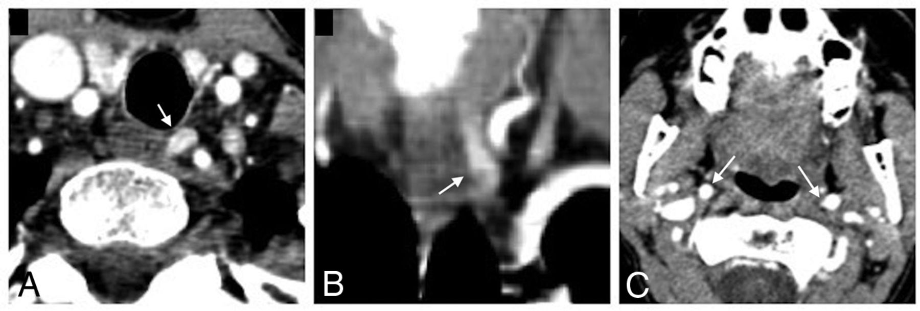
In case 1, 4D CT revealed an ectopically located right superior parathyroid adenoma in the retropharyngeal space (14 mm × 9 mm × 5 mm), posterior to the larynx at the level of the piriform sinuses, slightly right of midline (Fig 1A, -B). The adenoma was MIBI avid on SPECT/CT. The bilateral carotid arteries appeared medialized such that they nearly “kissed” immediately posterior to the oropharynx and larynx (Fig 1C, -D). The patient underwent a focused parathyroidectomy. After the right thyroid lobe and thyroid cartilage were mobilized and retracted, the bilateral internal carotid arteries were noted to be very medialized as expected. Posterior to the larynx in the retropharyngeal space, just anterior to the spine, a well-defined parathyroid adenoma was identified and resected.

FIG 1.
  • Download figure
  • Open in new tab
  • Download powerpoint
FIG 1.

Case 1. Contrast-enhanced CT scan shows axial (A) and coronal (B) images of the retropharyngeal parathyroid adenoma (arrow) and axial (C) and coronal (D) images of the nearly “kissing” carotid arteries (arrow).

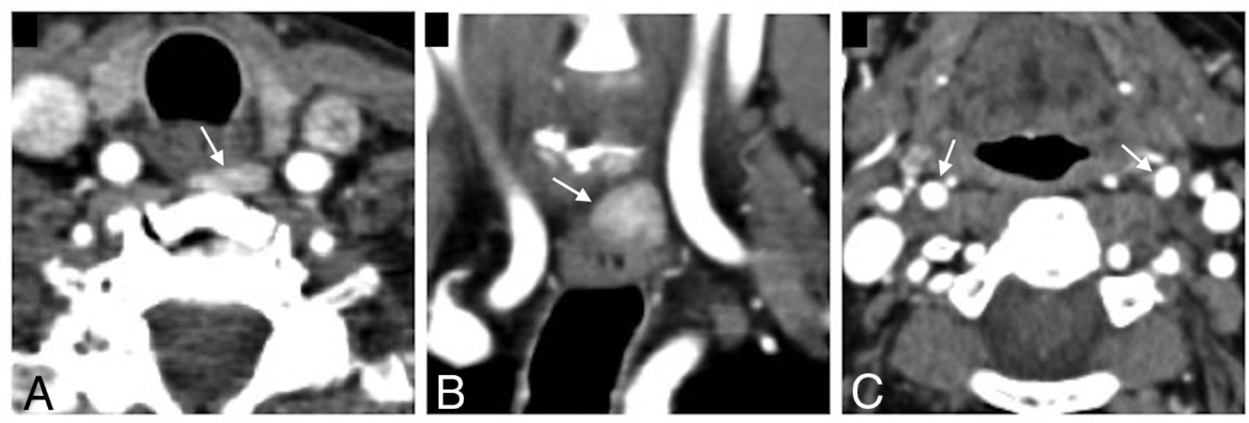
In case 2, 4D CT revealed a 6 mm × 9 mm × 20 mm enhancing paraesophageal nodule, consistent with a left upper parathyroid adenoma (Fig 2). The adenoma was MIBI avid on SPECT/CT. The adenoma was approximately 9 mm posterior to the lower pole of the left thyroid lobe and immediately anteromedial to the left carotid artery. The patient underwent a focused parathyroidectomy. The left thyroid lobe and thyroid cartilage were mobilized and retracted anteromedially. In the retropharyngeal space, posterior to the left lower pole, a well-defined parathyroid adenoma was identified, dissected, and excised.

FIG 2.
  • Download figure
  • Open in new tab
  • Download powerpoint
FIG 2.

Case 2. Contrast-enhanced CT scan shows axial (A) and coronal (B) images of the paraesophageal adenoma (arrow) and an axial (C) image of the bilateral internal carotid arteries (arrow) at the level of the oropharynx.

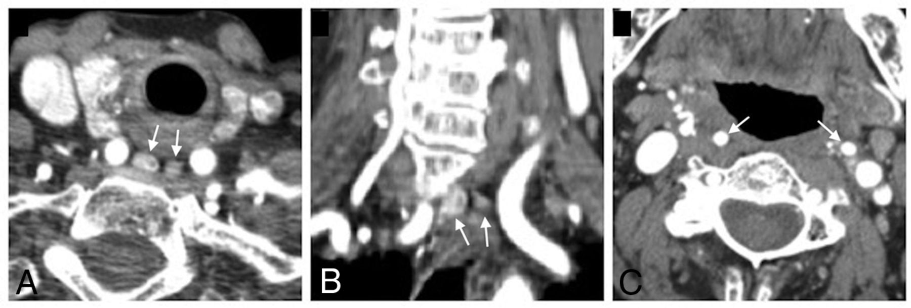
In case 3, the patient had undergone 2 neck explorations with resection of right upper and left lower adenomas 7 years and 2 years, respectively, before presenting to our institution for work-up of persistent hyperparathyroidism. An ectopic parathyroid adenoma (21 mm × 14 mm × 6 mm) was identified on 4D CT in the left retropharyngeal space immediately posterior to the esophagus and medial to the left common carotid artery (Fig 3), with corresponding increased MIBI uptake on SPECT/CT. The bilateral common carotid arteries appeared to be retropharyngeal and medialized with the adenoma in between the 2 arteries. The patient underwent a third parathyroidectomy. The left thyroid lobe was mobilized and retracted anteriorly, at which point the left common carotid artery was noted to be significantly medial, preventing easy identification of the parathyroid adenoma. After switching to a “backdoor” lateral approach (retraction of the sternohyoid and sternothyroid muscles anteriorly and more posterior dissection anterior to the sternocleidomastoid muscle), a classic parathyroid adenoma was accessed and identified in the retropharyngeal space posterior to the esophagus and medial to the left common carotid artery. The adenoma was dissected and excised.

FIG 3.
  • Download figure
  • Open in new tab
  • Download powerpoint
FIG 3.

Case 3. Contrast-enhanced CT scan shows axial (A) and coronal (B) images of the retropharyngeal parathyroid adenoma (arrow) between the common carotid arteries and an axial (C) image of the bilateral internal carotid arteries (arrow) at the level of the oropharynx.

In case 4, 4D CT revealed a left superior retroesophageal adenoma (12 × 12 × 5 mm), located immediately posterior to the left upper thyroid gland, 2 mm medial to the left common carotid artery (Fig 4). There was also an additional smaller hyperenhancing nodule adjacent to the right thyroid suggestive of a potential additional adenoma. The bilateral carotid arteries appeared to be in a relatively typical position. Because imaging suggested 2 potential adenomas, the patient underwent a bilateral neck exploration. Although imaging had suggested a left upper parathyroid adenoma, the left upper, left lower, and right lower glands all appeared to be of normal size and color in their usual positions. The retropharyngeal adenoma appeared to be an adenomatous right upper parathyroid gland, which was dissected and excised.

FIG 4.
  • Download figure
  • Open in new tab
  • Download powerpoint
FIG 4.

Case 4. Contrast-enhanced CT scan shows axial (A) and coronal (B) images of the retropharyngeal parathyroid adenoma (arrow) and an axial (C) image of the bilateral carotid arteries (arrow) at the level of the oropharynx.

In case 5, 4D CT revealed bilateral superior parathyroid adenomas (double adenomas) in the retropharyngeal space: right (8 × 7 × 14 mm) and left (5 × 4 × 8 mm) (Fig 5). Both adenomas were located immediately medial to medialized bilateral carotid arteries and posterior to the esophagus. The right superior adenoma was approximately 5 mm posterior to the right mid-thyroid gland, and the left superior adenoma was 12 mm posteromedial to the left mid-thyroid gland. On SPECT/CT, the larger right adenoma was MIBI avid, but the smaller left adenoma was not. The patient is currently taking cinacalcet (Sensipar) for medical management of PHPT and was scheduled for parathyroidectomy, but this was delayed because of the coronavirus 2019 pandemic. Of note, 6 months before her 4D CT, she underwent a left adrenalectomy for a left pheochromocytoma with elevated plasma metanephrines and normetanephrines. Given the patient’s age, both diseases were likely unrelated, sporadic, and not attributable to a genetic predisposition.

FIG 5.
  • Download figure
  • Open in new tab
  • Download powerpoint
FIG 5.

Case 5. Contrast-enhanced CT scan shows axial (A) and coronal (B) images of the double retropharyngeal parathyroid adenomas (arrow) and an axial (C) image of the bilateral carotid arteries (arrow) at the level of the oropharynx.

DISCUSSION

We have presented 5 patients with retropharyngeal parathyroid adenomas with varying anatomy of the carotid arteries. After applying the previously discussed grading system, all of these patients had at least 1 ICA that was grade II or higher, suggesting at least a moderate risk during pharyngeal procedures.5 Fortunately, all 4 patients who underwent surgery experienced no complications from intubation or the operation itself. To our knowledge, we are the first to report on a possible association between ectopic adenomas and medial deviations of the carotid arteries.

Theoretically, it is possible that these variants are related secondary to anomalous embryonic development in the neck. The parathyroid glands originate from the third (inferior glands) and fourth (superior glands) pharyngeal pouches, and the common carotid arteries and proximal ICAs derive from the third pharyngeal arch.7 Unsurprisingly, ectopic parathyroid glands are often found within structures that derive from neighboring embryologic tissues and descend into the neck, such as the thyroid, thymus, and carotid sheath.18 Other common locations for ectopic glands include the aforementioned retropharyngeal space, the tracheoesophageal groove, the thyro-thymic ligament, and elsewhere in the mediastinum.11

Clinically, this series highlights the importance of imaging for operative planning in parathyroid surgery. Advances in preoperative imaging have made it possible to localize disease and perform a focused parathyroidectomy in many patients, whereas the previous standard was a bilateral neck exploration.15 However, the utility of imaging in PHPT is dependent on the experience of the radiologist, who must be trained and experienced in parathyroid imaging to accurately interpret preoperative scans. Furthermore, when the radiologist suspects a retropharyngeal adenoma, it is especially critical to communicate to the surgeon any aberrant carotid anatomy that is at increased risk of injury when dissecting into the retropharyngeal space.

In summary, our experience with 5 patients raises the question of whether some association exists between retropharyngeal ectopic parathyroid adenomas and abnormal carotid artery anatomy, which could have implications for the radiologist and the parathyroid surgeon. Radiologists and surgeons alike should be aware of variations in carotid anatomy, and we agree with Pfeiffer et al5 on the importance of using a universal grading system to describe these variations objectively and relate them to operative risk.

Footnotes

  • Disclosures: James Lee—UNRELATED: Board Membership: Medscape, Summus; Expert Testimony: Various firms on individual basis.

References

  1. 1.↵
    1. Ozgur Z,
    2. Celik S,
    3. Govsa F, et al
    . A study of the course of the internal carotid artery in the parapharyngeal space and its clinical importance. Eur Arch Otorhinolaryngol 2007;264:1483–89 doi:10.1007/s00405-007-0398-6 pmid:17638001
    CrossRefPubMed
  2. 2.↵
    1. Togay-Isikay C,
    2. Kim J,
    3. Betterman K, et al
    . Carotid artery tortuosity, kinking, coiling: stroke risk factor, marker, or curiosity? Acta Neurol Belg 2005;105:68–72 pmid:16076059
    PubMed
  3. 3.↵
    1. Munoz A,
    2. De Vergas J,
    3. Crespo J
    . Imaging and clinical findings in patients with aberrant course of the cervical internal carotid arteries. Open Neuroimag J 2010;4:174–81 doi:10.2174/1874440001004010174 pmid:21283647
    CrossRefPubMed
  4. 4.↵
    1. Paulsen F,
    2. Tillmann B,
    3. Christofides C, et al
    . Curving and looping of the internal carotid artery in relation to the pharynx: frequency, embryology and clinical implications. J Anat 2000;197(Pt 3):373–81 doi:10.1046/j.1469-7580.2000.19730373.x pmid:11117624
    CrossRefPubMed
  5. 5.↵
    1. Pfeiffer J,
    2. Becker C,
    3. Ridder GJ
    . Aberrant extracranial internal carotid arteries: new insights, implications, and demand for a clinical grading system. Head Neck 2016;38:E687–93 doi:10.1002/hed.24071 pmid:25899075
    CrossRefPubMed
  6. 6.↵
    1. Sai Kiran NA,
    2. Kiran Kumar VA,
    3. Sivaraju L, et al
    . Management issues in a case of congenital craniovertebral junction anomaly with aberrant retropharyngeal midline course of bilateral cervical internal carotid arteries at C1-C2. World Neurosurg 2018;114:94–98 doi:10.1016/j.wneu.2018.03.012 pmid:29545218
    CrossRefPubMed
  7. 7.↵
    1. Mnatsakanian A,
    2. Minutello K,
    3. Anatomy BB
    . Head and Neck, Retropharyngeal Space. StatPearls Publishing; Updated 2020 Apr 3: https://www.ncbi.nlm.nih.gov/books/NBK537044. Accessed 2020 May 5
  8. 8.↵
    1. Wasserman JM,
    2. Sclafani SJ,
    3. Goldstein NA
    . Intraoperative evaluation of a pulsatile oropharyngeal mass during adenotonsillectomy. Int J Pediatr Otorhinolaryngol 2006;70:371–75 doi:10.1016/j.ijporl.2005.07.002 pmid:16112205
    CrossRefPubMed
  9. 9.↵
    1. Windfuhr JP,
    2. Stow NW,
    3. Landis BN
    . Beware of kissing carotids. ANZ J Surg 2010;80:668–69 doi:10.1111/j.1445-2197.2010.05421.x pmid:20840422
    CrossRefPubMed
  10. 10.↵
    1. Basekim C,
    2. Gungor A,
    3. Kizilkaye E
    . Unilateral aberrant retropharyngeal internal carotid artery. Eurorad 2005:Case 4282. https://www.eurorad.org/case/4282
  11. 11.↵
    1. Taterra D,
    2. Wong LM,
    3. Vikse J, et al
    . The prevalence and anatomy of parathyroid glands: a meta-analysis with implications for parathyroid surgery. Langenbecks Arch Surg 2019;404:63–70 doi:10.1007/s00423-019-01751-8 pmid:30762091
    CrossRefPubMed
  12. 12.↵
    1. Phitayakorn R,
    2. McHenry CR
    . Incidence and location of ectopic abnormal parathyroid glands. Am J Surg 2006;191:418–23 doi:10.1016/j.amjsurg.2005.10.049 pmid:16490559
    CrossRefPubMed
  13. 13.↵
    1. McIntyre CJ,
    2. Allen JL,
    3. Constantinides VA, et al
    . Patterns of disease in patients at a tertiary referral centre requiring reoperative parathyroidectomy. Ann R Coll Surg Engl 2015;97:598–602 doi:10.1308/rcsann.2015.0039 pmid:26444799
    CrossRefPubMed
  14. 14.↵
    1. Wilhelm SM,
    2. Wang TS,
    3. Ruan DT, et al
    . The American Association of Endocrine Surgeons Guidelines for Definitive Management of Primary Hyperparathyroidism. JAMA Surg 2016;151:959–68 doi:10.1001/jamasurg.2016.2310 pmid:27532368
    CrossRefPubMed
  15. 15.↵
    1. Bunch PM,
    2. Kelly HR
    . Preoperative imaging techniques in primary hyperparathyroidism: a review. JAMA Otolaryngol Head Neck Surg 2018;144:929–37 doi:10.1001/jamaoto.2018.1671 pmid:30193297
    CrossRefPubMed
  16. 16.↵
    1. Bilezikian JP,
    2. Brandi ML,
    3. Eastell R, et al
    . Guidelines for the management of asymptomatic primary hyperparathyroidism: summary statement from the Fourth International Workshop. J Clin Endocrinol Metab 2014;99:3561–69 doi:10.1210/jc.2014-1413 pmid:25162665
    CrossRefPubMed
  17. 17.↵
    1. Yeh R,
    2. Tay YD,
    3. Tabacco G, et al
    . Diagnostic performance of 4D CT and sestamibi SPECT/CT in localizing parathyroid adenomas in primary hyperparathyroidism. Radiology 2019;291:469–76 doi:10.1148/radiol.2019182122 pmid:30835187
    CrossRefPubMed
  18. 18.↵
    1. Okuda I,
    2. Nakajima Y,
    3. Miura D, et al
    . Diagnostic localization of ectopic parathyroid lesions: developmental consideration. Jpn J Radiol 2010;28:707–13 doi:10.1007/s11604-010-0492-3 pmid:21191734
    CrossRefPubMed
  • Received June 6, 2020.
  • Accepted after revision November 24, 2020.
  • © 2021 by American Journal of Neuroradiology
PreviousNext
Back to top
Advertisement
Print
Download PDF
Email Article

Thank you for your interest in spreading the word on American Journal of Neuroradiology.

NOTE: We only request your email address so that the person you are recommending the page to knows that you wanted them to see it, and that it is not junk mail. We do not capture any email address.

Enter multiple addresses on separate lines or separate them with commas.
Variations in the Course of the Carotid Arteries in Patients with Retropharyngeal Parathyroid Adenomas
(Your Name) has sent you a message from American Journal of Neuroradiology
(Your Name) thought you would like to see the American Journal of Neuroradiology web site.
CAPTCHA
This question is for testing whether or not you are a human visitor and to prevent automated spam submissions.
Cite this article
G.K. Steinl, R. Yeh, C.M. McManus, J.A. Lee, J.H. Kuo
Variations in the Course of the Carotid Arteries in Patients with Retropharyngeal Parathyroid Adenomas
American Journal of Neuroradiology Feb 2021, DOI: 10.3174/ajnr.A6995

Citation Manager Formats

  • BibTeX
  • Bookends
  • EasyBib
  • EndNote (tagged)
  • EndNote 8 (xml)
  • Medlars
  • Mendeley
  • Papers
  • RefWorks Tagged
  • Ref Manager
  • RIS
  • Zotero
0 Responses
Respond to this article
Share
Bookmark this article
Variations in the Course of the Carotid Arteries in Patients with Retropharyngeal Parathyroid Adenomas
G.K. Steinl, R. Yeh, C.M. McManus, J.A. Lee, J.H. Kuo
American Journal of Neuroradiology Feb 2021, DOI: 10.3174/ajnr.A6995
del.icio.us logo Twitter logo Facebook logo Mendeley logo
  • Tweet Widget
  • Facebook Like
  • Google Plus One
Purchase

Jump to section

  • Article
    • Abstract
    • ABBREVIATIONS:
    •  
    • DISCUSSION
    • Footnotes
    • References
  • Figures & Data
  • Info & Metrics
  • Responses
  • References
  • PDF

Related Articles

  • No related articles found.
  • PubMed
  • Google Scholar

Cited By...

  • Are Retropharyngeal Parathyroid Adenomas Associated with Retropharyngeal Carotid Arteries?
  • Crossref (1)
  • Google Scholar

This article has been cited by the following articles in journals that are participating in Crossref Cited-by Linking.

  • Are Retropharyngeal Parathyroid Adenomas Associated with Retropharyngeal Carotid Arteries?
    G.H. Kadam, S.G. Dua
    American Journal of Neuroradiology 2021

More in this TOC Section

Head and Neck Imaging

  • ASL Sensitivity for Head and Neck Paraganglioma
  • Post SRS Peritumoral Hyperintense Signal of VSs
  • Contrast Enhanced Pituitary CISS/FIESTA
Show more Head and Neck Imaging

EXTRACRANIAL VASCULAR

  • Proximal Vertebral Artery Variants and Embryology
  • High-Risk Plaque Features in Carotid MRI
  • Nonstenotic Carotid Plaques and Stroke Review
Show more EXTRACRANIAL VASCULAR

Similar Articles

Advertisement

Indexed Content

  • Current Issue
  • Accepted Manuscripts
  • Article Preview
  • Past Issues
  • Editorials
  • Editor's Choice
  • Fellows' Journal Club
  • Letters to the Editor
  • Video Articles

Cases

  • Case Collection
  • Archive - Case of the Week
  • Archive - Case of the Month
  • Archive - Classic Case

More from AJNR

  • Trainee Corner
  • Imaging Protocols
  • MRI Safety Corner
  • Book Reviews

Multimedia

  • AJNR Podcasts
  • AJNR Scantastics

Resources

  • Turnaround Time
  • Submit a Manuscript
  • Submit a Video Article
  • Submit an eLetter to the Editor/Response
  • Manuscript Submission Guidelines
  • Statistical Tips
  • Fast Publishing of Accepted Manuscripts
  • Graphical Abstract Preparation
  • Imaging Protocol Submission
  • Evidence-Based Medicine Level Guide
  • Publishing Checklists
  • Author Policies
  • Become a Reviewer/Academy of Reviewers
  • News and Updates

About Us

  • About AJNR
  • Editorial Board
  • Editorial Board Alumni
  • Alerts
  • Permissions
  • Not an AJNR Subscriber? Join Now
  • Advertise with Us
  • Librarian Resources
  • Feedback
  • Terms and Conditions
  • AJNR Editorial Board Alumni

American Society of Neuroradiology

  • Not an ASNR Member? Join Now

© 2025 by the American Society of Neuroradiology All rights, including for text and data mining, AI training, and similar technologies, are reserved.
Print ISSN: 0195-6108 Online ISSN: 1936-959X

Powered by HighWire