Advertisement

Main menu

  • Home
  • Content
    • Current Issue
    • Accepted Manuscripts
    • Article Preview
    • Past Issue Archive
    • Video Articles
    • AJNR Case Collection
    • Case of the Week Archive
    • Case of the Month Archive
    • Classic Case Archive
  • Special Collections
    • AJNR Awards
    • Low-Field MRI
    • Alzheimer Disease
    • ASNR Foundation Special Collection
    • Photon-Counting CT
    • View All
  • Multimedia
    • AJNR Podcasts
    • AJNR SCANtastic
    • Trainee Corner
    • MRI Safety Corner
    • Imaging Protocols
  • For Authors
    • Submit a Manuscript
    • Submit a Video Article
    • Submit an eLetter to the Editor/Response
    • Manuscript Submission Guidelines
    • Statistical Tips
    • Fast Publishing of Accepted Manuscripts
    • Graphical Abstract Preparation
    • Imaging Protocol Submission
    • Author Policies
  • About Us
    • About AJNR
    • Editorial Board
    • Editorial Board Alumni
  • More
    • Become a Reviewer/Academy of Reviewers
    • Subscribers
    • Permissions
    • Alerts
    • Feedback
    • Advertisers
    • ASNR Home

User menu

  • Alerts
  • Log in

Search

  • Advanced search
American Journal of Neuroradiology

American Journal of Neuroradiology

ASHNR American Society of Functional Neuroradiology ASHNR American Society of Pediatric Neuroradiology ASSR
  • Alerts
  • Log in

Advanced Search

  • Home
  • Content
    • Current Issue
    • Accepted Manuscripts
    • Article Preview
    • Past Issue Archive
    • Video Articles
    • AJNR Case Collection
    • Case of the Week Archive
    • Case of the Month Archive
    • Classic Case Archive
  • Special Collections
    • AJNR Awards
    • Low-Field MRI
    • Alzheimer Disease
    • ASNR Foundation Special Collection
    • Photon-Counting CT
    • View All
  • Multimedia
    • AJNR Podcasts
    • AJNR SCANtastic
    • Trainee Corner
    • MRI Safety Corner
    • Imaging Protocols
  • For Authors
    • Submit a Manuscript
    • Submit a Video Article
    • Submit an eLetter to the Editor/Response
    • Manuscript Submission Guidelines
    • Statistical Tips
    • Fast Publishing of Accepted Manuscripts
    • Graphical Abstract Preparation
    • Imaging Protocol Submission
    • Author Policies
  • About Us
    • About AJNR
    • Editorial Board
    • Editorial Board Alumni
  • More
    • Become a Reviewer/Academy of Reviewers
    • Subscribers
    • Permissions
    • Alerts
    • Feedback
    • Advertisers
    • ASNR Home
  • Follow AJNR on Twitter
  • Visit AJNR on Facebook
  • Follow AJNR on Instagram
  • Join AJNR on LinkedIn
  • RSS Feeds

AJNR Awards, New Junior Editors, and more. Read the latest AJNR updates

Research ArticleHead and Neck Imaging
Open Access

Vestibular Implant Imaging

A. Hedjoudje, D.P. Schoo, B.K. Ward, J.P. Carey, C.C. Della Santina and M. Pearl
American Journal of Neuroradiology December 2020, DOI: https://doi.org/10.3174/ajnr.A6991
A. Hedjoudje
aFrom the Department of Otolaryngology–Head and Neck Surgery (A.H., D.P.S., B.K.W., J.P.C., C.C.D.S.)
bDivision of Interventional Neuroradiology (A.H., M.P.)
dNeuroradiology Unit (A.H.), Service of diagnostic and interventional imaging, Sion Hospital, Sion, Valais, Switzerland
  • Find this author on Google Scholar
  • Find this author on PubMed
  • Search for this author on this site
  • ORCID record for A. Hedjoudje
D.P. Schoo
aFrom the Department of Otolaryngology–Head and Neck Surgery (A.H., D.P.S., B.K.W., J.P.C., C.C.D.S.)
  • Find this author on Google Scholar
  • Find this author on PubMed
  • Search for this author on this site
  • ORCID record for D.P. Schoo
B.K. Ward
aFrom the Department of Otolaryngology–Head and Neck Surgery (A.H., D.P.S., B.K.W., J.P.C., C.C.D.S.)
  • Find this author on Google Scholar
  • Find this author on PubMed
  • Search for this author on this site
  • ORCID record for B.K. Ward
J.P. Carey
aFrom the Department of Otolaryngology–Head and Neck Surgery (A.H., D.P.S., B.K.W., J.P.C., C.C.D.S.)
  • Find this author on Google Scholar
  • Find this author on PubMed
  • Search for this author on this site
  • ORCID record for J.P. Carey
C.C. Della Santina
aFrom the Department of Otolaryngology–Head and Neck Surgery (A.H., D.P.S., B.K.W., J.P.C., C.C.D.S.)
cDepartment of Biomedical Engineering (C.C.D.S.), Johns Hopkins University School of Medicine, Baltimore, Maryland
eLabyrinth Devices (C.C.D.S.), Baltimore, Maryland
  • Find this author on Google Scholar
  • Find this author on PubMed
  • Search for this author on this site
  • ORCID record for C.C. Della Santina
M. Pearl
bDivision of Interventional Neuroradiology (A.H., M.P.)
  • Find this author on Google Scholar
  • Find this author on PubMed
  • Search for this author on this site
  • ORCID record for M. Pearl
  • Article
  • Figures & Data
  • Info & Metrics
  • Responses
  • References
  • PDF
Loading

Article Figures & Data

Figures

  • Tables
  • FIG 1.
    • Download figure
    • Open in new tab
    • Download powerpoint
    FIG 1.

    A, The MVI stimulator comprises 3 fixation magnets, an inductive coil link, electrical current stimulator circuitry, a stimulation electrode array, a stimulation reference electrode, and a recording reference electrode. The electrode array includes a 3-electrode shank for the posterior canal (B, E3–E5), a forked subarray with 2 shanks for the horizontal (C, E6–E8) and anterior (C, E9–E11) canals, and a stimulation reference electrode (D). eCAP indicates electrically evoked compound action potential. Reprinted with permission from Labyrinth Devices, LLC, 2019.

  • FIG 2.
    • Download figure
    • Open in new tab
    • Download powerpoint
    FIG 2.

    Method for generating MSCT (A and B) and FPCT (C and D) MPR. Two planes are generated. The first plane is approximately tangential to the thin segments of the superior and horizontal SCCs at their junctions with their ampullae and includes the 6 electrode contacts of the forked array inserted into the superior and horizontal ampullae. The second plane is in the posterior plane of the SCC and includes the 3 electrode contacts of the linear array implanted in the posterior canal and the tip of braided platinum/iridium wire inserted into the common crus. Section thickness was set to 2 mm to include all electrode contacts on 1 image for both planes. Window width and contrast level were adjusted as needed to optimize the visibility of electrode contacts. A 3D representation of the vestibular lumen and vestibular nerve is added in transparency (E and F) to help visualize the anatomy.
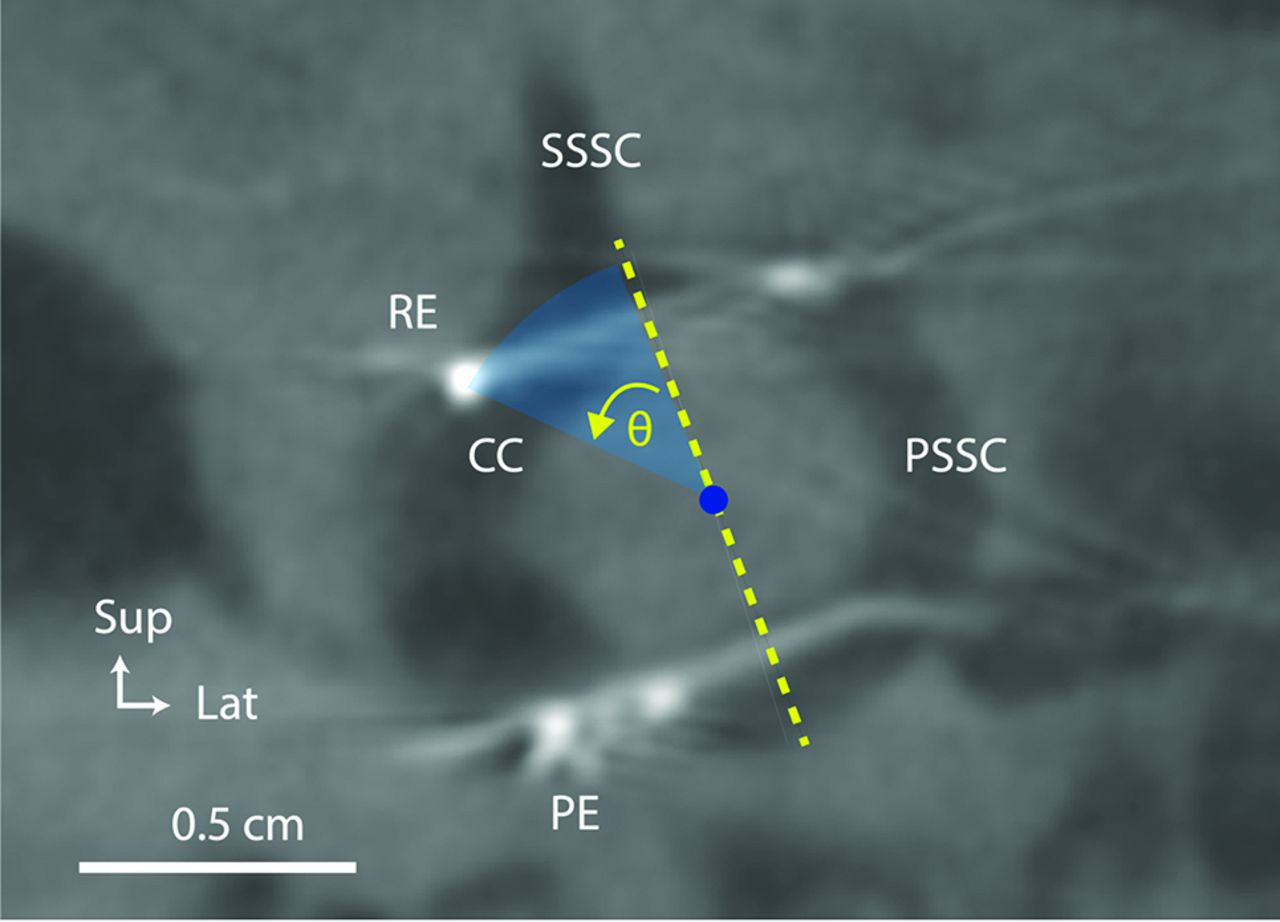
  • FIG 3.
    • Download figure
    • Open in new tab
    • Download powerpoint
    FIG 3.

    Subject: Participant 7. Method for calculating the angle (θ) of the angular insertion depth of the common crus reference electrode. CC indicates common crus of the implanted labyrinth; PE, posterior electrode array; PSCC, posterior semicircular canal; SSCC, superior semicircular canal; RE, reference electrode; Sup, superior; Lat, lateral.
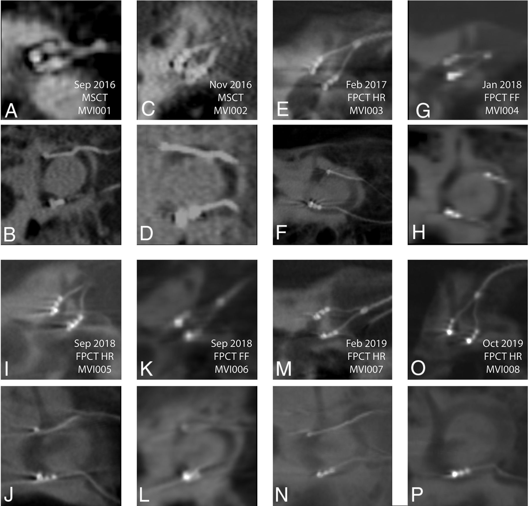
  • FIG 4.
    • Download figure
    • Open in new tab
    • Download powerpoint
    FIG 4.

    MSCT (A–D) and FPCT (E–P) multiplanar reconstructions for all participants. A and B, Participant 1 MSCT. C and D, Participant 2 MSCT. E and F, Participant 3 FPCT. G and H, Participant 4 FPCT. I and J, Participant 5 FPCT. K and L, Participant 6 FPCT, M and N, Participant 7 FPCT. O and P, Participant 8 FPCT. In every panel, the top of the image is superior and the left edge of the image is anteromedial. Sep indicates September; Feb, February; Oct, October; Nov, November; Jan, January.

  • FIG 5.
    • Download figure
    • Open in new tab
    • Download powerpoint
    FIG 5.

    A, Lateral scout view showing electrode arrays visible through the external auditory canals (yellow arrowhead) and cephalic edges of the infraorbital rims (yellow diamond). Those palpable landmarks define the Reid plane (yellow line) and the plane of horizontal canals (red line). The plane of the horizontal semicircular canals, the standard “axial” plane for temporal bone CT reconstructions, is at a ∼20° pitch from the Reid plane.17 By supporting the head on a firm wedge to pitch the head forward from supine (and flexing the neck until the Reid plane is pitched ∼20° nose toward chest from Earth vertical), one can minimize scatter artifacts from the stimulator cannister to the inner ear by keeping them on opposite sides of a separation plane perpendicular to the gantry rotation axis (B).

Tables

  • Figures
    • View popup
    Table 1:

    Demographic information for participants with vestibular implants

    ParticipantsDate ImplantedDate ImagedAge (yr),a SexImaging ProtocolImplant SideReference Location
    112 Aug 2016Sep 201662, MMSCTLeftCC
    24 Nov 2016Nov 201657, MMSCTLeftCC
    33 Feb 2017Feb 201763, FFPCT, HR modeLeftCC
    415 Dec 2017Jan 201862, FFPCT, FF modeLeftCC
    524 Aug 2018Sep 201851, FFPCT, HR modeRightCC
    631 Aug 2018Sep 201866, FFPCT, FF modeRightCC
    714 Jan 2019Feb 201953, FFPCT, HR modeLeftCC
    813 Sep 2019Oct 201955, MFPCT, HR modeRightSP
    • Note:—CC indicates common crus of the implanted labyrinth; SP, in a subperiosteal pocket outside the temporal bone; Aug, August; Jan, January; Sep, September; Dec, December; Feb, February; Nov, November.

    • ↵a Age in years at time of implantation.

    • View popup
    Table 2:

    Common crus reference electrode angular depth and insertion length calculations and locations

    Participant No.CC Insertion IntendedInsertion Depth AngleInsertion Length (mm)Anatomic Location of the Reference Electrode Tip
    1Yes–3°39Common crus
    2Yes+26°66Superior canal
    3Yes–30°42Posterior canal
    4Yes–57°21Posterior canal
    5Yes+21°46Common crus
    6Yes–2°41Posterior canal
    7Yes+4551Common crus
    8NoNANAOutside temporal bone
    • Note:—NA indicates not appliable.

PreviousNext
Back to top
Advertisement
Print
Download PDF
Email Article

Thank you for your interest in spreading the word on American Journal of Neuroradiology.

NOTE: We only request your email address so that the person you are recommending the page to knows that you wanted them to see it, and that it is not junk mail. We do not capture any email address.

Enter multiple addresses on separate lines or separate them with commas.
Vestibular Implant Imaging
(Your Name) has sent you a message from American Journal of Neuroradiology
(Your Name) thought you would like to see the American Journal of Neuroradiology web site.
CAPTCHA
This question is for testing whether or not you are a human visitor and to prevent automated spam submissions.
Cite this article
A. Hedjoudje, D.P. Schoo, B.K. Ward, J.P. Carey, C.C. Della Santina, M. Pearl
Vestibular Implant Imaging
American Journal of Neuroradiology Dec 2020, DOI: 10.3174/ajnr.A6991

Citation Manager Formats

  • BibTeX
  • Bookends
  • EasyBib
  • EndNote (tagged)
  • EndNote 8 (xml)
  • Medlars
  • Mendeley
  • Papers
  • RefWorks Tagged
  • Ref Manager
  • RIS
  • Zotero
0 Responses
Respond to this article
Share
Bookmark this article
Vestibular Implant Imaging
A. Hedjoudje, D.P. Schoo, B.K. Ward, J.P. Carey, C.C. Della Santina, M. Pearl
American Journal of Neuroradiology Dec 2020, DOI: 10.3174/ajnr.A6991
del.icio.us logo Twitter logo Facebook logo Mendeley logo
  • Tweet Widget
  • Facebook Like
  • Google Plus One
Purchase

Jump to section

  • Article
    • Abstract
    • ABBREVIATIONS:
    • MATERIALS AND METHODS
    • RESULTS
    • DISCUSSION
    • CONCLUSIONS
    • Acknowledgments
    • Footnotes
    • References
  • Figures & Data
  • Info & Metrics
  • Responses
  • References
  • PDF

Related Articles

  • No related articles found.
  • PubMed
  • Google Scholar

Cited By...

  • No citing articles found.
  • Crossref (5)
  • Google Scholar

This article has been cited by the following articles in journals that are participating in Crossref Cited-by Linking.

  • A prosthesis utilizing natural vestibular encoding strategies improves sensorimotor performance in monkeys
    Kantapon Pum Wiboonsaksakul, Dale C. Roberts, Charles C. Della Santina, Kathleen E. Cullen, Peter Thier
    PLOS Biology 2022 20 9
  • New Frontiers in Managing the Dizzy Patient
    Desi P. Schoo, Bryan K. Ward
    Otolaryngologic Clinics of North America 2021 54 5
  • Prosthetic Stimulation of the Vestibular Nerve Can Evoke Robust Eye and Head Movements Despite Prior Labyrinthectomy
    Kantapon Pum Wiboonsaksakul, Charles C. Della Santina, Kathleen E. Cullen
    Otology & Neurotology 2023 44 10
  • Vestibular Implant Surgery
    Desi P. Schoo, Bryan K. Ward, Margaret R. Chow, Andrianna I. Ayiotis, Celia Fernández Brillet, Peter J. Boutros, Kelly E. Lane, Claudia N. Lee, Brian J. Morris, John P. Carey, Charles C. Della Santina
    The Laryngoscope 2024 134 4
  • Surdités de perception de l’enfant
    Natalie Loundon
    Contraste 2023 N° 58 2

More in this TOC Section

  • ASL Sensitivity for Head and Neck Paraganglioma
  • Post SRS Peritumoral Hyperintense Signal of VSs
  • Contrast Enhanced Pituitary CISS/FIESTA
Show more Head and Neck Imaging

Similar Articles

Advertisement

Indexed Content

  • Current Issue
  • Accepted Manuscripts
  • Article Preview
  • Past Issues
  • Editorials
  • Editor's Choice
  • Fellows' Journal Club
  • Letters to the Editor
  • Video Articles

Cases

  • Case Collection
  • Archive - Case of the Week
  • Archive - Case of the Month
  • Archive - Classic Case

More from AJNR

  • Trainee Corner
  • Imaging Protocols
  • MRI Safety Corner
  • Book Reviews

Multimedia

  • AJNR Podcasts
  • AJNR Scantastics

Resources

  • Turnaround Time
  • Submit a Manuscript
  • Submit a Video Article
  • Submit an eLetter to the Editor/Response
  • Manuscript Submission Guidelines
  • Statistical Tips
  • Fast Publishing of Accepted Manuscripts
  • Graphical Abstract Preparation
  • Imaging Protocol Submission
  • Evidence-Based Medicine Level Guide
  • Publishing Checklists
  • Author Policies
  • Become a Reviewer/Academy of Reviewers
  • News and Updates

About Us

  • About AJNR
  • Editorial Board
  • Editorial Board Alumni
  • Alerts
  • Permissions
  • Not an AJNR Subscriber? Join Now
  • Advertise with Us
  • Librarian Resources
  • Feedback
  • Terms and Conditions
  • AJNR Editorial Board Alumni

American Society of Neuroradiology

  • Not an ASNR Member? Join Now

© 2025 by the American Society of Neuroradiology All rights, including for text and data mining, AI training, and similar technologies, are reserved.
Print ISSN: 0195-6108 Online ISSN: 1936-959X

Powered by HighWire