Advertisement

Main menu

  • Home
  • Content
    • Current Issue
    • Accepted Manuscripts
    • Article Preview
    • Past Issue Archive
    • Video Articles
    • AJNR Case Collection
    • Case of the Week Archive
    • Case of the Month Archive
    • Classic Case Archive
  • Special Collections
    • AJNR Awards
    • Low-Field MRI
    • Alzheimer Disease
    • ASNR Foundation Special Collection
    • Photon-Counting CT
    • View All
  • Multimedia
    • AJNR Podcasts
    • AJNR SCANtastic
    • Trainee Corner
    • MRI Safety Corner
    • Imaging Protocols
  • For Authors
    • Submit a Manuscript
    • Submit a Video Article
    • Submit an eLetter to the Editor/Response
    • Manuscript Submission Guidelines
    • Statistical Tips
    • Fast Publishing of Accepted Manuscripts
    • Graphical Abstract Preparation
    • Imaging Protocol Submission
    • Author Policies
  • About Us
    • About AJNR
    • Editorial Board
    • Editorial Board Alumni
  • More
    • Become a Reviewer/Academy of Reviewers
    • Subscribers
    • Permissions
    • Alerts
    • Feedback
    • Advertisers
    • ASNR Home

User menu

  • Alerts
  • Log in

Search

  • Advanced search
American Journal of Neuroradiology

American Journal of Neuroradiology

ASHNR American Society of Functional Neuroradiology ASHNR American Society of Pediatric Neuroradiology ASSR
  • Alerts
  • Log in

Advanced Search

  • Home
  • Content
    • Current Issue
    • Accepted Manuscripts
    • Article Preview
    • Past Issue Archive
    • Video Articles
    • AJNR Case Collection
    • Case of the Week Archive
    • Case of the Month Archive
    • Classic Case Archive
  • Special Collections
    • AJNR Awards
    • Low-Field MRI
    • Alzheimer Disease
    • ASNR Foundation Special Collection
    • Photon-Counting CT
    • View All
  • Multimedia
    • AJNR Podcasts
    • AJNR SCANtastic
    • Trainee Corner
    • MRI Safety Corner
    • Imaging Protocols
  • For Authors
    • Submit a Manuscript
    • Submit a Video Article
    • Submit an eLetter to the Editor/Response
    • Manuscript Submission Guidelines
    • Statistical Tips
    • Fast Publishing of Accepted Manuscripts
    • Graphical Abstract Preparation
    • Imaging Protocol Submission
    • Author Policies
  • About Us
    • About AJNR
    • Editorial Board
    • Editorial Board Alumni
  • More
    • Become a Reviewer/Academy of Reviewers
    • Subscribers
    • Permissions
    • Alerts
    • Feedback
    • Advertisers
    • ASNR Home
  • Follow AJNR on Twitter
  • Visit AJNR on Facebook
  • Follow AJNR on Instagram
  • Join AJNR on LinkedIn
  • RSS Feeds

AJNR Awards, New Junior Editors, and more. Read the latest AJNR updates

Research ArticleHead and Neck Imaging
Open Access

Anosmia in COVID-19 Associated with Injury to the Olfactory Bulbs Evident on MRI

M.F.V.V. Aragão, M.C. Leal, O.Q. Cartaxo Filho, T.M. Fonseca and M.M. Valença
American Journal of Neuroradiology June 2020, DOI: https://doi.org/10.3174/ajnr.A6675
M.F.V.V. Aragão
aFrom the Universidade Federal de Pernambuco (M.F.V.V.A., M.C.L., M.M.V.), Recife, Brazil
bCentro Diagnostico Multimagem (M.F.V.V.A.), Recife, Brazil
  • Find this author on Google Scholar
  • Find this author on PubMed
  • Search for this author on this site
  • ORCID record for M.F.V.V. Aragão
M.C. Leal
aFrom the Universidade Federal de Pernambuco (M.F.V.V.A., M.C.L., M.M.V.), Recife, Brazil
cReal Hospital de Beneficencia Portuguesa (M.C.L., O.Q.C.F., T.M.F.), Recife, Brazil.
  • Find this author on Google Scholar
  • Find this author on PubMed
  • Search for this author on this site
  • ORCID record for M.C. Leal
O.Q. Cartaxo Filho
cReal Hospital de Beneficencia Portuguesa (M.C.L., O.Q.C.F., T.M.F.), Recife, Brazil.
  • Find this author on Google Scholar
  • Find this author on PubMed
  • Search for this author on this site
  • ORCID record for O.Q. Cartaxo Filho
T.M. Fonseca
cReal Hospital de Beneficencia Portuguesa (M.C.L., O.Q.C.F., T.M.F.), Recife, Brazil.
  • Find this author on Google Scholar
  • Find this author on PubMed
  • Search for this author on this site
  • ORCID record for T.M. Fonseca
M.M. Valença
aFrom the Universidade Federal de Pernambuco (M.F.V.V.A., M.C.L., M.M.V.), Recife, Brazil
  • Find this author on Google Scholar
  • Find this author on PubMed
  • Search for this author on this site
  • ORCID record for M.M. Valença
  • Article
  • Figures & Data
  • Supplemental
  • Info & Metrics
  • Responses
  • References
  • PDF
Loading

Abstract

SUMMARY: Patients with coronavirus disease 2019 (COVID-19) may have symptoms of anosmia or partial loss of the sense of smell, often accompanied by changes in taste. We report 5 cases (3 with anosmia) of adult patients with COVID-19 in whom injury to the olfactory bulbs was interpreted as microbleeding or abnormal enhancement on MR imaging. The patients had persistent headache (n = 4) or motor deficits (n = 1). This olfactory bulb injury may be the mechanism by which the Severe Acute Respiratory Syndrome coronavirus 2 causes olfactory dysfunction.

ABBREVIATIONS:

COVID-19
coronavirus disease 2019
SARS-CoV-2
Severe Acute Respiratory Syndrome coronavirus 2

Coronavirus has the human respiratory system as its main target but also has neuroinvasive capabilities and can spread from the respiratory tract to the CNS.1-3 Therefore, patients with coronavirus disease 19 (COVID-19) may present with neurologic symptomatology with repercussions on imaging examinations,4⇓⇓⇓⇓⇓⇓⇓⇓⇓⇓⇓⇓⇓-18 and these have been described in association with ischemic infarct,8,9 hemorrhage,11 acute hemorrhagic necrotizing encephalopathy,10 cerebral venous thrombosis,13 and diffuse leukoencephalopathy with microhemorrhage.15

Transmission from person to person occurs mainly by direct contact or droplets spread by coughing or sneezing by an infected individual with Severe Acute Respiratory Syndrome coronavirus 2 (SARS-CoV-2).5,19 Symptoms of COVID-19 usually appear after an incubation period of about 5 days. The most common symptoms are fever, cough, fatigue, headache, and dyspnea.5,19,20 In the most severe cases, patients may develop pneumonia, acute respiratory failure, distress syndrome, and acute heart problems.5,19,20

Anosmia or partial loss of the sense of smell, usually accompanied by changes in taste, is a frequent symptom that helps in the diagnosis of COVID-19.21-28 It is often a transitory phenomenon, lasting just a few weeks.21 However, the mechanism by which anosmia occurs has not yet been established.29

The hypothesis is that the virus enters the central nervous system through the first neurons of the olfactory pathway, also called olfactory sensory neurons, located in the olfactory mucosa. The olfactory mucosa is a specialized neuroepithelium located in the highest portion of the nasal cavity in direct contact with the external environment below to the cribriform plate.1 Therefore, the virus crosses the cribriform plate to reach the olfactory bulbs, which contain the second olfactory neurons.1,30

There are currently only 2 reports evaluating olfactory bulb imaging, and they are discordant.18,31 The first report showed bilateral inflammatory obstruction of the olfactory clefts that was confirmed on MR imaging of the nasal cavity, but no anomalies of the olfactory bulbs and tracts.31 The second study reported a case with anosmia evaluated with 3D-CISS T2WI, which demonstrated severe enlargement and an abnormal high signal intensity on T2, being interpreted as bilateral olfactory bulb edema and also olfactory cleft mild edema.18 The control MR imaging (D24) showed a reduction in the volume of the bulbs.18

To our knowledge, no other report has evaluated the characteristics of the olfactory bulb, especially using fat-suppressed T1WI. Also, no report evaluates and shows the presence of bleeding or a break in the blood-brain barrier in the olfactory bulbs and tracts as the possible pathophysiology of olfactory neuropathy associated with COVID-19.

Thus, in this study, the authors demonstrate by MR imaging that a possible mechanism by which the SARS-CoV-2 causes olfactory dysfunction is by affecting, intracranially, the olfactory bulbs by a likely microvascular phenomenon.

MATERIALS AND METHODS

This retrospective study was approved by the institutional review board of the ethics committee of Universidade Federal de Pernambuco, Brazil. Informed consent was waived.

All scans were initially analyzed by the institution’s own neuroradiologists. Subsequently, all images were reviewed independently by 2 neuroradiologists (M.F.V.V.A. and O.Q.C.F, who were certified by the Ministry of Education and Culture of Brazil and the Brazilian College of Radiology) with 30 and 18 years, respectively, of neuroradiology experience, with no discordant results. MR imaging was indicated mainly because of a persistent incapacitating headache.

The intensity of the olfactory bulbs is defined as normal when the bulbs have the same cortical intensity, as typically seen in healthy controls. Abnormal olfactory bulb intensity is defined when the bulb is more hyperintense than the cortex on T1WI and STIR.

After gadolinium injection on T1WI, enhancement of the olfactory bulbs is defined when they become more hyperintense in comparison with their intensity on pregadolinium T1WI. However, when there is only the postgadolinium T1WI and the bulb is more hyperintense than the normal cortex, these features represent olfactory bulb intensity abnormality and may be an enhancement or microbleeding (methemoglobin), as interpreted in the present study. Microbleeding (methemoglobin) in the olfactory bulb is considered when there is a hyperintense olfactory bulb, compared with the normal cortex or the normal contralateral bulb, on pregadolinium fat-suppressed T1WI.

Brain MR imaging of patients with COVID-19 was evaluated from April 1, 2020, to May 18, 2020. Five patients were included in this study because their brain MRIs assessed their olfactory bulbs appropriately, with at least 2 sequences with thin slices examining the anterior cranial fossa. All 5 patients were evaluated with 2 coronal sequences with thin slices: postcontrast fat-suppressed T1WI and STIR. Only 1 patient also had thin-slice pregadolinium fat-suppressed T1WI.

The brain MRIs of the patients were performed on two 1.5T machines with the main technical parameters of the sequences described as follows. The coronal fat-suppressed T1WI (spectral presaturation with inversion recovery) parameters were, respectively, on both MR imaging machines the following: TR/TE = 561–605/15–9 ms, matrix = 256–88, FOV = 190–150 mm, thickness = 3 mm, 3.5-mm section, coronal orientation, bandwidth = 181–96.6 Hz, time = 3.39–4.49 minutes, NEX = 1–3.

The coronal STIR sequence had the following parameters in each MR imaging machine, respectively: TR/TE = 4000–2650/51–90 ms, TI = 180 ms, matrix = 256–224, FOV = 190–150 mm, thickness = 3–3.45 mm, section orientation = coronal, bandwidth = 190–232.4 Hz, time = 2.46–3.42 minutes, and NEX = 1–2.

RESULTS

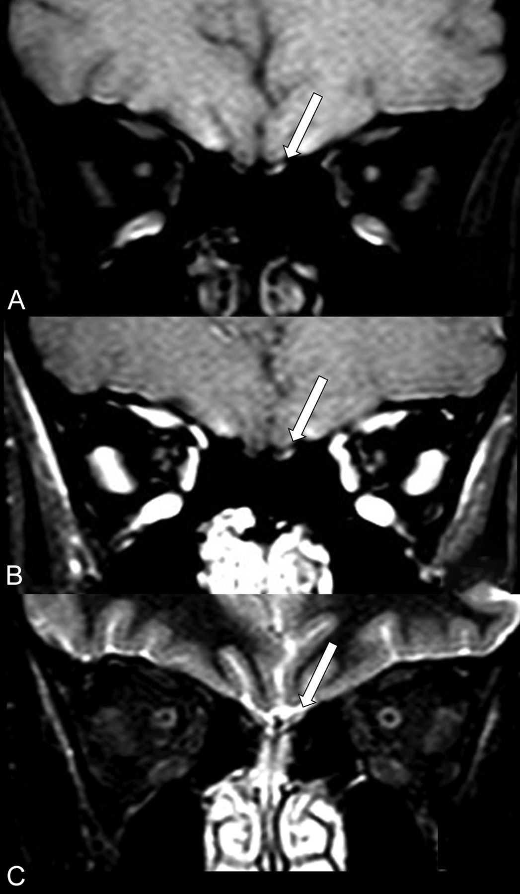
All 5 patients with COVID-19 (On-line Table) had fever, headache, and cough. The medical indications for the performance of MR imaging were persistent headache (n = 4) or motor deficit (n = 1). All 5 patients had injury to the olfactory bulbs demonstrated by MR imaging with the following sequences: coronal pre-contrast (Fig 1A) and post-contrast fat suppression T1WI (Figs 1B and 2A, -D) and coronal STIR (Fig 1C).

FIG 1.
  • Download figure
  • Open in new tab
  • Download powerpoint
FIG 1.

MR imaging shows probably microbleeding (methemoglobin) in the left olfactory bulb of a patient (case 1) with COVID-19 and anosmia. The left olfactory bulb (long arrows) has partial hyperintensity on precontrast fat-suppressed T1WI (A) and also on postcontrast fat-suppressed T1WI (B) and STIR (C).

FIG 2.
  • Download figure
  • Open in new tab
  • Download powerpoint
FIG 2.

The coronal postcontrast fat-suppressed T1WI shows hyperintensity suggestive of enhancement or methemoglobin in the olfactory bulbs of 4 patients with COVID-19 (A–D; cases 2–5) compared with a healthy patient with normal olfactory bulbs (E and F). The coronal postcontrast fat-suppressed T1WI in 3 patients with COVID-19 (A–C; cases 2–4) shows that both olfactory bulbs (long arrows) are small oval images that are hyperintense with contrast, having signal intensity higher than the intensity of the cortex. D, A patient (case 5) with COVID-19 shows hyperintensity only on the left bulb (long arrow), the right olfactory bulb being normal (short arrow). In a healthy 60-year-old man, the coronal T2WI (E) and the postcontrast fat-suppressed T1WI (F) demonstrate normal olfactory bulbs (long arrows), which are isointense to the cortex and normally hypointense on postgadolinium sequence (F).

The only patient who had pregadolinium fat-suppressed T1WI (case 1) showed a small hyperintensity in the left olfactory bulb (Fig 1A), which remained hyperintense on the postgadolinium sequence (Fig 1B) and also on STIR (Fig 1C). This finding was suggestive of a small area of methemoglobin in the left olfactory bulb in this patient with anosmia.

In the 4 patients who did not have a pregadolinium sequence, we did not have information about anosmia in 1 patient (Fig 2A; case 2), and 2 of them with anosmia (Fig 2B, -C; Cases 3 and 4) showed hyperintensity suggestive of enhancement of both olfactory bulbs following gadolinium injection. However, only in the patient with COVID-19 without clinical anosmia (case 5) was there a suggestive enhancement in the left olfactory bulb (Fig 2D). The case 5 had a negative result of the CSF real-time RT-PCR for SARS-CoV-2. The differential diagnosis in these cases is mainly microbleeding (methemoglobin) because the pregadolinium sequence was not performed. Coronal STIR of the anterior cranial fossa did not show any abnormality in the olfactory bulbs in these 4 patients.

MR imaging of a healthy individual was used as a comparative control (Fig 2E, -F) to demonstrate that the normal olfactory bulbs do not enhance and are isointense to the cerebral cortex.

DISCUSSION

This case series demonstrates abnormal intensity of the olfactory bulbs in 5 adult patients with COVID-19, three of whom had anosmia. In 1 patient (case 1), the abnormal intensity could represent microbleeding (methemoglobin). However, in the other 4 patients, it could represent abnormal enhancement or microbleeding (methemoglobin) because they only underwent the sequence after injection of gadolinium in fat-suppressed T1WI.

Previously, it was demonstrated, using an experimental mouse model, that the SARS-CoV could travel from the nose to the olfactory bulb.32 Regarding the SAR-CoV infection, there was a time delay of about 60 hours from the time of nasal infection until the detection of the virus in the olfactory bulb.1,32

The literature has already reported that some other viruses can use the olfactory nerve as a shortcut into the CNS, such as influenza A virus, herpesviruses, poliovirus, paramyxoviruses, vesicular stomatitis virus, rabies virus, parainfluenza virus, adenoviruses, Japanese encephalitis virus, West Nile virus, chikungunya virus, La Crosse virus, mouse hepatitis virus, and bunya viruses.30 van Riel et al30 have reported that “Viral infection of the CNS can lead to damage from infection of nerve cells per se, from the immune response, or from a combination of both. Clinical consequences range from nervous dysfunction in the absence of histopathological changes to severe meningoencephalitis and neurodegenerative disease.” However, to our knowledge, no study evaluated and documented, by brain MR imaging, the abnormalities such as microbleeding and/or enhancement in the olfactory bulbs occurring in these other kinds of viruses.

Probably, the impairment of olfactory function is much more frequent in COVID-19 because, strictly speaking, unilateral anosmia can only be detected through a detailed physical examination. The patient hardly perceives unilateral anosmia.

Recognizing this hypersignal in the olfactory bulbs on the thin slices of pre- and/or postgadolinium fat-suppressed T1WI, identified in this study, may help to suggest or support the etiologic diagnosis of COVID-19 during and after this new pandemic.

Thus, we suggest, henceforth, including in the routine brain MR imaging protocol at least a sequence with coronal thin-slice pre- and/or postgadolinium fat-suppressed T1WI in the anterior fossa of the cranium. This feature will be more important in cases of refractory headache associated or not with other symptoms and signs such as fever and anosmia.

The weakness of this work is that it is a retrospective study with only a few cases in which it was possible to evaluate the olfactory bulbs. Brain MR imaging of patients with COVID-19 has not been routinely scheduled to adequately evaluate the olfactory bulbs because other neurologic complications were being investigated. The distortion at the air-tissue interface in fat-suppressed T1WI makes the findings somewhat difficult to interpret, but it seems that the images are true abnormal lesions along the olfactory bulbs. Future prospective studies geared to evaluating the olfactory bulbs with a larger sample size will be needed to confirm our findings.

In conclusion, the authors demonstrated by MR imaging that a possible mechanism by which the SARS-CoV-2 causes olfactory dysfunction is by affecting, intracranially, the olfactory bulbs, with development of a microvascular phenomenon and injury such as microbleeding and/or a blood-brain barrier break. This seems to be the first time that a neuroimaging study has documented this type of olfactory bulb injury in patients with COVID-19.

Indicates open access to non-subscribers at www.ajnr.org

References

  1. 1.↵
    1. Fodoulian L,
    2. Tuberosa J,
    3. Rossier D, et al
    . SARS-CoV-2 receptor and entry genes are expressed by sustentacular cells in the human olfactory neuroepithelium. bioRxiv 2020. https://www.biorxiv.org/content/10.1101/2020.03.31.013268v1. Accessed May 12, 2020
  2. 2.
    1. Desforges M,
    2. Le Coupanec A,
    3. Dubeau P, et al
    . Human coronaviruses and other respiratory viruses: underestimated opportunistic pathogens of the central nervous system? Viruses 2019;12:14 doi:10.3390/v12010014 pmid:31861926
    CrossRefPubMed
  3. 3.↵
    1. Morris M,
    2. Zohrabian VM
    . Neuroradiologists, be mindful of the neuroinvasive potential of COVID-19. AJNR Am J Neuroradiol 2020 Apr 30. [Epub ahead of print] doi:10.3174/ajnr.A6551 pmid:32354715
    FREE Full Text
  4. 4.↵
    1. Montalvan V,
    2. Lee J,
    3. Bueso T, et al
    . Neurological manifestations of COVID-19 and other coronavirus infections: a systematic review. Clin Neurol Neurosurg 2020;194:10592 doi:10.1016/j.clineuro.2020.105921 pmid:32422545
    CrossRefPubMed
  5. 5.↵
    1. Whittaker A,
    2. Anson M,
    3. Harky A
    . Neurological Manifestations of COVID-19: A review. Acta Neurol Scand 2020;142:14–22 doi:10.1111/ane.13266 pmid:32412088
    CrossRefPubMed
  6. 6.↵
    1. Ahmad I,
    2. Rathore FA
    . Neurological manifestations and complications of COVID-19: a literature review. J Clin Neurosci 2020 May 6. [Epub ahead of print] doi:10.1016/j.jocn.2020.05.017 pmid:32409215
    CrossRefPubMed
  7. 7.↵
    1. Asadi-Pooya AA,
    2. Simani L
    . Central nervous system manifestations of COVID-19: a systematic review. J Neurol Sci 2020;413:116832 doi:10.1016/j.jns.2020.116832 pmid:32299017
    CrossRefPubMed
  8. 8.↵
    1. Goldberg MF,
    2. Cerejo R,
    3. Tayal AH
    . Cerebrovascular disease in COVID-19. AJNR Am J Neuroradiol 2020 May 14. [Epub ahead of print] doi:10.3174/ajnr.A6588 pmid:32409316
    CrossRefPubMed
  9. 9.↵
    1. Aggarwal G,
    2. Lippi G,
    3. Michael Henry B
    . Cerebrovascular disease is associated with an increased disease severity in patients with coronavirus disease 2019 (COVID-19): a pooled analysis of published literature. Int J Stroke 2020;15:385–89 doi:10.1177/1747493020921664 pmid:32310015
    CrossRefPubMed
  10. 10.↵
    1. Poyiadji N,
    2. Shahin G,
    3. Noujaim D, et al
    . COVID-19-associated acute hemorrhagic necrotizing encephalopathy: CT and MRI features. Radiology 2020 March 31. [Epub ahead of print] doi:10.1148/radiol.2020201187 pmid:32228363
    CrossRefPubMed
  11. 11.↵
    1. Franceschi AM,
    2. Ahmed O,
    3. Giliberto L, et al
    . Hemorrhagic posterior reversible encephalopathy syndrome as a manifestation of COVID-19 infection. AJNR Am J Neuroradiol 2020 May 21. [Epub ahead of print] doi:10.3174/ajnr.A6595 pmid:32439646
    CrossRefPubMed
  12. 12.↵
    1. Poillon G,
    2. Obadia M,
    3. Perrin M, et al
    . Cerebral venous thrombosis associated with COVID-19 infection: causality or coincidence? J Neuroradiol 2020 May 10. [Epub ahead of print] doi:10.1016/j.neurad.2020.05.003 pmid:32437707
    CrossRefPubMed
  13. 13.↵
    1. Garaci F,
    2. Di Giuliano F,
    3. Picchi E, et al
    . Venous cerebral thrombosis in COVID-19 patient. J Neurol Sci 2020;414:116871 doi:10.1016/j.jns.2020.116871 pmid:32422428
    CrossRefPubMed
  14. 14.↵
    1. Hughes C,
    2. Nichols T,
    3. Pike M, et al
    . Cerebral venous sinus thrombosis as a presentation of COVID-19. Eur J Case Rep Intern Med 2020;7:001691 doi:10.12890/2020_001691 pmid:32399457
    CrossRefPubMed
  15. 15.↵
    1. Radmanesh A,
    2. Derman A,
    3. Lui YW, et al
    . COVID-19-associated diffuse leukoencephalopathy and microhemorrhages. Radiology 2020 May 21. [Epub ahead of print] doi:10.1148/radiol.2020202040 pmid:32437314
    CrossRefPubMed
  16. 16.↵
    1. Mahammedi A,
    2. Saba L,
    3. Vagal A, et al
    . Imaging in neurological disease of hospitalized COVID-19 patients: an Italian multicenter retrospective observational study. Radiology 2020 May 21. [Epub ahead of print] doi:10.1148/radiol.2020201933 pmid:32437313
    CrossRefPubMed
  17. 17.↵
    1. Karimi-Galougahi M,
    2. Yousefi-Koma A,
    3. Bakhshayeshkaram M, et al
    . 18FDG PET/CT scan reveals hypoactive orbitofrontal cortex in anosmia of COVID-19. Acad Radiol 2020 May 3. [Epub ahead of print] doi:10.1016/j.acra.2020.04.030 pmid:32386948
    CrossRefPubMed
  18. 18.↵
    1. Laurendon T,
    2. Radulesco T,
    3. Mugnier J, et al
    . Bilateral transient olfactory bulbs edema during COVID-19-related anosmia. Neurology 2020 May 22. [Epub ahead of print] doi:10.1212/WNL.0000000000009850 pmid:32444492
    CrossRefPubMed
  19. 19.↵
    1. Weiss P,
    2. Murdoch DR
    . Clinical course and mortality risk of severe COVID-19. Lancet 2020;395:1014–15 doi:10.1016/S0140-6736(20)30633-4 pmid:32197108
    CrossRefPubMed
  20. 20.↵
    1. Zhou F,
    2. Yu T,
    3. Du R, et al
    . Clinical course and risk factors for mortality of adult inpatients with COVID-19 in Wuhan, China: a retrospective cohort study. Lancet 2020;395:1054–62 doi:10.1016/S0140-6736(20)30566-3 pmid:32192581
    CrossRefPubMed
  21. 21.↵
    1. Hopkins C,
    2. Surda P,
    3. Whitehead E, et al
    . Early recovery following new onset anosmia during the COVID-19 pandemic: an observational cohort study. J Otolaryngol Head Neck Surg 2020;49:26 doi:10.1186/s40463-020-00423-8 pmid:32366299
    CrossRefPubMed
  22. 22.
    1. Kaye R,
    2. Chang CW,
    3. Kazahaya K, et al
    . Anosmia reporting tool: initial findings. Otolaryngol Head Neck Surg 2020 Apr 28. [Epub ahead of print] doi:10.1177/0194599820922992 pmid:32340555
    CrossRefPubMed
  23. 23.
    1. Gane SB,
    2. Kelly C,
    3. Hopkins C
    . Isolated sudden onset anosmia in COVID-19 infection: a novel syndrome? Rhinology 2020;58:299–301 doi:10.4193/Rhin20.114] pmid:32240279
    CrossRefPubMed
  24. 24.
    1. Ghiasvand F,
    2. SeyedAlinaghi S
    . Isolated anosmia as a presentation of COVID-19: an experience in a referral hospital. Infect Disord Drug Targets 2020 May 20. [Epub ahead of print] doi:10.2174/1871526520999200520173216 pmid:32436835
    CrossRefPubMed
  25. 25.
    1. Heidari F,
    2. Karimi E,
    3. Firouzifar M, et al
    . Anosmia as a prominent symptom of COVID-19 infection. Rhinology 2020;58:302–03 doi:10.4193/Rhin20.140 pmid:32319971
    CrossRefPubMed
  26. 26.
    1. Lechien JR,
    2. Barillari MR,
    3. Jouffe L, et al
    . Anosmia Is a key symptom of COVID-19 infection and should be used as a diagnostic tool. Ear Nose Throat J 2020 May 21. [Epub ahead of print] doi:10.1177/0145561320925191 pmid:32436441
    CrossRefPubMed
  27. 27.
    1. Villalba NL,
    2. Maouche Y,
    3. Ortiz MB, et al
    . Anosmia and dysgeusia in the absence of other respiratory diseases: should COVID-19 infection be considered? Eur J Case Rep Intern Med 2020;7:001641 doi:10.12890/2020_001641 pmid:32309267
    CrossRefPubMed
  28. 28.↵
    1. Marinosci A,
    2. Landis BN,
    3. Calmy A
    . Possible link between anosmia and COVID-19: sniffing out the truth. Eur Arch Otorhinolaryngol 2020 Apr 17. [Epub ahead of print] doi:10.1007/s00405-020-05966-0 pmid:32303881
    CrossRefPubMed
  29. 29.↵
    1. Vaira LA,
    2. Salzano G,
    3. Fois AG, et al
    . Potential pathogenesis of ageusia and anosmia in COVID-19 patients. Int Forum Allergy Rhinol 2020 Apr 27. [Epub ahead of print] doi:10.1002/alr.22593 pmid:32342636
    CrossRefPubMed
  30. 30.↵
    1. van Riel D,
    2. Verdijk R,
    3. Kuiken T
    . The olfactory nerve: a shortcut for influenza and other viral diseases into the central nervous system. J Pathol 2015;235:277–87 doi:10.1002/path.4461 pmid:25294743
    CrossRefPubMed
  31. 31.↵
    1. Eliezer M,
    2. Hautefort C,
    3. Hamel AL, et al
    . Sudden and complete olfactory loss function as a possible symptom of COVID-19. JAMA Otolaryngol Head Neck Surg 2020 Apr 8. [Epub ahead of print] doi:10.1001/jamaoto.2020.0832 pmid:32267483
    CrossRefPubMed
  32. 32.↵
    1. Netland J,
    2. Meyerholz DK,
    3. Moore S, et al
    . Severe acute respiratory syndrome coronavirus infection causes neuronal death in the absence of encephalitis in mice transgenic for human ACE2. J Virol 2008;82:7264–75 doi:10.1128/JVI.00737-08 pmid:18495771
    Abstract/FREE Full Text
  • Received May 20, 2020.
  • Accepted after revision June 1, 2020.
  • © 2020 by American Journal of Neuroradiology
View Abstract
PreviousNext
Back to top
Advertisement
Print
Download PDF
Email Article

Thank you for your interest in spreading the word on American Journal of Neuroradiology.

NOTE: We only request your email address so that the person you are recommending the page to knows that you wanted them to see it, and that it is not junk mail. We do not capture any email address.

Enter multiple addresses on separate lines or separate them with commas.
Anosmia in COVID-19 Associated with Injury to the Olfactory Bulbs Evident on MRI
(Your Name) has sent you a message from American Journal of Neuroradiology
(Your Name) thought you would like to see the American Journal of Neuroradiology web site.
CAPTCHA
This question is for testing whether or not you are a human visitor and to prevent automated spam submissions.
Cite this article
M.F.V.V. Aragão, M.C. Leal, O.Q. Cartaxo Filho, T.M. Fonseca, M.M. Valença
Anosmia in COVID-19 Associated with Injury to the Olfactory Bulbs Evident on MRI
American Journal of Neuroradiology Jun 2020, DOI: 10.3174/ajnr.A6675

Citation Manager Formats

  • BibTeX
  • Bookends
  • EasyBib
  • EndNote (tagged)
  • EndNote 8 (xml)
  • Medlars
  • Mendeley
  • Papers
  • RefWorks Tagged
  • Ref Manager
  • RIS
  • Zotero
0 Responses
Respond to this article
Share
Bookmark this article
Anosmia in COVID-19 Associated with Injury to the Olfactory Bulbs Evident on MRI
M.F.V.V. Aragão, M.C. Leal, O.Q. Cartaxo Filho, T.M. Fonseca, M.M. Valença
American Journal of Neuroradiology Jun 2020, DOI: 10.3174/ajnr.A6675
del.icio.us logo Twitter logo Facebook logo Mendeley logo
  • Tweet Widget
  • Facebook Like
  • Google Plus One
Purchase

Jump to section

  • Article
    • Abstract
    • ABBREVIATIONS:
    • MATERIALS AND METHODS
    • RESULTS
    • DISCUSSION
    • References
  • Figures & Data
  • Supplemental
  • Info & Metrics
  • Responses
  • References
  • PDF

Related Articles

  • PubMed
  • Google Scholar

Cited By...

  • Effects of COVID-19 on the Human Central Olfactory System: A Natural Pre-Post Experiment
  • SARS-CoV-2 Airway Infection Results in Time-dependent Sensory Abnormalities in a Hamster Model
  • Effects of Covid-19 on the human central olfactory system: a natural pre-post experiment
  • Quantitative Analysis of the Olfactory System in COVID-19: An MR Imaging Study
  • Susceptibility Artifacts in the Anterior Cranial Fossa Mimicking Hemorrhage in Patients with Anosmia
  • Reply:
  • Virtual Biopsy: A Reality Thanks to Advances in Radiology
  • COVID-19-related anosmia is associated with viral persistence and inflammation in human olfactory epithelium and brain infection in hamsters
  • Argentine Registry of neurological manifestations due to coronavirus-19 (COVID-19)
  • A Novel Olfactory Self-Test Effectively Screens for COVID-19
  • Seeing What We Expect to See in COVID-19
  • Reply:
  • COVID-19-associated olfactory dysfunction reveals SARS-CoV-2 neuroinvasion and persistence in the olfactory system
  • Crossref (68)
  • Google Scholar

This article has been cited by the following articles in journals that are participating in Crossref Cited-by Linking.

  • Anosmia in COVID-19: Underlying Mechanisms and Assessment of an Olfactory Route to Brain Infection
    Rafal Butowt, Christopher S. von Bartheld
    The Neuroscientist 2021 27 6
  • The Conundrum of ‘Long-COVID-19ʹ: A Narrative Review
    Mandeep Garg, Muniraju Maralakunte, Suruchi Garg, Sahajal Dhooria, Inderpaul Sehgal, Ashu Seith Bhalla, Rajesh Vijayvergiya, Sandeep Grover, Vikas Bhatia, Priya Jagia, Ashish Bhalla, Vikas Suri, Manoj Goyal, Ritesh Agarwal, Goverdhan Dutt Puri, Manavjit Singh Sandhu
    International Journal of General Medicine 2021 Volume 14
  • Anosmia and dysgeusia in SARS-CoV-2 infection: incidence and effects on COVID-19 severity and mortality, and the possible pathobiology mechanisms - a systematic review and meta-analysis
    Endang Mutiawati, Marhami Fahriani, Sukamto S. Mamada, Jonny Karunia Fajar, Andri Frediansyah, Helnida Anggun Maliga, Muhammad Ilmawan, Talha Bin Emran, Youdiil Ophinni, Ichsan Ichsan, Nasrul Musadir, Ali A. Rabaan, Kuldeep Dhama, Syahrul Syahrul, Firzan Nainu, Harapan Harapan
    F1000Research 2021 10
  • Mechanism of Anosmia Caused by Symptoms of COVID-19 and Emerging Treatments
    Raziyeh Najafloo, Jila Majidi, Alimohamad Asghari, Mina Aleemardani, Seyed Kamran Kamrava, Sara Simorgh, Amelia Seifalian, Zohreh Bagher, Alexander M. Seifalian
    ACS Chemical Neuroscience 2021 12 20
  • Ultramicronized Palmitoylethanolamide and Luteolin Supplement Combined with Olfactory Training to Treat Post-COVID-19 Olfactory Impairment: A Multi-Center Double-Blinded Randomized Placebo- Controlled Clinical Trial
    Arianna Di Stadio, Luca D’Ascanio, Luigi Angelo Vaira, Elena Cantone, Pietro De Luca, Cristina Cingolani, Gaetano Motta, Giacomo De Riu, Federica Vitelli, Giuseppe Spriano, Marco De Vincentiis, Angelo Camaioni, Ignazio La Mantia, Fabio Ferreli, Michael J. Brenner
    Current Neuropharmacology 2022 20 10
  • Postmortem Assessment of Olfactory Tissue Degeneration and Microvasculopathy in Patients With COVID-19
    Cheng-Ying Ho, Mohammad Salimian, Julia Hegert, Jennifer O’Brien, Sun Gyeong Choi, Heather Ames, Meaghan Morris, John C. Papadimitriou, Joseph Mininni, Peter Niehaus, Allen Burke, Leyla Canbeldek, Jonathan Jacobs, Autumn LaRocque, Kavi Patel, Kathryn Rice, Ling Li, Robert Johnson, Alexandra LeFevre, Thomas Blanchard, Ciara M. Shaver, Ann Moyer, Cinthia Drachenberg
    JAMA Neurology 2022 79 6
  • Elucidating the Neuropathologic Mechanisms of SARS-CoV-2 Infection
    Mar Pacheco-Herrero, Luis O. Soto-Rojas, Charles R. Harrington, Yazmin M. Flores-Martinez, Marcos M. Villegas-Rojas, Alfredo M. León-Aguilar, Paola A. Martínez-Gómez, B. Berenice Campa-Córdoba, Ricardo Apátiga-Pérez, Carolin N. Corniel-Taveras, Jesabelle de J. Dominguez-García, Víctor Manuel Blanco-Alvarez, José Luna-Muñoz
    Frontiers in Neurology 2021 12
  • Structural and metabolic brain abnormalities in COVID-19 patients with sudden loss of smell
    Maxime Niesen, Nicola Trotta, Antoine Noel, Tim Coolen, Georges Fayad, Gil Leurkin-Sterk, Isabelle Delpierre, Sophie Henrard, Niloufar Sadeghi, Jean-Christophe Goffard, Serge Goldman, Xavier De Tiège
    European Journal of Nuclear Medicine and Molecular Imaging 2021 48 6
  • Persistent Headache and Persistent Anosmia Associated With COVID‐19
    Pedro Augusto Sampaio Rocha‐Filho, Lara Voss
    Headache: The Journal of Head and Face Pain 2020 60 8
  • Headache, anosmia, ageusia and other neurological symptoms in COVID-19: a cross-sectional study
    Pedro Augusto Sampaio Rocha-Filho, Pedro Mota Albuquerque, Larissa Clementino Leite Sá Carvalho, Mylana Dandara Pereira Gama, João Eudes Magalhães
    The Journal of Headache and Pain 2022 23 1

More in this TOC Section

  • ASL Sensitivity for Head and Neck Paraganglioma
  • Post SRS Peritumoral Hyperintense Signal of VSs
  • Contrast Enhanced Pituitary CISS/FIESTA
Show more Head and Neck Imaging

Similar Articles

Advertisement

Indexed Content

  • Current Issue
  • Accepted Manuscripts
  • Article Preview
  • Past Issues
  • Editorials
  • Editor's Choice
  • Fellows' Journal Club
  • Letters to the Editor
  • Video Articles

Cases

  • Case Collection
  • Archive - Case of the Week
  • Archive - Case of the Month
  • Archive - Classic Case

More from AJNR

  • Trainee Corner
  • Imaging Protocols
  • MRI Safety Corner
  • Book Reviews

Multimedia

  • AJNR Podcasts
  • AJNR Scantastics

Resources

  • Turnaround Time
  • Submit a Manuscript
  • Submit a Video Article
  • Submit an eLetter to the Editor/Response
  • Manuscript Submission Guidelines
  • Statistical Tips
  • Fast Publishing of Accepted Manuscripts
  • Graphical Abstract Preparation
  • Imaging Protocol Submission
  • Evidence-Based Medicine Level Guide
  • Publishing Checklists
  • Author Policies
  • Become a Reviewer/Academy of Reviewers
  • News and Updates

About Us

  • About AJNR
  • Editorial Board
  • Editorial Board Alumni
  • Alerts
  • Permissions
  • Not an AJNR Subscriber? Join Now
  • Advertise with Us
  • Librarian Resources
  • Feedback
  • Terms and Conditions
  • AJNR Editorial Board Alumni

American Society of Neuroradiology

  • Not an ASNR Member? Join Now

© 2025 by the American Society of Neuroradiology All rights, including for text and data mining, AI training, and similar technologies, are reserved.
Print ISSN: 0195-6108 Online ISSN: 1936-959X

Powered by HighWire