
Article Figures & Data
Figures
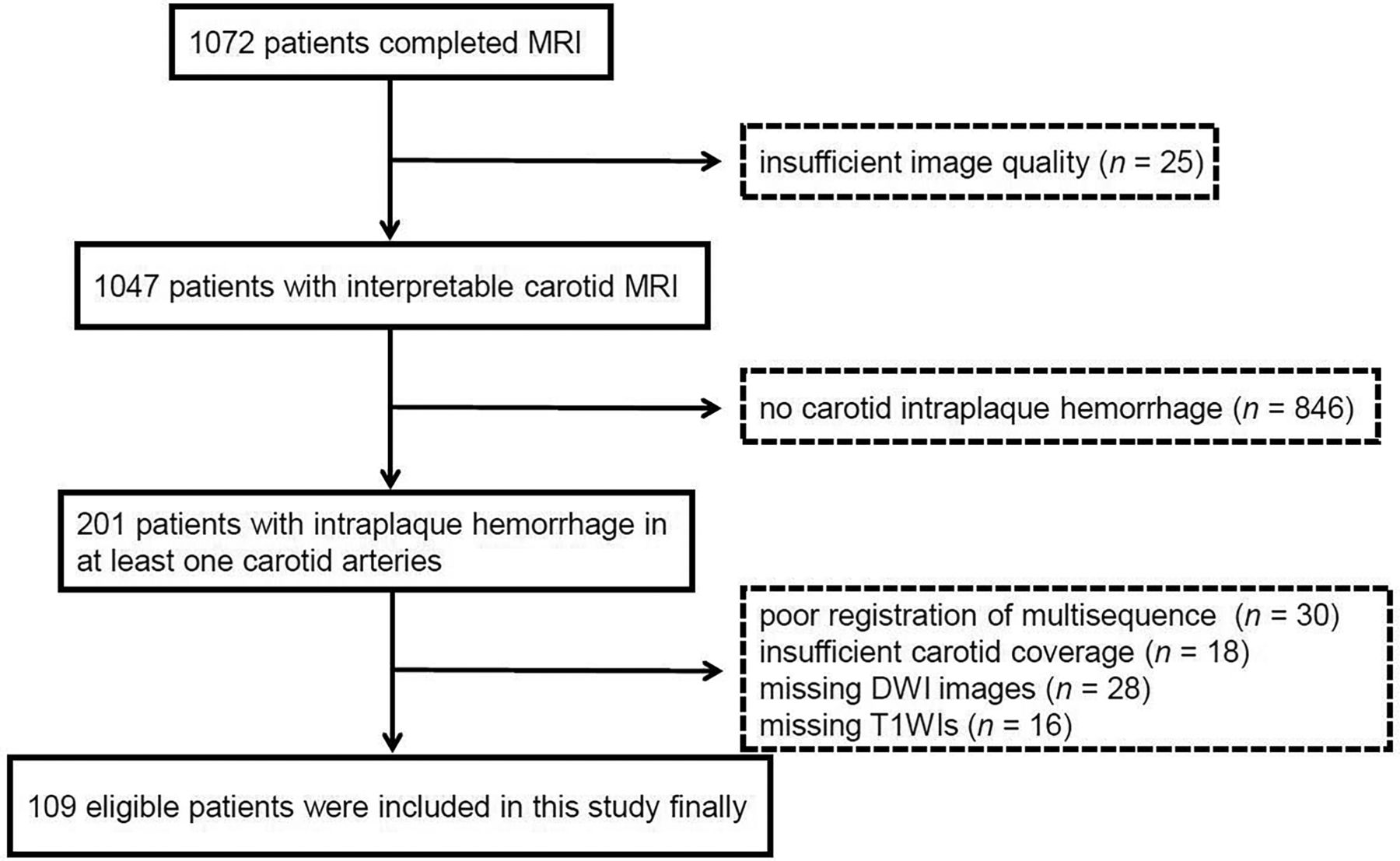
- FIG 1.
Flow chart of the study sample.
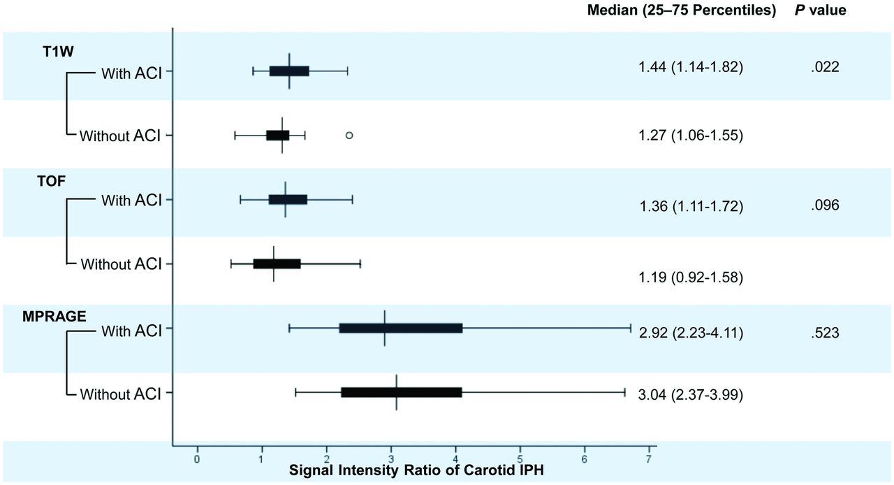
- FIG 2.
Boxplots of the SIR of carotid IPH on different T1-weighted MR imaging sequences in patients with and without ACI. Patients with ipsilateral ACI lesions had significantly higher SIRs of carotid IPH on T1-weighted images than those without.
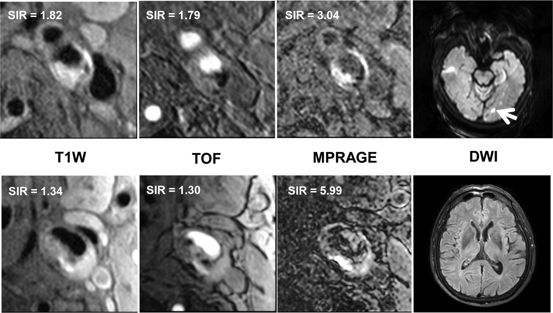
- FIG 3.
Comparison of the SIR of carotid IPH on T1-weighted MR imaging sequences, including T1-weighted, TOF, and MPRAGE, between patients with ACI (4 images above, hyperintense on DWI as the arrow indicates) and without ACI (4 images below, no abnormality on DWI). It shows that the patient with the higher SIR on T1-weighted images had ACI lesions in the ipsilateral hemisphere.


Tables
Carotid Artery Imaging Brain Imaging T1WI T2WI TOF MPRAGE DWIa T1WI Sequence TSE TSE FFE FFE EPI FFE TR (ms) 800 4800 20 8.8 2724 308 TE (ms) 10 50 4.9 5.3 86 4.6 Flip angle 90° 90° 20° 15° 90° 90° FOV (cm2) 14 × 14 14 × 14 14 × 14 14 × 14 23 × 23 23 × 23 Matrix size 256 × 256 256 × 256 256 × 256 256 × 256 128 × 126 400 × 256 Slice thickness (mmb) 2 2 1 1 5.5 5.5 Mean ± SD or (No.) (%) P Valuea Patients with ACI (n = 40) Patients without ACI (n = 69) Age (yr) 64.2 ± 10.2 68.3 ± 9.5 .035 Sex, male 37 (92.5) 59 (85.5) .366 Body mass index (kg/m2) 25.1 ± 3.6 24.0 ± 2.7 .117 History of smoking 30 (75.0) 38 (55.1) .038 History of hypertension 33 (82.5) 58 (84.1) .833 Systolic blood pressure (mm Hg) 144.2 ± 20.9 143.9 ± 21.3 .840 Diastolic blood pressure (mm Hg) 85.2 ± 9.2 85.8 ± 14.1 .720 History of hyperlipidemia 21 (52.5) 39 (56.5) .684 LDL (mmol/L) 2.7 ± 1.0 3.0 ± 1.0 .182 HDL (mmol/L) 1.1 ± 0.2 1.1 ± 0.2 .780 TC (mmol/L) 4.3 ± 1.2 4.7 ± 1.1 .161 TG (mmol/L) 1.9 ± 1.2 2.0 ± 1.1 .519 History of diabetes mellitus 15 (37.5) 25 (36.2) .895 History of coronary heart disease 11 (27.5) 14 (20.3) .388 Statin use 15 (37.5) 25 (36.2) .895 Antihypertension medication use 28 (70.0) 49 (71.0) .911 TIA 12 (30.0) 32 (46.4) .108 Recent stroke 28 (70.0) 37 (53.6) .108 Note:—LDL indicates low-density lipoprotein cholesterol; HDL, high-density lipoprotein cholesterol; TC, total cholesterol; TG, triglycerides; TIA, transient ischemia attack.
↵a P values were calculated by the Mann-Whitney U test or Fisher exact test.
- Table 3:
Comparison of carotid plaque morphologic and compositional characteristics between patients with and without ipsilateral ACI lesions
Mean ± SD. (No.) (%) or Median (25–75 Percentiles) P Valuea Patients with ACI (n = 40) Patients without ACI (n = 69) Mean lumen area (mm2) 35.5 ± 13.5 36.5 ± 12.4 .555 Mean wall area (mm2) 48.3 ± 14.9 41.5 ± 11.1 .028 Mean total vessel area (mm2) 83.8 ± 20.6 78.0 ± 18.6 .172 Mean wall thickness (mm) 1.9 ± 0.6 1.6 ± 0.4 .010 Mean normalized wall index (%) 57.9 ± 11.3 53.6 ± 9.1 .087 Total luminal occlusion 21 (52.5%) 11 (15.9%) <.001 Plaque volume (mm2) 1240.7 ± 535.2 1064.4 ± 414.1 .159 Presence of calcification 33 (82.5) 60 (87.0) .526 Presence of FCR 14 (35.0) 23 (33.3) .859 Volume of calcification (mm3)b 35.8 (12.4–87.3) 36.2 (15.8–79.6) .794 Percentage calcification volume (%)b 3.1 (1.3–6.4) 3.5 (1.9–7.7) .312 Volume of LRNC (mm3)b 312.7 (174.1–625.3) 227.7 (122.1–351.4) .038 Percentage LRNC volume (%)b 29.4 (17.9–42.1) 23.6 (14.0–34.0) .039 Volume of IPH (mm3)b 93.2 (31.4–201.2) 56.4 (21.9–104.9) .016 Percentage IPH volume (%)b 7.9 (3.0–17.0) 5.4 (2.5–10.1) .074 IPH length (mm) 12.0 (4.5–17.5) 10.0 (4.0–14.0) .101 - Table 4:
Association between signal intensity ratio of carotid IPH and the presence of ipsilateral ACI lesions
SIRIPH-to-muscle on MR Image Presence of ACI Univariate Regression Multivariate Model 1a Multivariate Model 2a Multivariate Model 3a OR 95% CI Pb OR 95% CI Pb OR 95% CI Pb OR 95% CI Pb T1-weighted 4.08 1.34–12.40 .013 3.34 1.08–10.31 .036 3.12 0.96–10.11 .058 1.57 0.48–5.20 .458 TOF 1.98 0.80–4.93 .141 2.54 0.94–6.82 .065 2.70 0.90–8.13 .077 1.21 0.37–3.93 .756 MPRAGE 0.86 0.51–1.45 .561 0.85 0.48–1.48 .557 0.98 0.53–1.79 .941 0.56 0.28–1.12 .101 Note:—SIRIPH-to-muscle indicates the maximum signal intensity of carotid IPH/the mean signal intensity of the sternocleidomastoid muscle.
↵a Multivariate model 1 was adjusted for age, sex, and history of smoking; model 2 was further adjusted using model 1 and total luminal occlusion of the carotid artery. Model 3 was further adjusted using model 1 and the volume of carotid IPH and LRNC.
↵b Associations between patients with and without ipsilateral ACI lesions were assessed using the OR values.


