
Article Figures & Data
Figures
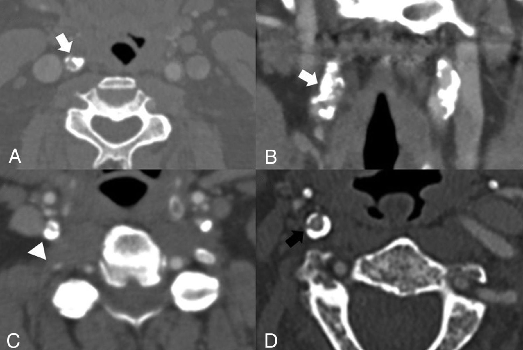
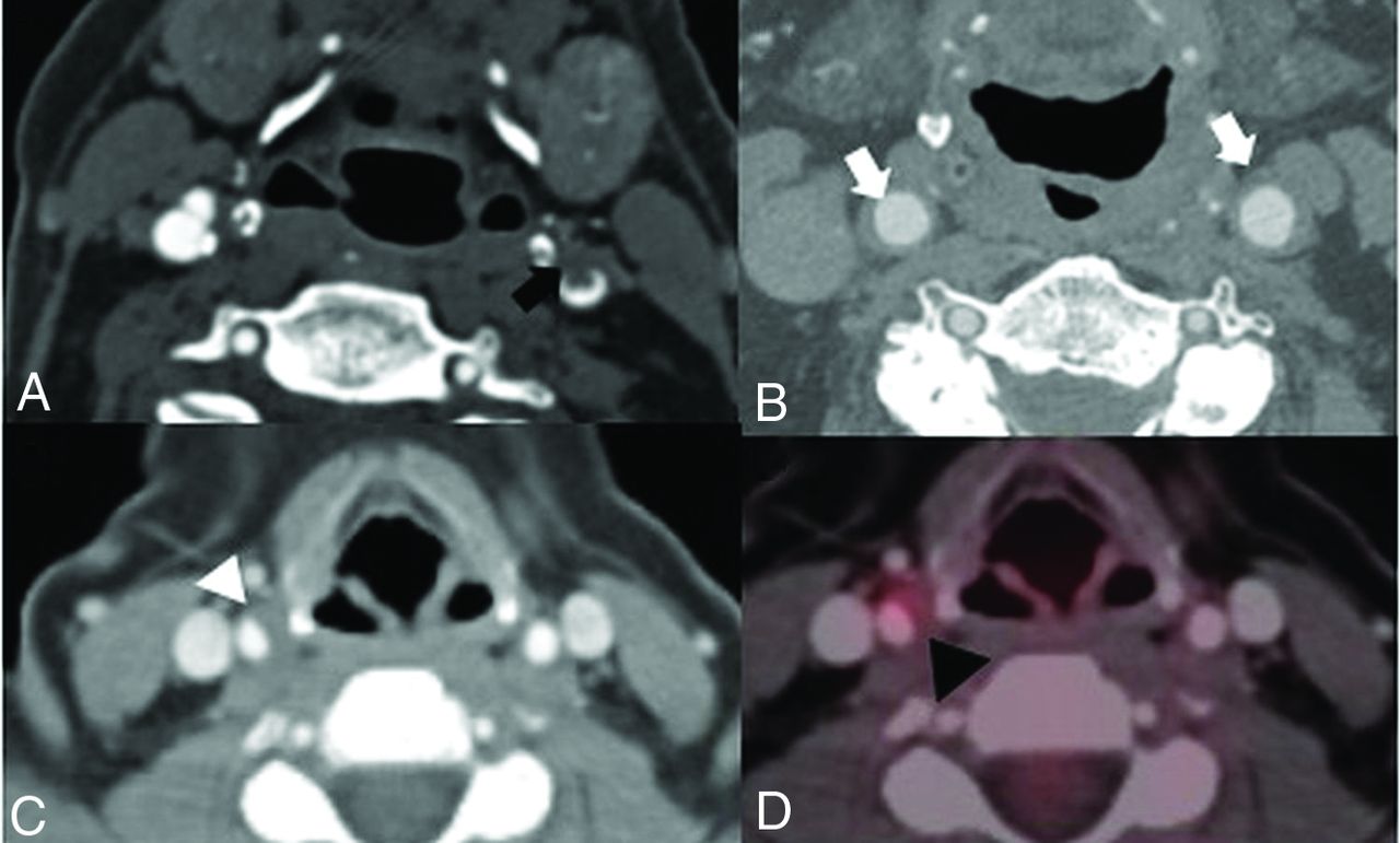
- FIG 1.
Examples of densely calcified plaques. A and B, A 73-year-old asymptomatic man with a densely calcific plaque resulting in 70% stenosis of the proximal right ICA by the NASCET criteria (white arrows). Note the relative lack of soft or fibrofatty plaque. C, A 50-year-old man presenting with right-sided weakness found to have left MCA vascular territory infarction and with incidental note of severe (approximately 80%) stenosis of the proximal right ICA secondary to a densely calcified plaque (arrowhead). Note the presence of a calcified plaque in the left ICA. Additional plaque was noted more proximally in the left ICA (not shown). D, An asymptomatic 81-year-old man found to have circumferential densely calcified plaque around his proximal right ICA (black arrow), with approximately 40% stenosis by the NASCET criteria.
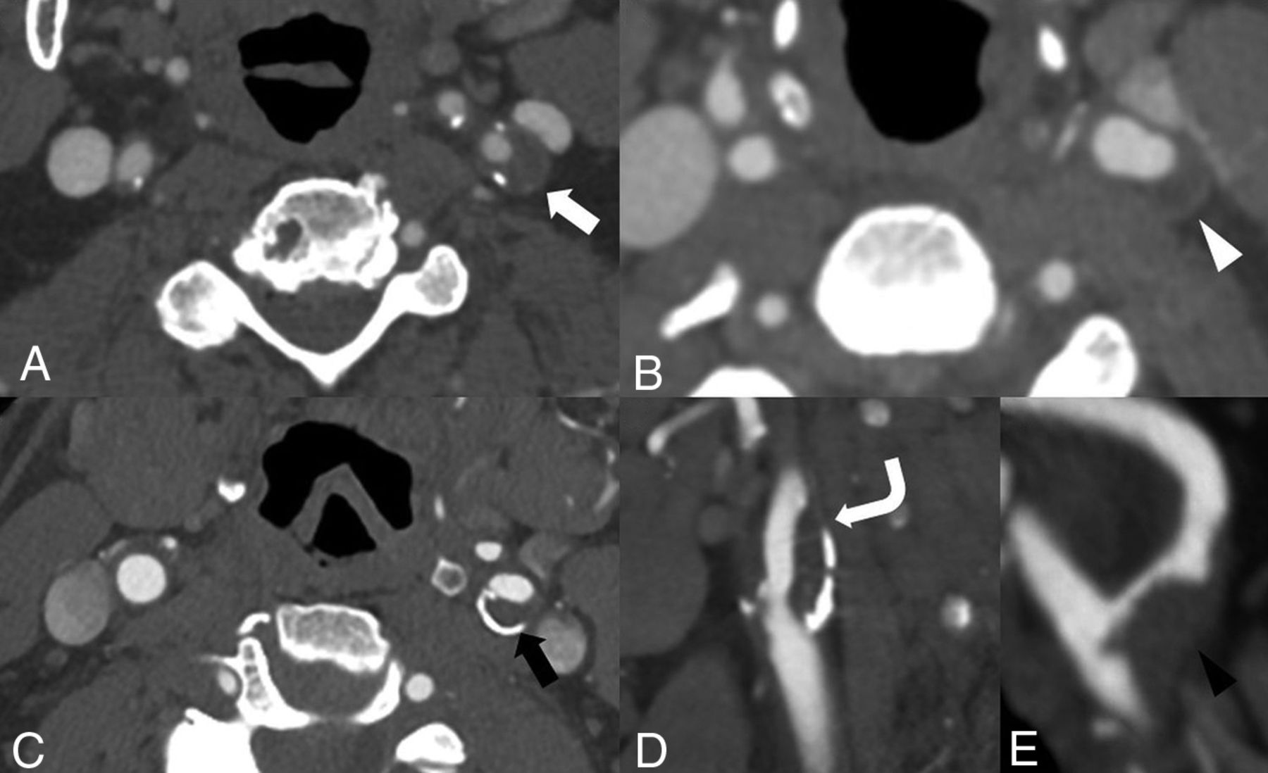
- FIG 2.
Soft plaque. A, 68-year-old man with a left-sided visual deficit found to have a large predominantly soft plaque (white arrow) in the proximal left ICA without significant stenosis by the NASCET criteria. B, A 74-year-old woman with right-sided weakness with a predominantly soft plaque (white arrowhead) at the left carotid bifurcation and proximal left ICA, again without significant stenosis by the NASCET criteria. C, An 82-year-old man with a large soft plaque with peripheral calcifications (black arrow) in the proximal left ICA with <50% stenosis by the NASCET criteria. D, Sagittal reconstruction of the same patient as in C demonstrates a large soft plaque component (curved arrow) extending throughout the proximal left ICA. E, Sagittal reconstruction demonstrates a large, irregular soft plaque narrowing the proximal right ICA (black arrowhead) of a 71-year-old man.
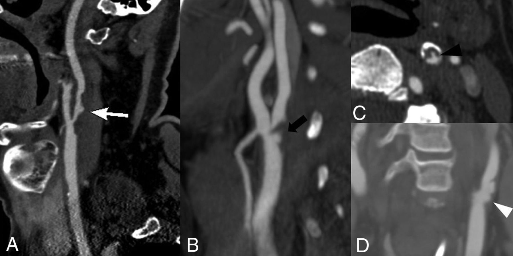
- FIG 3.
Plaque surface morphology and ulceration. A, A 69-year-old woman with a large soft plaque with an irregular surface and focal plaque ulceration (white arrow) with the plaque narrowing the proximal left ICA. B, A 62-year-old man with focal soft plaque at the right carotid bifurcation and proximal ICA with a large plaque ulceration (black arrow). C, A 73-year-old man with a predominant soft plaque with peripheral calcification narrowing the left ICA with focal plaque ulceration (black arrowhead) extending into the soft plaque. D, A 67-year-old woman with irregular, ulcerated plaque (white arrowhead) best seen on the coronal reconstruction. There is no significant associated luminal narrowing with this irregular, ulcerated plaque.
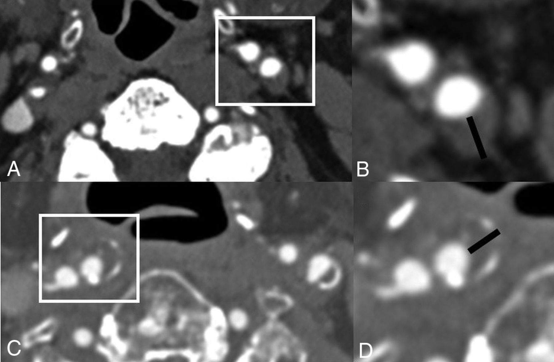
- FIG 4.
Plaque thickness measurements. A and B, A 78-year-old man with predominantly soft plaque without significant narrowing of the proximal left ICA, however, with a large soft plaque component measuring up to 5mm in thickness. Despite the lack of significant narrowing, the thickness of the plaque increases the risk of ipsilateral cerebrovascular ischemia. C and D, An 84-year-old man with a large predominantly soft plaque with peripheral calcifications without significant stenosis by the NASCET criteria but with total plaque thickness of 4mm, again increasing the risk despite the lack of significant stenosis.
- FIG 5.
Plaque inflammation. A, An 81-year-old man presenting with left MCA territory infarction with a large soft plaque (black arrow) in the distal left common carotid artery with associated intraluminal thrombus. B, A 67-year-old man with diffuse inflammatory thickening of the distal common carotid artery walls (white arrows). C and D, A 54-year-old woman with mucinous cystadenoma who had a contrast-enhanced PET/CT demonstrating a large eccentric soft plaque in the distal right common carotid artery (white arrowhead) with associated increased FDG avidity with a maximum standard uptake value of 6.3 (black arrowhead).


Tables
Standard definitions of plaque features
Plaque Characteristic Imaging Definition Histopathologic Correlate Calcified plaque Plaque with increased attenuation of >130 HU Plaque calcification Soft plaque Low-attenuation plaque, around 40–50 HU Intraplaque hemorrhage and lipid-rich necrotic core Plaque ulceration Extension of contrast material beyond the vascular lumen of the plaque, usually of at least 1 mm Plaque surface irregularity and ulceration Total plaque thickness Linear measurement of greatest axial dimension of plaque Plaque thickness Carotid plaque enhancement Enhancement of plaque after administration of contrast Plaque neovascularity


