
Article Figures & Data
Figures
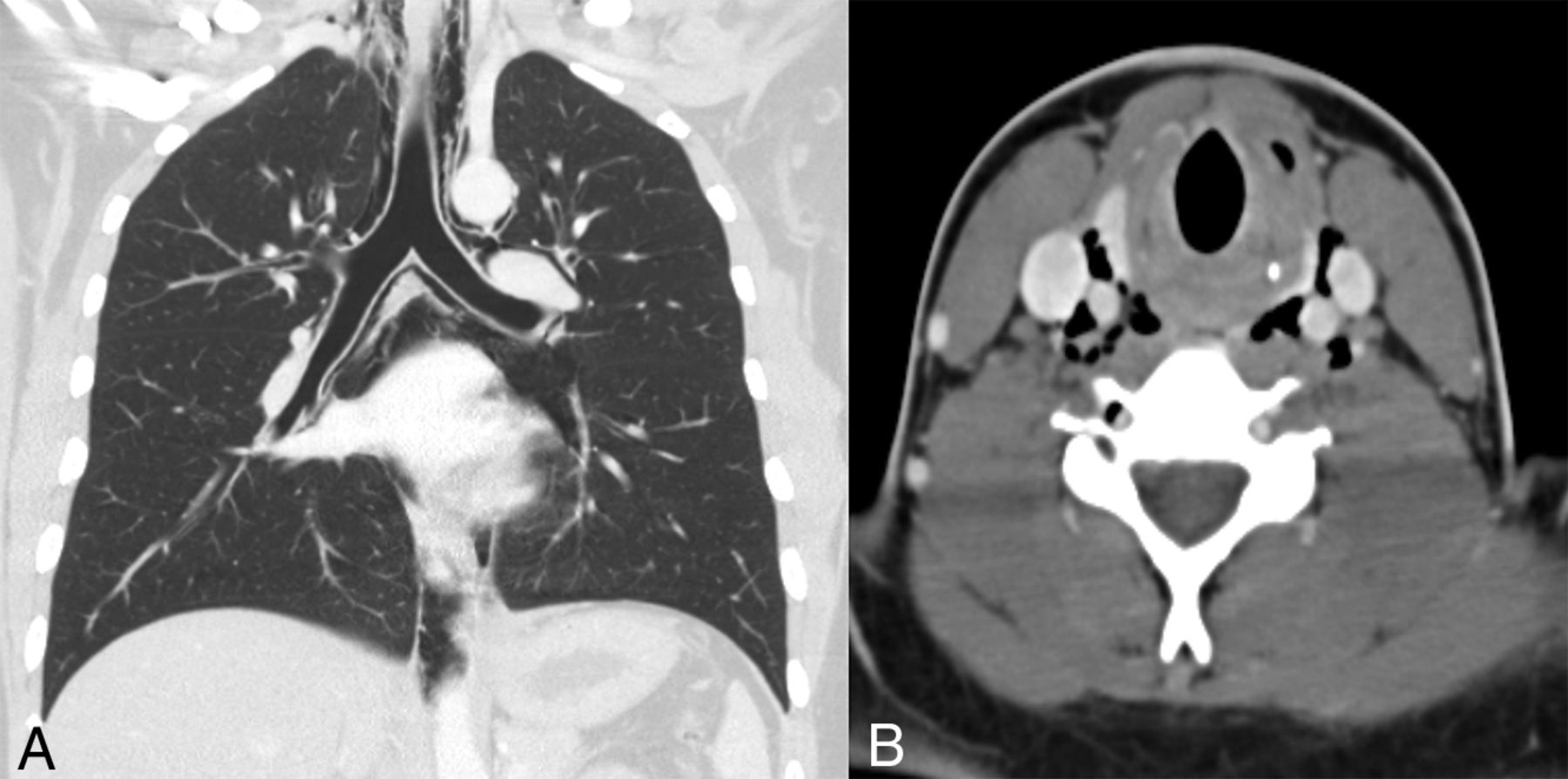
- Fig 1.
Spontaneous pneumomediastinum. A 19-year-old man with a history of cyclic vomiting syndrome presented to the emergency department after several bouts of severe vomiting with subsequent neck and chest pain. Chest (A) and neck (B) CT images show pneumomediastinum with air dissecting superiorly into both carotid spaces. An esophagram (not shown) demonstrated no evidence of esophageal perforation. The patient was observed for 3 days and discharged uneventfully.
- Fig 2.
Jugular phlebectasia. A 72-year-old woman with headaches had a neck CTA performed to evaluate vascular disease. Axial (A) and coronal (B) CTA images show a large right jugular venous aneurysm (arrows). This was deemed to be benign without need for treatment.
- Fig 3.
Inducible jugular phlebectasia. A 3-year-old boy presented with intermittent right-neck swelling. Sonographic images of the right neck with (A) and without (B) the use of the Valsalva maneuver show significant enlargement of the right internal jugular vein with Valsalva. After additional work-up to exclude any associated cardiovascular or genetic abnormality, the family was reassured that this was consistent with benign jugular phlebectasia.
- Fig 4.
Laryngocele. A 54-year-old with recent viral upper respiratory infection underwent a contrast-enhanced neck CT. Axial contrast images from this patient show mixed internal and external laryngoceles (A, solid and dashed arrows, respectively). Axial contrast-enhanced CT in an asymptomatic 69-year-old (B) shows a fluid-filled internal laryngocele (arrowhead), which was subsequently resected to confirm the diagnosis.
- Fig 5.
Spontaneous orbital floor fracture. A 58-year-old woman with chronic sinusitis who presented with sudden left periorbital swelling that started after forcefully blowing her nose. Coronal noncontrast CT images with bone (A and B) and soft-tissue (C) windows demonstrate an open-door-type left orbital floor fracture and orbital/periorbital emphysema. After ophthalmology consultation, which excluded any evidence of orbital muscle entrapment or compartment syndrome, she was discharged with a short course of antibiotics without incident.
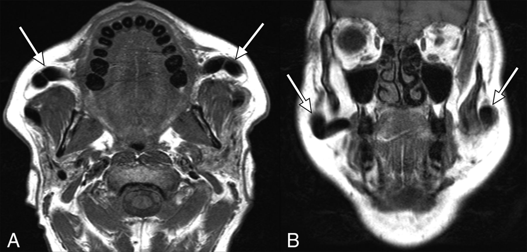
- Fig 6.
Pneumoparotid. A 57-year-old man was referred to the Ear, Nose and Throat department for chronic bilateral parotid swelling and pain, having been previously treated with antibiotics for parotitis with no symptom improvement. The patient reported periodically massaging his parotid glands, which expressed bubbly saliva into his mouth and temporarily alleviated his symptoms. Axial T1-weighted noncontrast axial (A) and coronal (B) MR images demonstrate marked dilation of the parotid ducts bilaterally (arrows). On further questioning, the patient recalled that he had been chronically pursing his lips and putting pressure inside his mouth. He was advised to avoid this in the future and to return should his symptoms worsen.
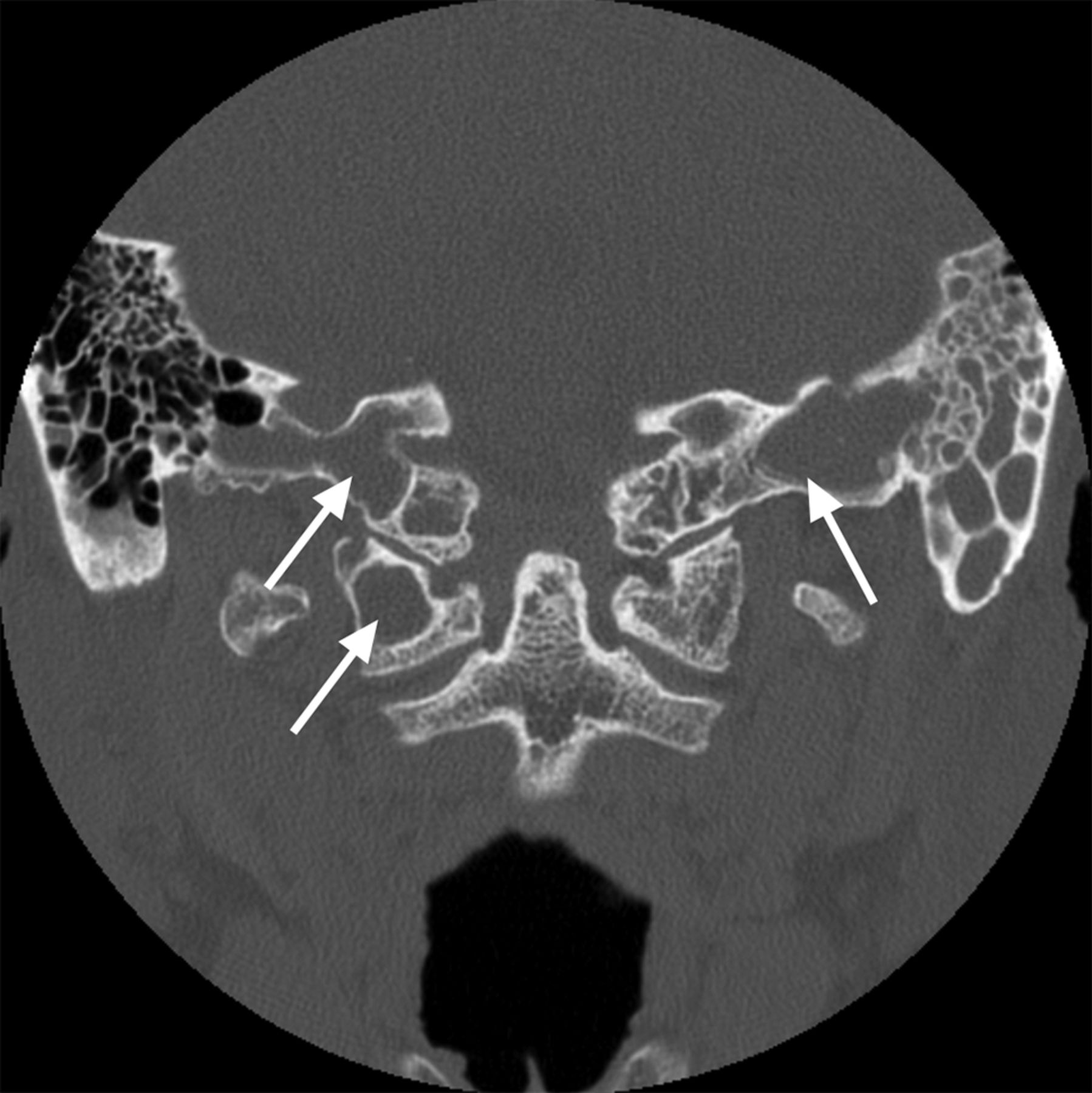
- Fig 7.
Hyperpneumatization of the skull base. A 47-year-old woman was referred to the Ear, Nose and Throat department for chronic allergies that caused her to habitually clear her throat. Coronal noncontrast CT of the skull base shows hyperpneumatization of the temporal bones, C1 ring, and dens, which are secondarily opacified (arrows).
- Fig 8.
Orbital varix. A 70-year-old woman who presented due to an orbital mass incidentally discovered on an outside brain MR imaging. Axial (A and C) and coronal (B and D) CT images of the orbits without (A and B) and with (C and D) a Valsalva maneuver demonstrate inducible enlargement of a lobular structure in the right orbit (arrows), consistent with a varix. The patient subsequently reported right-eye fullness when bending over. No additional treatment was pursued.
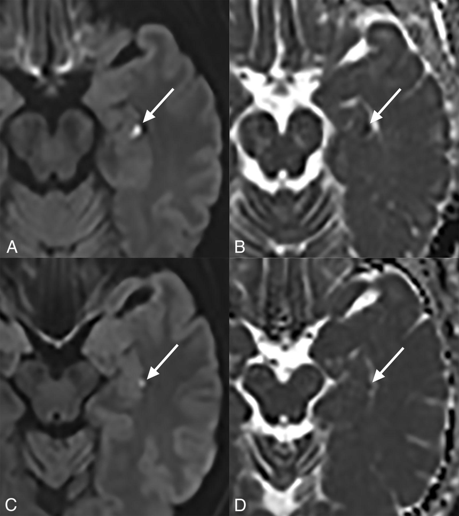
- Fig 9.
Transient global amnesia. A 58-year-old man who presented with retrograde amnesia. He had gone to the gym and completed his usual workout routine including bench-pressing earlier that day. When he returned home, he had no memory of being at the gym despite having recorded his exercises in his daily workout log. An axial diffusion-weighted MR image (A) and a corresponding ADC map (B) show a focus of diffusion restriction in the left hippocampus (arrows). The same findings (arrows) are demonstrated on axial diffusion-weighted (C) and ADC (D) images from a 57-year-old man with mixed retrograde/anterograde amnesia following sexual intercourse.
- Fig 10.
Spontaneous pneumocephalus. A 27-year-old ex-Marine with a recent history of clear rhinorrhea for 1 week presented with acute, severe head pressure that started during weight-lifting. Sagittal CT (A) shows a giant osteoma extending superiorly from the ethmoid air cells (arrow). There is extensive subarachnoid pneumocephalus (A and B).


Tables
Pressure Changes Cardiovascular Effects Increased intrathoracic pressure Decreased thoracic venous return Increased extrathoracic airway pressure Distention of extrathoracic venous system Increased middle ear pressure Decreased stroke volume Increased intracranial pressure Decreased cardiac output Opening of the Eustachian tube Peripheral vascular constriction Neck Skull Base/Face Orbit/Head Jugular phlebectasia Nontraumatic orbital floor fracture Orbital varix Laryngocele Pneumoparotid Transient global amnesia Spontaneous pneumomediastinum Hyperpneumatization of the skull base Spontaneous pneumocephalus


Jump to section
Related Articles
Cited By...
- No citing articles found.