Advertisement
  • Main menu
  • User menu
  • Search
  • English ▼
    • English
    • Afrikaans
    • Albanian
    • Amharic
    • Arabic
    • Armenian
    • Azerbaijani
    • Basque
    • Belarusian
    • Bengali
    • Bosnian
    • Bulgarian
    • Catalan
    • Cebuano
    • Chichewa
    • Chinese (Simplified)
    • Chinese (Traditional)
    • Corsican
    • Croatian
    • Czech
    • Danish
    • Dutch
    • Esperanto
    • Estonian
    • Filipino
    • Finnish
    • French
    • Frisian
    • Galician
    • Georgian
    • German
    • Greek
    • Gujarati
    • Haitian Creole
    • Hausa
    • Hawaiian
    • Hebrew
    • Hindi
    • Hmong
    • Hungarian
    • Icelandic
    • Igbo
    • Indonesian
    • Irish
    • Italian
    • Japanese
    • Javanese
    • Kannada
    • Kazakh
    • Khmer
    • Korean
    • Kurdish (Kurmanji)
    • Kyrgyz
    • Lao
    • Latin
    • Latvian
    • Lithuanian
    • Luxembourgish
    • Macedonian
    • Malagasy
    • Malay
    • Malayalam
    • Maltese
    • Maori
    • Marathi
    • Mongolian
    • Myanmar (Burmese)
    • Nepali
    • Norwegian
    • Pashto
    • Persian
    • Polish
    • Portuguese
    • Punjabi
    • Romanian
    • Russian
    • Samoan
    • Scottish Gaelic
    • Serbian
    • Sesotho
    • Shona
    • Sindhi
    • Sinhala
    • Slovak
    • Slovenian
    • Somali
    • Spanish
    • Sudanese
    • Swahili
    • Swedish
    • Tajik
    • Tamil
    • Telugu
    • Thai
    • Turkish
    • Ukrainian
    • Urdu
    • Uzbek
    • Vietnamese
    • Welsh
    • Xhosa
    • Yiddish
    • Yoruba
    • Zulu

Main menu

  • Home
  • Content
    • Current Issue
    • Accepted Manuscripts
    • Article Preview
    • Past Issue Archive
    • Video Articles
    • AJNR Case Collection
    • Case of the Week Archive
    • Case of the Month Archive
    • Classic Case Archive
  • Special Collections
    • Advancing NeuroMRI with High-Relaxivity Contrast Agents
    • AJNR Awards
    • Low-Field MRI
    • Alzheimer Disease
    • ASNR Foundation Special Collection
    • Photon-Counting CT
    • View All
  • Multimedia
    • AJNR Podcasts
    • AJNR SCANtastic
    • Trainee Corner
    • MRI Safety Corner
    • Imaging Protocols
  • For Authors
    • Submit a Manuscript
    • Submit a Video Article
    • Submit an eLetter to the Editor/Response
    • Manuscript Submission Guidelines
    • Statistical Tips
    • Fast Publishing of Accepted Manuscripts
    • Graphical Abstract Preparation
    • Imaging Protocol Submission
    • Author Policies
  • About Us
    • About AJNR
    • Editorial Board
    • Editorial Board Alumni
  • More
    • Become a Reviewer/Academy of Reviewers
    • Subscribers
    • Permissions
    • Alerts
    • Feedback
    • Advertisers
    • ASNR Home

User menu

  • Alerts
  • Log in

Search

  • Advanced search
American Journal of Neuroradiology

American Journal of Neuroradiology

ASHNR American Society of Functional Neuroradiology ASHNR American Society of Pediatric Neuroradiology ASSR
  • Alerts
  • Log in

Advanced Search

English ▼
  • English
  • Afrikaans
  • Albanian
  • Amharic
  • Arabic
  • Armenian
  • Azerbaijani
  • Basque
  • Belarusian
  • Bengali
  • Bosnian
  • Bulgarian
  • Catalan
  • Cebuano
  • Chichewa
  • Chinese (Simplified)
  • Chinese (Traditional)
  • Corsican
  • Croatian
  • Czech
  • Danish
  • Dutch
  • Esperanto
  • Estonian
  • Filipino
  • Finnish
  • French
  • Frisian
  • Galician
  • Georgian
  • German
  • Greek
  • Gujarati
  • Haitian Creole
  • Hausa
  • Hawaiian
  • Hebrew
  • Hindi
  • Hmong
  • Hungarian
  • Icelandic
  • Igbo
  • Indonesian
  • Irish
  • Italian
  • Japanese
  • Javanese
  • Kannada
  • Kazakh
  • Khmer
  • Korean
  • Kurdish (Kurmanji)
  • Kyrgyz
  • Lao
  • Latin
  • Latvian
  • Lithuanian
  • Luxembourgish
  • Macedonian
  • Malagasy
  • Malay
  • Malayalam
  • Maltese
  • Maori
  • Marathi
  • Mongolian
  • Myanmar (Burmese)
  • Nepali
  • Norwegian
  • Pashto
  • Persian
  • Polish
  • Portuguese
  • Punjabi
  • Romanian
  • Russian
  • Samoan
  • Scottish Gaelic
  • Serbian
  • Sesotho
  • Shona
  • Sindhi
  • Sinhala
  • Slovak
  • Slovenian
  • Somali
  • Spanish
  • Sudanese
  • Swahili
  • Swedish
  • Tajik
  • Tamil
  • Telugu
  • Thai
  • Turkish
  • Ukrainian
  • Urdu
  • Uzbek
  • Vietnamese
  • Welsh
  • Xhosa
  • Yiddish
  • Yoruba
  • Zulu
  • Home
  • Content
    • Current Issue
    • Accepted Manuscripts
    • Article Preview
    • Past Issue Archive
    • Video Articles
    • AJNR Case Collection
    • Case of the Week Archive
    • Case of the Month Archive
    • Classic Case Archive
  • Special Collections
    • Advancing NeuroMRI with High-Relaxivity Contrast Agents
    • AJNR Awards
    • Low-Field MRI
    • Alzheimer Disease
    • ASNR Foundation Special Collection
    • Photon-Counting CT
    • View All
  • Multimedia
    • AJNR Podcasts
    • AJNR SCANtastic
    • Trainee Corner
    • MRI Safety Corner
    • Imaging Protocols
  • For Authors
    • Submit a Manuscript
    • Submit a Video Article
    • Submit an eLetter to the Editor/Response
    • Manuscript Submission Guidelines
    • Statistical Tips
    • Fast Publishing of Accepted Manuscripts
    • Graphical Abstract Preparation
    • Imaging Protocol Submission
    • Author Policies
  • About Us
    • About AJNR
    • Editorial Board
    • Editorial Board Alumni
  • More
    • Become a Reviewer/Academy of Reviewers
    • Subscribers
    • Permissions
    • Alerts
    • Feedback
    • Advertisers
    • ASNR Home
  • Follow AJNR on Twitter
  • Visit AJNR on Facebook
  • Follow AJNR on Instagram
  • Join AJNR on LinkedIn
  • RSS Feeds

AJNR Awards, New Junior Editors, and more. Read the latest AJNR updates


Improved Turnaround Times | Median time to first decision: 12 days

Research ArticleNeurointervention
Open Access

Safety and Efficacy of Transvenous Embolization of Ruptured Brain Arteriovenous Malformations as a Last Resort: A Prospective Single-Arm Study

Y. He, Y. Ding, W. Bai, T. Li, F.K. Hui, W.-J. Jiang and J. Xue
American Journal of Neuroradiology September 2019, DOI: https://doi.org/10.3174/ajnr.A6197
Y. He
aFrom the Department of Interventional Neuroradiology (Y.H., W.B., T.L., J.X.), Henan Provincial People’s Hospital, Zhengzhou University People’s Hospital, Henan University People’s Hospital, and Henan Provincial Neurointerventional Engineering Research Center, Zhengzhou, China
  • Find this author on Google Scholar
  • Find this author on PubMed
  • Search for this author on this site
  • ORCID record for Y. He
Y. Ding
bDepartment of Radiology (Y.D.), Mayo Clinic, Rochester, Minnesota
  • Find this author on Google Scholar
  • Find this author on PubMed
  • Search for this author on this site
  • ORCID record for Y. Ding
W. Bai
aFrom the Department of Interventional Neuroradiology (Y.H., W.B., T.L., J.X.), Henan Provincial People’s Hospital, Zhengzhou University People’s Hospital, Henan University People’s Hospital, and Henan Provincial Neurointerventional Engineering Research Center, Zhengzhou, China
  • Find this author on Google Scholar
  • Find this author on PubMed
  • Search for this author on this site
  • ORCID record for W. Bai
T. Li
aFrom the Department of Interventional Neuroradiology (Y.H., W.B., T.L., J.X.), Henan Provincial People’s Hospital, Zhengzhou University People’s Hospital, Henan University People’s Hospital, and Henan Provincial Neurointerventional Engineering Research Center, Zhengzhou, China
  • Find this author on Google Scholar
  • Find this author on PubMed
  • Search for this author on this site
  • ORCID record for T. Li
F.K. Hui
cDepartment of Radiology (F.K.H.), Johns Hopkins Hospital, Baltimore, Maryland
  • Find this author on Google Scholar
  • Find this author on PubMed
  • Search for this author on this site
  • ORCID record for F.K. Hui
W.-J. Jiang
dDepartment of Vascular Neurosurgery (W.-J.J.), the PLA Rocket Force General Hospital, Beijing, China.
  • Find this author on Google Scholar
  • Find this author on PubMed
  • Search for this author on this site
  • ORCID record for W.-J. Jiang
J. Xue
aFrom the Department of Interventional Neuroradiology (Y.H., W.B., T.L., J.X.), Henan Provincial People’s Hospital, Zhengzhou University People’s Hospital, Henan University People’s Hospital, and Henan Provincial Neurointerventional Engineering Research Center, Zhengzhou, China
  • Find this author on Google Scholar
  • Find this author on PubMed
  • Search for this author on this site
  • ORCID record for J. Xue
  • Article
  • Figures & Data
  • Supplemental
  • Info & Metrics
  • Responses
  • References
  • PDF
Loading

Article Figures & Data

Figures

  • Tables
  • Fig 1.
    • Download figure
    • Open in new tab
    • Download powerpoint
    Fig 1.

    A 31-year-old man with intraparenchymal hemorrhage. Selective DSA of the left ICA (anteroposterior [A] and lateral [B] views) demonstrates that the AVM located at the frontal lobe is fed by the branches and perforators of anterior cerebral artery, MCA, and ICA and drains a single venous outlet via the cortical vein to the superior sagittal sinus (SSS). The high-resolution MR imaging shows that there is no severe stenosis or valvelike chordae in the connection part of the draining vein and superior sagittal sinus (C, white arrow). The nidus cast is through the transvenous embolization (D, unsubtracted image of the DSA), and the AVM is completely angiographically obliterated at the end of the operation (anteroposterior [E] and lateral [F] views) and at the 5-month follow-up (anteroposterior [G] and lateral [H] views).

  • Fig 2.
    • Download figure
    • Open in new tab
    • Download powerpoint
    Fig 2.

    A 28-year-old man with intraparenchymal and intraventricular hemorrhage. Ventriculostomy, decompressive craniectomy, and transarterial embolization were performed at the local hospital. Three months later, the selective DSA of the left ICA (anteroposterior [A] and lateral [B] views, both white arrows referring to the nidus) demonstrates that the parietal lobe and basal ganglia arteriovenous malformation are fed by the branches of the MCA and drain a single venous outlet via the deep vein to the straight sinus. The nidus cast was through transvenous embolization (C, unsubtracted image of the DSA), but there is a small residual AVM (anteroposterior [D] and lateral [E] views at the median arterial phase) with drainage via a cortical vein (F, white arrow) to the superior sagittal sinus, which appeared at the late arterial phase of DSA. Thirteen-month angiography follow-up confirms the complete occlusion of the residual AVM (anteroposterior [G] and lateral [H] views at the median arterial phase and lateral view [I] at the late arterial phase).
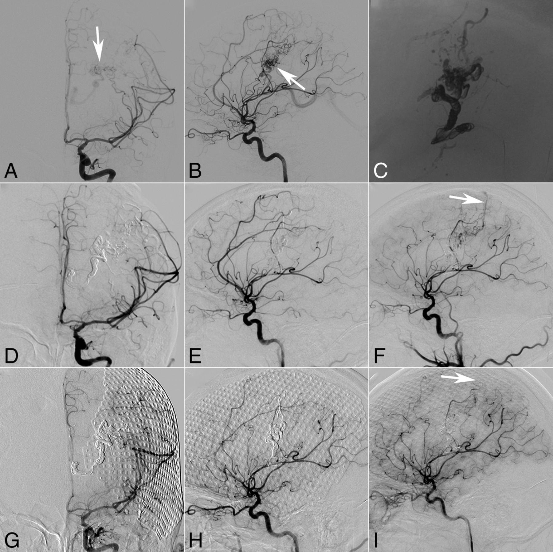
  • Fig 3.
    • Download figure
    • Open in new tab
    • Download powerpoint
    Fig 3.

    A 28-year-old man with intraventricular hemorrhage. Selective DSA of the right ICA (anteroposterior [A] and lateral [B] views) demonstrates that the AVM with an intranidal aneurysm (B, white arrow) is fed by perforators of the MCA and ICA and drains a single venous outlet via the deep vein to the straight sinus. Axial MR image indicates a basal ganglia arteriovenous malformation (C, white arrow) with the intranidus aneurysm next to the ventricle (D, white arrow). At the end of the operation, the AVM does not appear at the last angiography (anteroposterior [E] and lateral [F] views), but the cast image shows that the aneurysm does not have complete penetration by the embolic agent (G, white arrow) after transarterial and transvenous embolization. Two days later, intraventricular hemorrhage occurred (H, CT).

  • Fig 4.
    • Download figure
    • Open in new tab
    • Download powerpoint
    Fig 4.

    An 8-year-old boy who presented with sudden headache and vomiting. CT shows intraventricular hemorrhage. Selective DSA of the left vertebral artery (anteroposterior [A] and lateral [B] views, white arrow) demonstrates that the AVM with an intranidus aneurysm (C, 3D reconstruction, white arrow) is fed by the perforators of the posterior cerebral artery and drains a single venous outlet via the deep vein to the straight sinus. Axial MR image indicates a diencephalon arteriovenous malformation (D, white arrow). Transarterial ethanol sclerotherapy (80% ethanol in iohexol, Omnipaque 300 [GE Healthcare, Piscataway, New Jersey]) was performed to occlude the aneurysm (E, white arrow, the injection course can be seen in the On-line Video). Both the immediate angiography after sclerotherapy and the 2-month follow-up angiography (anteroposterior [F] and lateral [G] views, white arrow) demonstrate occlusion of the aneurysm. At 2-month follow-up, transvenous embolization was performed under transarterial balloon blocking (H). The last angiography (anteroposterior [I] and lateral [J] views) shows complete occlusion of the AVM. The intraprocedure electroencephalography monitoring did not show an abnormality, but the patient presented with light coma or lethargy. The MR imaging performed 12 days after the operation shows multiple infarctions in the mesencephalon (K, white arrow) and thalamus (L, white arrows).

  • Fig 5.
    • Download figure
    • Open in new tab
    • Download powerpoint
    Fig 5.

    The good functional outcome (mRS  ≤ 2) ratios improved from 57.1% (12/21) before the operation to 66.7% (14/21) at 1-month follow-up and 100% (19/19) at 6-month follow-up, respectively.

Tables

  • Figures
    • View popup
    Table 1:

    Baseline characteristics of the 21 patients in this study

    VariableValue
    Age (yr)
     Mean29.9
     Median29
     Range8–59
     SD17.0
    Sex (No.) (%)
     Male14 (66.7)
     Female7 (33.3)
    mRS before embolization (No.) (%)
     0–212 (57.1)
     3–59 (42.9)
    Location (No.) (%)
     Deep18 (85.7)
     Superficial3 (14.3)
    Size (No.) (%)
     ≤3 cm12 (57.1)
     >3 cm9 (42.9)
    Eloquent (No.) (%)
     Yes15 (71.4)
     No6 (28.6)
    Venous pattern (No.) (%)
     Superficial11 (52.4)
     Deep9 (42.9)
     Deep (main) + superficial1 (4.8)
    No. of veins (%)
     Single20 (95.2)
     Multiple1 (4.8)
    Angioarchitecture (No.) (%)
     Aneurysms in the feeding artery or intranidus9 (42.9)
     Venous stenosis4 (19.0)
     Localized venous ectasia2 (9.5)
    Spetzler-Martin grade (No.) (%)
     I3 (14.3)
     II4 (19.0)
     III11 (52.4)
     IV3 (14.3)
     V0 (0)
    • View popup
    Table 2:

    Safety and efficacy outcomes

    VariableValue
    Procedure
     Patients (No.)21
     Patients with technically feasible AVMs (No.) (%)19 (90.5%)
     Procedure-related complications (No.) (%)6 (28.6)
      Transient4 (19.0)
      Permanent, nondisabling0 (0)
      Permanent, disabling1 (4.8)
      Death1 (4.8)
      Non-neurologic0 (0)
    Follow-up
     Immediate obliteration after procedure (No.) (%)
      In 19 patients with technically feasible AVMs16 (84.2)
      In all 21 patients16 (76.2)
     Imaging follow-up of patients (No.)14
      Follow-up time (median) (range)5.5 (3–15)
      Obliteration at follow-up (No.) (%)13 (92.9)
      Stable1 (7.1)
      Recanalization0 (0)
     Clinical follow-up of patients within 1 mo (No.)21
      Events6
      Stroke6
      Others0
     Clinical follow-up of patients beyond 1 mo (No.)20
      The latest follow-up time (median) (range)15 (2–26)
      Events (No.)1
      Epilepsy1
      Others0
      The latest mRS
       0–219
       3–51
       60
    • View popup
    Table 3:

    Relative analysis for the complications in 19 patients with technically feasible AVMs

    ComplicationP Value
    Variable+−
    Spetzler-Martin grade.801
     I–II15
     III46
     IV–V12
    Size.141
     ≤3 cm55
     >3 cm18
    Eloquent.605
     +58
     −15
    Deep venous drainage.057
     +54
     −19
    • Note:—+ indicates yes; −, no.

PreviousNext
Back to top
Advertisement
Print
Download PDF
Email Article
Cite this article
0 Responses
Respond to this article
Share
Bookmark this article
Safety and Efficacy of Transvenous Embolization of Ruptured Brain Arteriovenous Malformations as a Last Resort: A Prospective Single-Arm Study
Y. He, Y. Ding, W. Bai, T. Li, F.K. Hui, W.-J. Jiang, J. Xue
American Journal of Neuroradiology Sep 2019, DOI: 10.3174/ajnr.A6197
del.icio.us logo Twitter logo Facebook logo Mendeley logo
  • Tweet Widget
Purchase

Jump to section

  • Article
    • Abstract
    • Materials and Methods
    • Results
    • Discussion
    • Conclusions
    • Acknowledgments
    • Footnotes
    • References
  • Figures & Data
  • Supplemental
  • Info & Metrics
  • Responses
  • References
  • PDF

Related Articles

  • PubMed
  • Google Scholar

Cited By...

  • Experimental investigation of transvenous embolization of arteriovenous malformations using different in vivo models
  • Endovascular treatment in the multimodality management of brain arteriovenous malformations: report of the Society of NeuroInterventional Surgery Standards and Guidelines Committee
  • Experimental investigation of transvenous embolization of arteriovenous malformations using different in vivo models
  • Complete flow control using transient concurrent rapid ventricular pacing or intravenous adenosine and afferent arterial balloon occlusion during transvenous embolization of cerebral arteriovenous malformations: case series
  • Crossref
  • Google Scholar

This article has not yet been cited by articles in journals that are participating in Crossref Cited-by Linking.

More in this TOC Section

  • Prophylactic Endovascular Treatments for CBS
  • Smoking & Aneurysm Outcomes: A Meta-Analysis
  • “Buddy-wire anchoring” technique for TVE
Show more NEUROINTERVENTION

Similar Articles

Advertisement

Indexed Content

  • Current Issue
  • Accepted Manuscripts
  • Article Preview
  • Past Issues
  • Editorials
  • Editor's Choice
  • Fellows' Journal Club
  • Letters to the Editor
  • Video Articles

Cases

  • Case Collection
  • Archive - Case of the Week
  • Archive - Case of the Month
  • Archive - Classic Case

More from AJNR

  • Trainee Corner
  • Imaging Protocols
  • MRI Safety Corner
  • Book Reviews

Multimedia

  • AJNR Podcasts
  • AJNR Scantastics

Resources

  • Turnaround Time
  • Submit a Manuscript
  • Submit a Video Article
  • Submit an eLetter to the Editor/Response
  • Manuscript Submission Guidelines
  • Statistical Tips
  • Fast Publishing of Accepted Manuscripts
  • Graphical Abstract Preparation
  • Imaging Protocol Submission
  • Evidence-Based Medicine Level Guide
  • Publishing Checklists
  • Author Policies
  • Become a Reviewer/Academy of Reviewers
  • News and Updates

About Us

  • About AJNR
  • Editorial Board
  • Editorial Board Alumni
  • Alerts
  • Permissions
  • Not an AJNR Subscriber? Join Now
  • Advertise with Us
  • Librarian Resources
  • Feedback
  • Terms and Conditions
  • AJNR Editorial Board Alumni

American Society of Neuroradiology

  • Not an ASNR Member? Join Now

© 2025 by the American Society of Neuroradiology All rights, including for text and data mining, AI training, and similar technologies, are reserved.
Print ISSN: 0195-6108 Online ISSN: 1936-959X

Powered by HighWire
Email this Article

Thank you for your interest in spreading the word on American Journal of Neuroradiology.

NOTE: We only request your email address so that the person you are recommending the page to knows that you wanted them to see it, and that it is not junk mail. We do not capture any email address.

Enter multiple addresses on separate lines or separate them with commas.
Safety and Efficacy of Transvenous Embolization of Ruptured Brain Arteriovenous Malformations as a Last Resort: A Prospective Single-Arm Study
(Your Name) has sent you a message from American Journal of Neuroradiology
(Your Name) thought you would like to see the American Journal of Neuroradiology web site.
CAPTCHA
This question is for testing whether or not you are a human visitor and to prevent automated spam submissions.
Cite this article
Y. He, Y. Ding, W. Bai, T. Li, F.K. Hui, W.-J. Jiang, J. Xue
Safety and Efficacy of Transvenous Embolization of Ruptured Brain Arteriovenous Malformations as a Last Resort: A Prospective Single-Arm Study
American Journal of Neuroradiology Sep 2019, DOI: 10.3174/ajnr.A6197

Citation Manager Formats

  • BibTeX
  • Bookends
  • EasyBib
  • EndNote (tagged)
  • EndNote 8 (xml)
  • Medlars
  • Mendeley
  • Papers
  • RefWorks Tagged
  • Ref Manager
  • RIS
  • Zotero

We use cookies on this site to enhance your user experience. By clicking any link on this page you are giving your consent for us to set cookies.