Advertisement

Main menu

  • Home
  • Content
    • Current Issue
    • Accepted Manuscripts
    • Article Preview
    • Past Issue Archive
    • Video Articles
    • AJNR Case Collection
    • Case of the Week Archive
    • Case of the Month Archive
    • Classic Case Archive
  • Special Collections
    • AJNR Awards
    • Low-Field MRI
    • Alzheimer Disease
    • ASNR Foundation Special Collection
    • Photon-Counting CT
    • View All
  • Multimedia
    • AJNR Podcasts
    • AJNR SCANtastic
    • Trainee Corner
    • MRI Safety Corner
    • Imaging Protocols
  • For Authors
    • Submit a Manuscript
    • Submit a Video Article
    • Submit an eLetter to the Editor/Response
    • Manuscript Submission Guidelines
    • Statistical Tips
    • Fast Publishing of Accepted Manuscripts
    • Graphical Abstract Preparation
    • Imaging Protocol Submission
    • Author Policies
  • About Us
    • About AJNR
    • Editorial Board
    • Editorial Board Alumni
  • More
    • Become a Reviewer/Academy of Reviewers
    • Subscribers
    • Permissions
    • Alerts
    • Feedback
    • Advertisers
    • ASNR Home

User menu

  • Alerts
  • Log in

Search

  • Advanced search
American Journal of Neuroradiology

American Journal of Neuroradiology

ASHNR American Society of Functional Neuroradiology ASHNR American Society of Pediatric Neuroradiology ASSR
  • Alerts
  • Log in

Advanced Search

  • Home
  • Content
    • Current Issue
    • Accepted Manuscripts
    • Article Preview
    • Past Issue Archive
    • Video Articles
    • AJNR Case Collection
    • Case of the Week Archive
    • Case of the Month Archive
    • Classic Case Archive
  • Special Collections
    • AJNR Awards
    • Low-Field MRI
    • Alzheimer Disease
    • ASNR Foundation Special Collection
    • Photon-Counting CT
    • View All
  • Multimedia
    • AJNR Podcasts
    • AJNR SCANtastic
    • Trainee Corner
    • MRI Safety Corner
    • Imaging Protocols
  • For Authors
    • Submit a Manuscript
    • Submit a Video Article
    • Submit an eLetter to the Editor/Response
    • Manuscript Submission Guidelines
    • Statistical Tips
    • Fast Publishing of Accepted Manuscripts
    • Graphical Abstract Preparation
    • Imaging Protocol Submission
    • Author Policies
  • About Us
    • About AJNR
    • Editorial Board
    • Editorial Board Alumni
  • More
    • Become a Reviewer/Academy of Reviewers
    • Subscribers
    • Permissions
    • Alerts
    • Feedback
    • Advertisers
    • ASNR Home
  • Follow AJNR on Twitter
  • Visit AJNR on Facebook
  • Follow AJNR on Instagram
  • Join AJNR on LinkedIn
  • RSS Feeds

AJNR Awards, New Junior Editors, and more. Read the latest AJNR updates

Research ArticlePediatric Neuroimaging
Open Access

Evaluating Tissue Contrast and Detecting White Matter Injury in the Infant Brain: A Comparison Study of Synthetic Phase-Sensitive Inversion Recovery

D.Y. Kim, W.S. Jung, J.W. Choi, J. Choung and H.G. Kim
American Journal of Neuroradiology July 2019, DOI: https://doi.org/10.3174/ajnr.A6135
D.Y. Kim
aFrom the Department of Radiology (D.Y.K., W.S.J., J.W.C., H.G.K.)
  • Find this author on Google Scholar
  • Find this author on PubMed
  • Search for this author on this site
  • ORCID record for D.Y. Kim
W.S. Jung
aFrom the Department of Radiology (D.Y.K., W.S.J., J.W.C., H.G.K.)
  • Find this author on Google Scholar
  • Find this author on PubMed
  • Search for this author on this site
  • ORCID record for W.S. Jung
J.W. Choi
aFrom the Department of Radiology (D.Y.K., W.S.J., J.W.C., H.G.K.)
  • Find this author on Google Scholar
  • Find this author on PubMed
  • Search for this author on this site
  • ORCID record for J.W. Choi
J. Choung
bBiomedical Informatics (J.C.), Ajou University School of Medicine, Suwon, Republic of Korea
cOffice of Biostatistics (J.C.), Ajou Research Institute for Innovative Medicine, Ajou University Medical Center, Suwon, Republic of Korea
  • Find this author on Google Scholar
  • Find this author on PubMed
  • Search for this author on this site
  • ORCID record for J. Choung
H.G. Kim
aFrom the Department of Radiology (D.Y.K., W.S.J., J.W.C., H.G.K.)
dDepartment of Radiology (H.G.K.), Eunpyeong St. Mary's Hospital, College of Medicine, The Catholic University of Korea, Seoul, Republic of Korea.
  • Find this author on Google Scholar
  • Find this author on PubMed
  • Search for this author on this site
  • ORCID record for H.G. Kim
  • Article
  • Figures & Data
  • Info & Metrics
  • Responses
  • References
  • PDF
Loading

Article Figures & Data

Figures

  • Tables
  • Fig 1.
    • Download figure
    • Open in new tab
    • Download powerpoint
    Fig 1.

    Representative synthetic PSIR image showing the drawn ROI along with cortical GM (A), uWM (B), and mWM (C).
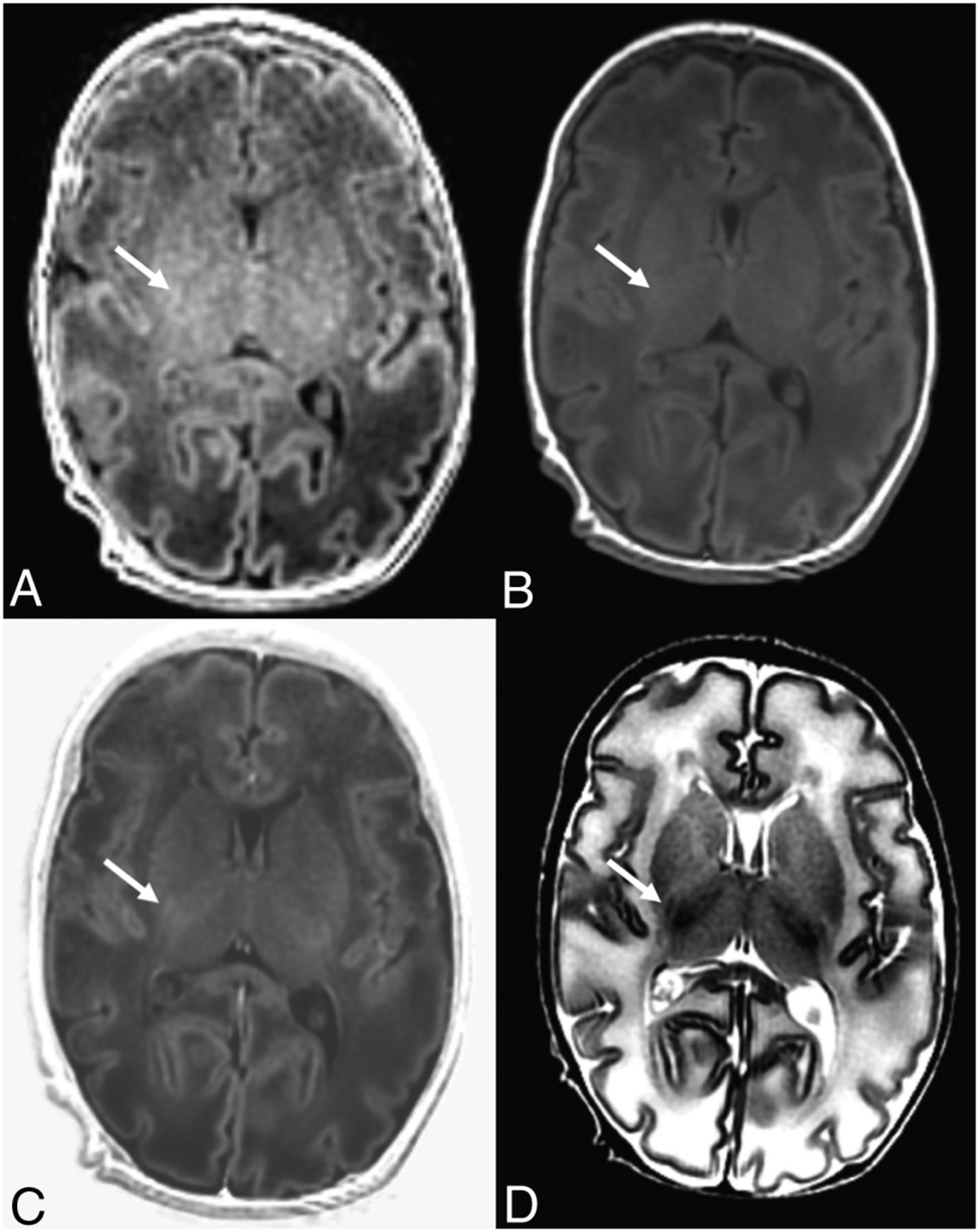
  • Fig 2.
    • Download figure
    • Open in new tab
    • Download powerpoint
    Fig 2.

    A 30-day-old neonate. There is limited myelination at the posterior limb of the internal capsule (arrow), showing higher signal intensity on the FSPGR (A), synthetic T1WI (B), and synthetic PSIR (C) images compared with the uWM regions. D, On the T2WI, the myelinated posterior limb of internal capsule (arrow) shows lower signal intensity compared with the uWM regions.
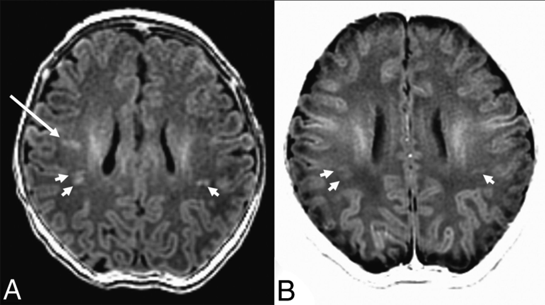
  • Fig 3.
    • Download figure
    • Open in new tab
    • Download powerpoint
    Fig 3.

    A 7-day-old neonate who underwent brain imaging due to an apnea event. There are multifocal high signal intensity lesions in the WM, which are readily detectable on both the FSPGR (A) and synthetic PSIR (B) images (short arrows). However, some of the lesions only appear on the FSPGR (long arrow) and not on the synthetic PSIR image.

Tables

  • Figures
    • View popup
    Table 1:

    Contrast comparison of the FSPGR, synthetic T1WI, and synthetic PSIR sequences (n = 91)a

    FSPGRT1WIbPSIRbP ValueFSPGR vs T1WIb (P Values)T1WIb vs PSIRb (P Values)FSPGR vs PSIRb (P Values)
    uWM-mWM0.249 (0.101)0.182 (0.062)0.506 (0.211)<.001<.001<.001<.001
    uWM-GM0.124 (0.117)0.099 (0.067)0.393 (0.309)<.001.454<.001<.001
    • ↵a Data are median (interquartile range) unless otherwise indicated.

    • ↵b Synthetic images.

    • View popup
    Table 2:

    Contrast comparison between the T2WI and synthetic PSIR sequences (n = 86)a

    T2WIPSIRbP Value
    uWM-mWM0.226 (0.106)0.516 (0.214)<.001
    uWM-GM0.176 (0.081)0.386 (0.327)<.001
    • ↵a Data are median (interquartile range) unless otherwise indicated.

    • ↵b Synthetic image.

    • View popup
    Table 3:

    Image quality and myelination degree assessment by the 2 radiologistsa

    Radiologist 1Radiologist 2
    FSPGRT1WIbPSIRbP ValueFSPGRT1WIbPSIRbP Value
    Image quality
        Diagnostic quality0/6/200/9/170/0/26.0060/1/250/0/260/1/25.603
        GM-WM0/9/170/16/100/0/26<.0010/3/231/23/20/2/24<.001
    Myelination
        CP0/6/200/20/60/0/26<.0010/1/252/21/30/0/26<.001
        PLIC2/16/83/18/52/15/9.4521/22/31/22/30/1/25<.001
        ALIC21/0/521/5/021/0/5.51322/4/022/4/021/1/4.460
        Frontal WM22/4/026/0/022/4/0.11124/2/024/2/021/4/1.421
        Occipital WM21/5/022/4/021/5/0.91821/5/021/5/021/0/5.549
    • Note:—ALIC indicates anterior limb of the internal capsule; PLIC, posterior limb of the internal capsule.

    • ↵a Number of cases graded in 3-point scale are presented in order: Image quality, 1 = poor, 2 = moderate, and 3 = good; myelination, 1 = unmyelinated, 2 = intermediate, and 3 = fully myelinated.

    • ↵b Synthetic image.

    • View popup
    Table 4:

    Number of punctate WM lesions detected on FSPGR, synthetic T1WI, and synthetic PSIR in the 10 patients

    FSPGRT1WIaPSIRa
    Rt LobeLt LobeRt LobeLt LobeRt LobeLt Lobe
    Patients
        11213126129
        2210000
        3100000
        4200000
        5230000
        6460100
        7551201
        8300000
        9431111
        10010000
    Total672424
    Mean No. of lesions detected (SD)3.35 (3.62)1.20 (2.89)1.20 (3.24)
    P Values
    FSPGR vs T1WIa.002
    FSPGR vs PSIRa.001
    T1WIa vs PSIRa>.999
    • Note:—Rt indicates right; Lt, left; SD, standard deviation.

    • ↵a Synthetic image.

PreviousNext
Back to top
Advertisement
Print
Download PDF
Email Article

Thank you for your interest in spreading the word on American Journal of Neuroradiology.

NOTE: We only request your email address so that the person you are recommending the page to knows that you wanted them to see it, and that it is not junk mail. We do not capture any email address.

Enter multiple addresses on separate lines or separate them with commas.
Evaluating Tissue Contrast and Detecting White Matter Injury in the Infant Brain: A Comparison Study of Synthetic Phase-Sensitive Inversion Recovery
(Your Name) has sent you a message from American Journal of Neuroradiology
(Your Name) thought you would like to see the American Journal of Neuroradiology web site.
CAPTCHA
This question is for testing whether or not you are a human visitor and to prevent automated spam submissions.
Cite this article
D.Y. Kim, W.S. Jung, J.W. Choi, J. Choung, H.G. Kim
Evaluating Tissue Contrast and Detecting White Matter Injury in the Infant Brain: A Comparison Study of Synthetic Phase-Sensitive Inversion Recovery
American Journal of Neuroradiology Jul 2019, DOI: 10.3174/ajnr.A6135

Citation Manager Formats

  • BibTeX
  • Bookends
  • EasyBib
  • EndNote (tagged)
  • EndNote 8 (xml)
  • Medlars
  • Mendeley
  • Papers
  • RefWorks Tagged
  • Ref Manager
  • RIS
  • Zotero
0 Responses
Respond to this article
Share
Bookmark this article
Evaluating Tissue Contrast and Detecting White Matter Injury in the Infant Brain: A Comparison Study of Synthetic Phase-Sensitive Inversion Recovery
D.Y. Kim, W.S. Jung, J.W. Choi, J. Choung, H.G. Kim
American Journal of Neuroradiology Jul 2019, DOI: 10.3174/ajnr.A6135
del.icio.us logo Twitter logo Facebook logo Mendeley logo
  • Tweet Widget
  • Facebook Like
  • Google Plus One
Purchase

Jump to section

  • Article
    • Abstract
    • ABBREVIATIONS:
    • Materials and Methods
    • Results
    • Discussion
    • Conclusions
    • Acknowledgments
    • Footnotes
    • REFERENCES
  • Figures & Data
  • Info & Metrics
  • Responses
  • References
  • PDF

Related Articles

  • No related articles found.
  • PubMed
  • Google Scholar

Cited By...

  • No citing articles found.
  • Crossref (2)
  • Google Scholar

This article has been cited by the following articles in journals that are participating in Crossref Cited-by Linking.

  • Three-Dimensional Magnetic Resonance Fingerprinting in Neonates
    Nali Yu, Jee Young Kim, Dongyeob Han, So Young Kim, Hye Mi Lee, Dong-Hyun Kim, Hyun Gi Kim
    Investigative Radiology 2022 57 1
  • Time-saving synthetic magnetic resonance imaging protocols for pediatric neuroimaging: impact of echo train length and bandwidth on image quality
    So Mi Lee, Hyun-Hae Cho, Sun Kyoung You, Eunji Kim, Seungho Kim, Moon Jung Hwang, Seung Hyun Cho, Byunggeon Park, Jihoon Hong, Sung Won Youn, Yongmin Chang
    Pediatric Radiology 2022 52 12

More in this TOC Section

  • FRACTURE MR in Congenital Vertebral Anomalies
  • Neuroimaging Delineation and Progression of SLSMD
  • fetal brain development of 10 weeks gestation
Show more PEDIATRIC NEUROIMAGING

Similar Articles

Advertisement

Indexed Content

  • Current Issue
  • Accepted Manuscripts
  • Article Preview
  • Past Issues
  • Editorials
  • Editor's Choice
  • Fellows' Journal Club
  • Letters to the Editor
  • Video Articles

Cases

  • Case Collection
  • Archive - Case of the Week
  • Archive - Case of the Month
  • Archive - Classic Case

More from AJNR

  • Trainee Corner
  • Imaging Protocols
  • MRI Safety Corner
  • Book Reviews

Multimedia

  • AJNR Podcasts
  • AJNR Scantastics

Resources

  • Turnaround Time
  • Submit a Manuscript
  • Submit a Video Article
  • Submit an eLetter to the Editor/Response
  • Manuscript Submission Guidelines
  • Statistical Tips
  • Fast Publishing of Accepted Manuscripts
  • Graphical Abstract Preparation
  • Imaging Protocol Submission
  • Evidence-Based Medicine Level Guide
  • Publishing Checklists
  • Author Policies
  • Become a Reviewer/Academy of Reviewers
  • News and Updates

About Us

  • About AJNR
  • Editorial Board
  • Editorial Board Alumni
  • Alerts
  • Permissions
  • Not an AJNR Subscriber? Join Now
  • Advertise with Us
  • Librarian Resources
  • Feedback
  • Terms and Conditions
  • AJNR Editorial Board Alumni

American Society of Neuroradiology

  • Not an ASNR Member? Join Now

© 2025 by the American Society of Neuroradiology All rights, including for text and data mining, AI training, and similar technologies, are reserved.
Print ISSN: 0195-6108 Online ISSN: 1936-959X

Powered by HighWire