Advertisement

Main menu

  • Home
  • Content
    • Current Issue
    • Accepted Manuscripts
    • Article Preview
    • Past Issue Archive
    • Video Articles
    • AJNR Case Collection
    • Case of the Week Archive
    • Case of the Month Archive
    • Classic Case Archive
  • Special Collections
    • AJNR Awards
    • Low-Field MRI
    • Alzheimer Disease
    • ASNR Foundation Special Collection
    • Photon-Counting CT
    • View All
  • Multimedia
    • AJNR Podcasts
    • AJNR SCANtastic
    • Trainee Corner
    • MRI Safety Corner
    • Imaging Protocols
  • For Authors
    • Submit a Manuscript
    • Submit a Video Article
    • Submit an eLetter to the Editor/Response
    • Manuscript Submission Guidelines
    • Statistical Tips
    • Fast Publishing of Accepted Manuscripts
    • Graphical Abstract Preparation
    • Imaging Protocol Submission
    • Author Policies
  • About Us
    • About AJNR
    • Editorial Board
    • Editorial Board Alumni
  • More
    • Become a Reviewer/Academy of Reviewers
    • Subscribers
    • Permissions
    • Alerts
    • Feedback
    • Advertisers
    • ASNR Home

User menu

  • Alerts
  • Log in

Search

  • Advanced search
American Journal of Neuroradiology

American Journal of Neuroradiology

ASHNR American Society of Functional Neuroradiology ASHNR American Society of Pediatric Neuroradiology ASSR
  • Alerts
  • Log in

Advanced Search

  • Home
  • Content
    • Current Issue
    • Accepted Manuscripts
    • Article Preview
    • Past Issue Archive
    • Video Articles
    • AJNR Case Collection
    • Case of the Week Archive
    • Case of the Month Archive
    • Classic Case Archive
  • Special Collections
    • AJNR Awards
    • Low-Field MRI
    • Alzheimer Disease
    • ASNR Foundation Special Collection
    • Photon-Counting CT
    • View All
  • Multimedia
    • AJNR Podcasts
    • AJNR SCANtastic
    • Trainee Corner
    • MRI Safety Corner
    • Imaging Protocols
  • For Authors
    • Submit a Manuscript
    • Submit a Video Article
    • Submit an eLetter to the Editor/Response
    • Manuscript Submission Guidelines
    • Statistical Tips
    • Fast Publishing of Accepted Manuscripts
    • Graphical Abstract Preparation
    • Imaging Protocol Submission
    • Author Policies
  • About Us
    • About AJNR
    • Editorial Board
    • Editorial Board Alumni
  • More
    • Become a Reviewer/Academy of Reviewers
    • Subscribers
    • Permissions
    • Alerts
    • Feedback
    • Advertisers
    • ASNR Home
  • Follow AJNR on Twitter
  • Visit AJNR on Facebook
  • Follow AJNR on Instagram
  • Join AJNR on LinkedIn
  • RSS Feeds

AJNR Awards, New Junior Editors, and more. Read the latest AJNR updates

Research ArticleSpine Imaging and Spine Image-Guided Interventions
Open Access

Spontaneous Intracranial Hypotension: A Systematic Imaging Approach for CSF Leak Localization and Management Based on MRI and Digital Subtraction Myelography

R.I. Farb, P.J. Nicholson, P.W. Peng, E.M. Massicotte, C. Lay, T. Krings and K.G. terBrugge
American Journal of Neuroradiology March 2019, DOI: https://doi.org/10.3174/ajnr.A6016
R.I. Farb
aFrom the Department of Medical Imaging, Division of Neuroradiology (R.I.F., P.J.N., T.K., K.G.t.B.)
  • Find this author on Google Scholar
  • Find this author on PubMed
  • Search for this author on this site
  • ORCID record for R.I. Farb
P.J. Nicholson
aFrom the Department of Medical Imaging, Division of Neuroradiology (R.I.F., P.J.N., T.K., K.G.t.B.)
  • Find this author on Google Scholar
  • Find this author on PubMed
  • Search for this author on this site
  • ORCID record for P.J. Nicholson
P.W. Peng
bDepartment of Anesthesiology (P.W.P.), Department of Surgery
  • Find this author on Google Scholar
  • Find this author on PubMed
  • Search for this author on this site
  • ORCID record for P.W. Peng
E.M. Massicotte
cDivision of Neurosurgery (E.M.M.), Toronto Western Hospital, Toronto, Ontario, Canada
  • Find this author on Google Scholar
  • Find this author on PubMed
  • Search for this author on this site
  • ORCID record for E.M. Massicotte
C. Lay
dDepartment of Medicine and Division of Neurology, Women's College Hospital (C.L.) University of Toronto, Toronto, Ontario, Canada.
  • Find this author on Google Scholar
  • Find this author on PubMed
  • Search for this author on this site
  • ORCID record for C. Lay
T. Krings
aFrom the Department of Medical Imaging, Division of Neuroradiology (R.I.F., P.J.N., T.K., K.G.t.B.)
  • Find this author on Google Scholar
  • Find this author on PubMed
  • Search for this author on this site
  • ORCID record for T. Krings
K.G. terBrugge
aFrom the Department of Medical Imaging, Division of Neuroradiology (R.I.F., P.J.N., T.K., K.G.t.B.)
  • Find this author on Google Scholar
  • Find this author on PubMed
  • Search for this author on this site
  • ORCID record for K.G. terBrugge
  • Article
  • Figures & Data
  • Info & Metrics
  • Responses
  • References
  • PDF
Loading

Article Figures & Data

Figures

  • Tables
  • Fig 1.
    • Download figure
    • Open in new tab
    • Download powerpoint
    Fig 1.

    Spinal longitudinal extradural collections. A, Sagittal T2 FSE. B, Reformatted axial T2 SPACE images show SLECs (arrows) and displaced dura outlined by the CSF. C and D, Images similar to A and B of the same patient show similar findings in the lower thoracic region.

  • Fig 2.
    • Download figure
    • Open in new tab
    • Download powerpoint
    Fig 2.

    Type 1 CSF leak (SLEC-P). A, Schematic drawing shows the relationship of the intervertebral disc spur and a ventral dural tear. B, “Shoot though” lateral subtracted image of the thoracic spine DSM with the patient positioned prone on the table. The patient's head is toward the top of the image and feet at the bottom. The contrast material can be seen escaping from the ventral aspect of the thecal sac at the T7–8 level (arrow).
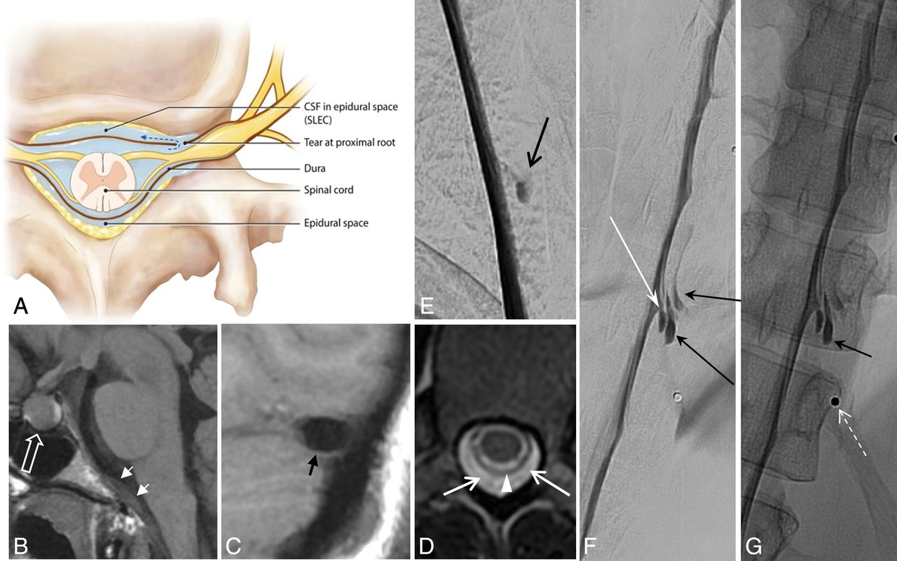
  • Fig 3.
    • Download figure
    • Open in new tab
    • Download powerpoint
    Fig 3.

    Type 2 CSF leak (SLEC-P). A, Schematic depiction of a proximal nerve root sleeve tear bridging the epidural and neural foraminal compartments. B–G, From a single patient. B, Sagittal T1WI of the brain shows the engorged pituitary gland (open white arrow) and dural thickening on the clivus (short white arrows). C, Sagittal T1WI of the brain shows a “positive venous distension sign” with a convex undersurface of the middle third of the dominant transverse sinus (short black arrow). D, T2-weighted axial MR image of the thoracic spine shows SLECs (white arrows) external to the dura (white arrowhead). E, Subtracted image from a prone thoracic DSM shows a posterolateral collection of contrast (black arrow). F and G, Subtracted and nonsubtracted images from a repeat right lateral decubitus DSM show contrast leaking into the extradural space (black arrows) from a tear along the proximal aspect of the right T11 root sleeve (long white arrow). Note the BB (nipple marker) placed on the skin for landmarking (dashed white arrow).
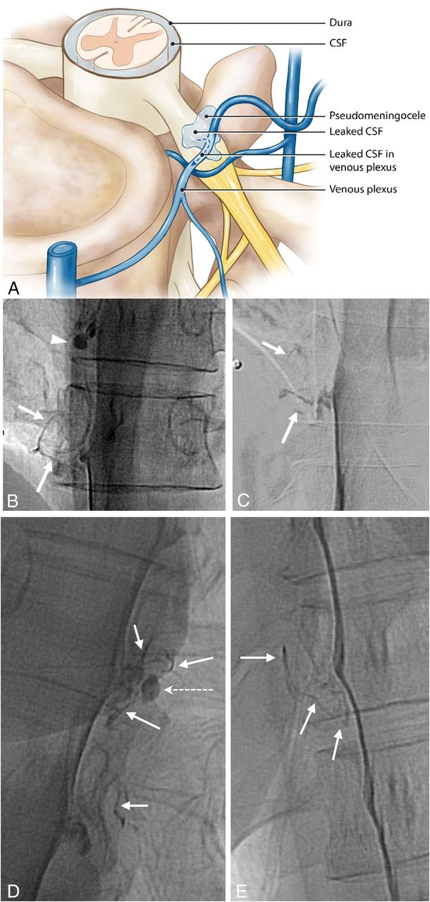
  • Fig 4.
    • Download figure
    • Open in new tab
    • Download powerpoint
    Fig 4.

    Type 3 CSF leak (SLEC-N). A, Schematic depiction of a CSF-to-venous fistula arising from a dural tear along the nerve root sleeve beyond the epidural compartment (see text). Nonsubtracted (B) and magnified, subtracted (C) images from separate left-side-down DSM runs in a patient negative for SLEC with SIH. A small vascular structure, in keeping with a tortuous vein of a CVF, can be seen coursing away from the root sleeve (arrows). An incidental normal diverticulum is also noted at the level above (arrowhead). D and E, Nonsubtracted images of decubitus DSMs of 2 other similarly presenting patients negative for SLEC demonstrating CVFs. Globular collections of contrast (dashed arrow) are commonly seen near the expected zone of origin of the vein, possibly representing a focal extravasation (pseudomeningocele) of contrast or a diverticulum from which the vein appears to arise.

  • Fig 5.
    • Download figure
    • Open in new tab
    • Download powerpoint
    Fig 5.

    Type 4 CSF leak (SLEC-N). A, Schematic depiction of a distal nerve root sleeve dural tear occurring beyond the epidural compartment extravasating into the surrounding fascial planes and loose connective tissue without loculation or fistulization. B, CT of the head. Sagittal reformat in a patient negative for SLEC demonstrates large low-density (bilateral) subdural hemorrhages (asterisk). Note the prominent “venous distension sign” (short arrow) despite the large subdural hemorrhages. C, Axial CT image obtained 10–20 minutes post-DSM shows subtle extravasated contrast in the region of the right C8 nerve (arrow). Note that on this nondynamic CT (slightly degraded due to beam-hardening artifacts associated with the shoulders), there is little to help distinguish this extravasated contrast from a normal diverticulum. D, Subtracted image from a right-side-down decubitus DSM shows extravasation of contrast (arrows) into the paraspinal tissues from a leak along the mid-to-distal right C8 nerve root sleeve.

  • Fig 6.
    • Download figure
    • Open in new tab
    • Download powerpoint
    Fig 6.

    Distribution of CSF leaks.

Tables

  • Figures
    • View popup
    Table 1:

    Prevalence of intracranial stigmata of hypotension

    VDSSagHygSDHGadPit
    SLEC-P
        No. of patients17151291717
        %817157438181
    SLEC-N
        No. of patients10107289
        %10010070208090
    • Note:—Hyg indicates positive subdural hygromas over the convexity; Pit, pituitary engorgement; Sag, sagging appearance of the brain stem and posterior fossa structures; VDS, positive venous distension sign; Gad, gadolinium enhancement of the pachymeninges; SDH, patients who displayed subdural hemorrhage over the cerebral convexities.

    • View popup
    Table 2:

    Stratification of patients with SIH by type, management, and outcome and demographics

    No. of PatientsMFAvg Age (yr)OP AvgOP RangeEffectively Treated with EBPNo. of Patients to SurgeryContinued Symptoms
    SLEC-P
        Type 1158746NANA4111 Back pain
        Type 240431NANA311 Mild headache, 1 back pain
        Not defined22053NANA20
    SLEC-N
        Type 3725528.40–12071 Awaiting surgery
        Type 4110560001
        Not found20258101000Continue unchanged
    • Note:—Avg indicates average; OP Avg, average CSF opening pressure; OP Range, the range of CSF opening pressures seen; NA, not applicable; M, male; F, female.

PreviousNext
Back to top
Advertisement
Print
Download PDF
Email Article

Thank you for your interest in spreading the word on American Journal of Neuroradiology.

NOTE: We only request your email address so that the person you are recommending the page to knows that you wanted them to see it, and that it is not junk mail. We do not capture any email address.

Enter multiple addresses on separate lines or separate them with commas.
Spontaneous Intracranial Hypotension: A Systematic Imaging Approach for CSF Leak Localization and Management Based on MRI and Digital Subtraction Myelography
(Your Name) has sent you a message from American Journal of Neuroradiology
(Your Name) thought you would like to see the American Journal of Neuroradiology web site.
CAPTCHA
This question is for testing whether or not you are a human visitor and to prevent automated spam submissions.
Cite this article
R.I. Farb, P.J. Nicholson, P.W. Peng, E.M. Massicotte, C. Lay, T. Krings, K.G. terBrugge
Spontaneous Intracranial Hypotension: A Systematic Imaging Approach for CSF Leak Localization and Management Based on MRI and Digital Subtraction Myelography
American Journal of Neuroradiology Mar 2019, DOI: 10.3174/ajnr.A6016

Citation Manager Formats

  • BibTeX
  • Bookends
  • EasyBib
  • EndNote (tagged)
  • EndNote 8 (xml)
  • Medlars
  • Mendeley
  • Papers
  • RefWorks Tagged
  • Ref Manager
  • RIS
  • Zotero
0 Responses
Respond to this article
Share
Bookmark this article
Spontaneous Intracranial Hypotension: A Systematic Imaging Approach for CSF Leak Localization and Management Based on MRI and Digital Subtraction Myelography
R.I. Farb, P.J. Nicholson, P.W. Peng, E.M. Massicotte, C. Lay, T. Krings, K.G. terBrugge
American Journal of Neuroradiology Mar 2019, DOI: 10.3174/ajnr.A6016
del.icio.us logo Twitter logo Facebook logo Mendeley logo
  • Tweet Widget
  • Facebook Like
  • Google Plus One
Purchase

Jump to section

  • Article
    • Abstract
    • ABBREVIATIONS:
    • Materials and Methods
    • Results
    • Discussion
    • Conclusions
    • Acknowledgments
    • Footnotes
    • REFERENCES
  • Figures & Data
  • Info & Metrics
  • Responses
  • References
  • PDF

Related Articles

  • No related articles found.
  • PubMed
  • Google Scholar

Cited By...

  • Volumetric response after closure of a spinal CSF leak in patients with spontaneous intracranial hypotension: a multicompartmental longitudinal study
  • Incidental asymptomatic spinal cerebrospinal fluid leaks: single-center experience, and a presentation of seven cases
  • Clinical and imaging outcomes of 100 patients with cerebrospinal fluid-venous fistulas treated by transvenous embolization
  • Spinal CSF Leaks: The Neuroradiologist Transforming Care
  • Skull Base CSF Leaks: Potential Underlying Pathophysiology and Evaluation of Brain MR Imaging Findings Associated with Spontaneous Intracranial Hypotension
  • Expounding on the Distinction between Lateral Dural Tears and Leaking Meningeal Diverticula in Spontaneous Intracranial Hypotension
  • Lateral Spinal CSF Leaks in Patients with Spontaneous Intracranial Hypotension: Radiologic-Anatomic Study of Different Variants
  • CT-guided percutaneous cyanoacrylate injection targeting the spinal cerebrospinal fluid leak: a potential therapeutic option for spontaneous intracranial hypotension
  • Lateral Decubitus Dynamic CT Myelography with Real-Time Bolus Tracking (dCTM-BT) for Evaluation of CSF-Venous Fistulas: Diagnostic Yield Stratified by Brain Imaging Findings
  • Transvenous embolization of cerebrospinal fluid-venous fistulas: Independent validation and feasibility of upper-extremity approach and using dual-microcatheter and balloon pressure cooker technique
  • Likelihood of Discovering a CSF Leak Based on Intracranial MRI Findings in Patients without a Spinal Longitudinal Extradural Collection: A New Probabilistic Scoring System
  • Conebeam CT as an Additional Tool in Digital Subtraction Myelography for the Detection of Spinal Lateral Dural Tears
  • Conebeam CT as an Additional Tool in Digitial Subtraction Myelography for the Detection of Spinal Lateral Dural Tears
  • Modified Dynamic CT Myelography for Type 1 and 2 CSF Leaks: A Procedural Approach
  • CSF Flow and Spinal Cord Motion in Patients With Spontaneous Intracranial Hypotension: A Phase Contrast MRI Study
  • Clinical and imaging outcomes of cerebrospinal fluid-venous fistula embolization
  • Utility of Dual-Energy CT to Improve Diagnosis of CSF Leaks on CT Myelography following Lateral Decubitus Digital Subtraction Myelography with Negative Findings
  • Spontaneous Spinal CSF Leaks Stratified by Age, Body Mass Index, and Spinal Level
  • Diffuse Calvarial Hyperostosis and Spontaneous Intracranial Hypotension: A Case-Control Study
  • Surgical Ligation of Spinal CSF-Venous Fistulas after Transvenous Embolization in Patients with Spontaneous Intracranial Hypotension
  • Same-Day Bilateral Decubitus CT Myelography for Detecting CSF-Venous Fistulas in Spontaneous Intracranial Hypotension
  • Multiple Spinal CSF Leaks in Spontaneous Intracranial Hypotension: Do They Exist?
  • A Novel Endovascular Therapy for CSF Hypotension Secondary to CSF-Venous Fistulas
  • Diagnostic Yield of Lateral Decubitus Digital Subtraction Myelogram Stratified by Brain MRI Findings
  • Spinal CSF-Venous Fistulas in Morbidly and Super Obese Patients with Spontaneous Intracranial Hypotension
  • Decubitus CT Myelography for CSF-Venous Fistulas: A Procedural Approach
  • Monro-Kellie Hypothesis: Increase of Ventricular CSF Volume after Surgical Closure of a Spinal Dural Leak in Patients with Spontaneous Intracranial Hypotension
  • Safety of Consecutive Bilateral Decubitus Digital Subtraction Myelography in Patients with Spontaneous Intracranial Hypotension and Occult CSF Leak
  • MR Myelography for the Detection of CSF-Venous Fistulas
  • Renal Excretion of Contrast on CT Myelography: A Specific Marker of CSF Leak
  • Lateral Decubitus Digital Subtraction Myelography: Tips, Tricks, and Pitfalls
  • Crossref (64)
  • Google Scholar

This article has been cited by the following articles in journals that are participating in Crossref Cited-by Linking.

  • A Novel Endovascular Therapy for CSF Hypotension Secondary to CSF-Venous Fistulas
    W. Brinjikji, L.E. Savastano, J.L.D. Atkinson, I. Garza, R. Farb, J.K. Cutsforth-Gregory
    American Journal of Neuroradiology 2021 42 5
  • Lateral Decubitus Digital Subtraction Myelography: Tips, Tricks, and Pitfalls
    D.K. Kim, W. Brinjikji, P.P. Morris, F.E. Diehn, V.T. Lehman, G.B. Liebo, J.M. Morris, J.T. Verdoorn, J.K. Cutsforth-Gregory, R.I. Farb, J.C Benson, C.M. Carr
    American Journal of Neuroradiology 2020 41 1
  • Decubitus CT Myelography for CSF-Venous Fistulas: A Procedural Approach
    M.D. Mamlouk, R.P. Ochi, P. Jun, P.Y. Shen
    American Journal of Neuroradiology 2021 42 1
  • Clinical and imaging outcomes of cerebrospinal fluid-venous fistula embolization
    Waleed Brinjikji, Ivan Garza, Mark Whealy, Narayan Kissoon, John L D Atkinson, Luis Savastano, Ajay Madhavan, Jeremy Cutsforth-Gregory
    Journal of NeuroInterventional Surgery 2022 14 10
  • MR Myelography for the Detection of CSF-Venous Fistulas
    J.L. Chazen, M.S. Robbins, S.B. Strauss, A.D. Schweitzer, J.P. Greenfield
    American Journal of Neuroradiology 2020 41 5
  • Clinical and imaging outcomes of 100 patients with cerebrospinal fluid-venous fistulas treated by transvenous embolization
    Waleed Brinjikji, Ajay Madhavan, Ivan Garza, Mark Whealy, Narayan Kissoon, Ian Mark, Pearse P Morris, Jared Verdoorn, John C Benson, John L D Atkinson, Hassan Kobeissi, Jeremy K Cutsforth-Gregory
    Journal of NeuroInterventional Surgery 2024 16 12
  • Same-Day Bilateral Decubitus CT Myelography for Detecting CSF-Venous Fistulas in Spontaneous Intracranial Hypotension
    L. Carlton Jones, P.J. Goadsby
    American Journal of Neuroradiology 2022 43 4
  • Spontaneous Spinal CSF Leaks Stratified by Age, Body Mass Index, and Spinal Level
    M.D. Mamlouk, P.Y. Shen, P. Jun, M.F. Sedrak
    American Journal of Neuroradiology 2022 43 7
  • CSF Flow and Spinal Cord Motion in Patients With Spontaneous Intracranial Hypotension
    Katharina Wolf, Niklas Luetzen, Hansjoerg Mast, Nico Kremers, Marco Reisert, Saúl Beltrán, Christian Fung, Jürgen Beck, Horst Urbach
    Neurology 2023 100 7
  • Monro-Kellie Hypothesis: Increase of Ventricular CSF Volume after Surgical Closure of a Spinal Dural Leak in Patients with Spontaneous Intracranial Hypotension
    T. Dobrocky, M. Rebsamen, C. Rummel, L. Häni, P. Mordasini, A. Raabe, C.T. Ulrich, J. Gralla, E.I. Piechowiak, J. Beck
    American Journal of Neuroradiology 2020 41 11

More in this TOC Section

  • Management Outcomes For VO Spine Biopsy
  • Cone Beam CT Myelography
  • Post-Procedural Brachial Neuritis Features
Show more Spine Imaging and Spine Image-Guided Interventions

Similar Articles

Advertisement

Indexed Content

  • Current Issue
  • Accepted Manuscripts
  • Article Preview
  • Past Issues
  • Editorials
  • Editor's Choice
  • Fellows' Journal Club
  • Letters to the Editor
  • Video Articles

Cases

  • Case Collection
  • Archive - Case of the Week
  • Archive - Case of the Month
  • Archive - Classic Case

More from AJNR

  • Trainee Corner
  • Imaging Protocols
  • MRI Safety Corner
  • Book Reviews

Multimedia

  • AJNR Podcasts
  • AJNR Scantastics

Resources

  • Turnaround Time
  • Submit a Manuscript
  • Submit a Video Article
  • Submit an eLetter to the Editor/Response
  • Manuscript Submission Guidelines
  • Statistical Tips
  • Fast Publishing of Accepted Manuscripts
  • Graphical Abstract Preparation
  • Imaging Protocol Submission
  • Evidence-Based Medicine Level Guide
  • Publishing Checklists
  • Author Policies
  • Become a Reviewer/Academy of Reviewers
  • News and Updates

About Us

  • About AJNR
  • Editorial Board
  • Editorial Board Alumni
  • Alerts
  • Permissions
  • Not an AJNR Subscriber? Join Now
  • Advertise with Us
  • Librarian Resources
  • Feedback
  • Terms and Conditions
  • AJNR Editorial Board Alumni

American Society of Neuroradiology

  • Not an ASNR Member? Join Now

© 2025 by the American Society of Neuroradiology All rights, including for text and data mining, AI training, and similar technologies, are reserved.
Print ISSN: 0195-6108 Online ISSN: 1936-959X

Powered by HighWire