
Article Figures & Data
Figures
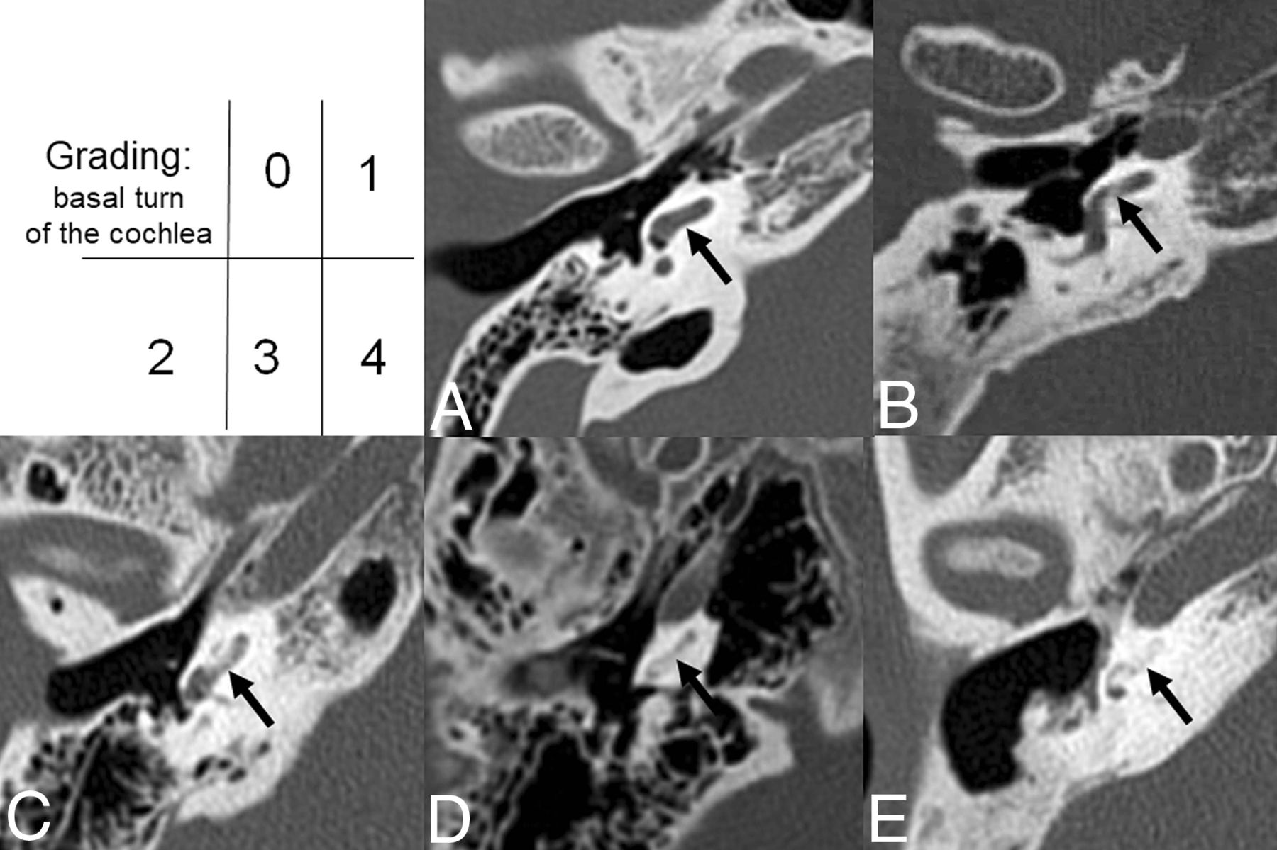
- Fig 1.
Example of LO mineralization grades (0–4) within the basal turn of the cochlea. Axial, noncontrast temporal bone images through the basal turn of the cochlea demonstrate various grades of mineralization/ossification. A, Grade 0, no evidence of mineralization/ossification. B, Grade 1, mineralization/ossification between 0% and 25%. C, Grade 2, mineralization/ossification between 25% and 50%. D, Grade 3, mineralization/ossification between 50% and 75%. E, Grade 4, Mineralization/ossification of >75%.
- Fig 2.
Example of LO mineralization grades (0–4) within the lateral semicircular canal. Axial, noncontrast temporal bone images through the lateral semicircular canals demonstrate various grades of mineralization/ossification. A, Grade 0, no evidence of mineralization/ossification. B, Grade 1, mineralization/ossification between 0% and 25%. C, Grade 2, mineralization/ossification between 25% and 50%. D, Grade 3, mineralization/ossification between 50% and 75%. E, Grade 4, mineralization/ossification of >75%.


Tables
- Table 1:
Distribution of labyrinthitis ossificans grade by membranous labyrinthine structuresa
No. Mean SD Overall Apical turn of cochlea 57 0.81 1.51 Middle turn of cochlea 57 0.86 1.46 Basal turn of cochlea 57 1.04 1.48 Vestibule 58 0.55 1.14 Lateral semicircular canal 58 1.81 1.37 Posterior semicircular canal 58 1.31 1.56 Superior semicircular canal 58 1.02 1.54 Right side Apical turn of cochlea 31 0.90 1.60 Middle turn of cochlea 31 0.94 1.48 Basal turn of cochlea 31 1.26 1.57 Vestibule 32 0.66 1.18 Lateral semicircular canal 32 2.03 1.40 Posterior semicircular canal 32 1.50 1.59 Superior semicircular canal 32 1.25 1.63 Left side Apical turn of cochlea 26 0.69 1.41 Middle turn of cochlea 26 0.77 1.45 Basal turn of cochlea 26 0.77 1.34 Vestibule 26 0.42 1.10 Lateral semicircular canal 26 1.54 1.30 Posterior semicircular canal 26 1.08 1.52 Superior semicircular canal 26 0.73 1.40 Note:—No. indicates the total number of patients.
↵a LO grades stratified by each structure in the membranous labyrinth and stratified for the left-versus-right ear.
Apical Turn of Cochlea Middle Turn of Cochlea Basal Turn of Cochlea Vestibule Lateral Semicircular Canal Posterior Semicircular Canal Superior Semicircular Canal Apical turn of cochlea – 0.839 .377 .371 .001 .071 .423 Middle turn of cochlea – .490 .256 .001 .109 .539 Basal turn of cochlea – .061 .011 .342 .966 Vestibule – <.001 .004 .069 Lateral semicircular canal – .060 .004 Posterior semicircular canal – .308 Superior semicircular canal – Note:—indicates analysis is based on 57 ears; data are P values.
Mean (SE) P Value No Chronic Otomastoiditis (n = 35) Chronic Otomastoiditis (n = 23) Apical turn of cochleab 0.79 (0.29) 0.82 (0.35) .947 Middle turn of cochleab 0.80 (0.28) 0.96 (0.34) .720 Basal turn of cochleab 0.98 (0.29) 1.23 (0.35) .578 Vestibule 0.44 (0.22) 0.76 (0.27) .347 Lateral semicircular canal 1.81 (0.23) 1.74 (0.28) .853 Posterior semicircular canal 1.13 (0.26) 1.54 (0.33) .328 Superior semicircular canal 0.75 (0.26) 1.39 (0.32) .136 Mean (SE) P Value No Meningitis (n = 52) Meningitis (n = 6) Apical turn of cochleab 0.84 (0.23) 0.50 (0.71) .657 Middle turn of cochleab 0.88 (0.23) 0.72 (0.70) .829 Basal turn of cochleab 1.11 (0.24) 0.81 (0.73) .697 Vestibule 0.63 (0.18) 0.04 (0.55) .317 Lateral semicircular canal 1.77 (0.19) 1.91 (0.56) .806 Posterior semicircular canal 1.22 (0.22) 1.96 (0.65) .296 Superior semicircular canal 1.0 (0.22) 0.94 (0.64) .925 Mean (SE) P Value No Sickle Cell Disease (n = 51) Sickle Cell Disease (n = 7) Apical turn of cochleab 0.79 (0.24) 0.91 (0.65) .858 Middle turn of cochleab 0.89 (0.23) 0.67 (0.63) .738 Basal turn of cochleab 1.15 (0.24) 0.50 (0.65) .349 Vestibule 0.63 (0.18) 0.09 (0.50) .316 Lateral semicircular canal 1.84 (0.19) 1.40 (0.51) .422 Posterior semicircular canal 1.32 (0.22) 1.11 (0.60) .747 Superior semicircular canal 1.09 (0.22) 0.37 (0.60) .261 Mean (SE) P Value No Trauma (n = 50) Trauma (n = 8) Apical turn of cochleab 0.75 (0.24) 1.15 (0.60) .535 Middle turn of cochleab 0.80 (0.23) 1.25 (0.58) .477 Basal turn of cochleab 1.0 (0.24) 1.58 (0.60) .372 Vestibule 0.59 (0.18) 0.44 (0.46) .763 Lateral semicircular canal 1.82 (0.19) 1.55 (0.48) .605 Posterior semicircular canal 1.36 (0.21) 0.50 (0.52) .130 Superior semicircular canal 1.03 (0.22) 0.66 (0.54) .528 Mean (SE) P Value No Surgery (n = 47) Surgery (n = 11) Apical turn of cochleab 0.74 (0.24) 1.07 (0.51) .553 Middle turn of cochleab 0.74 (0.23) 1.41 (0.49) .223 Basal turn of cochleab 0.84 (0.23) 2.07 (0.48) .027 Vestibule 0.30 (0.16) 1.67 (0.33) .001 Lateral semicircular canal 1.49 (0.15) 3.04 (0.32) <.001 Posterior semicircular canal 0.99 (0.20) 2.44 (0.43) .004 Superior semicircular canal 0.75 (0.22) 2.13 (0.45) .008 Mean (SE) P Value No Risk Factor (n = 11) Any Risk Factor (n = 47) Apical turn of cochleab 0.33 (0.49) 0.92 (0.24) .280 Middle turn of cochleab 0.22 (0.47) 1.03 (0.24) .132 Basal turn of cochleab 0.35 (0.48) 1.26 (0.24) .095 Vestibule 0.01 (0.37) 0.71 (0.19) .097 Lateral semicircular canal 1.29 (0.40) 1.90 (0.19) .181 Posterior semicircular canal 0.65 (0.46) 1.44 (0.23) .136 Superior semicircular canal 0.45 (0.47) 1.15 (0.23) .190


