Advertisement

Main menu

  • Home
  • Content
    • Current Issue
    • Accepted Manuscripts
    • Article Preview
    • Past Issue Archive
    • Video Articles
    • AJNR Case Collection
    • Case of the Week Archive
    • Case of the Month Archive
    • Classic Case Archive
  • Special Collections
    • AJNR Awards
    • Low-Field MRI
    • Alzheimer Disease
    • ASNR Foundation Special Collection
    • Photon-Counting CT
    • View All
  • Multimedia
    • AJNR Podcasts
    • AJNR SCANtastic
    • Trainee Corner
    • MRI Safety Corner
    • Imaging Protocols
  • For Authors
    • Submit a Manuscript
    • Submit a Video Article
    • Submit an eLetter to the Editor/Response
    • Manuscript Submission Guidelines
    • Statistical Tips
    • Fast Publishing of Accepted Manuscripts
    • Graphical Abstract Preparation
    • Imaging Protocol Submission
    • Author Policies
  • About Us
    • About AJNR
    • Editorial Board
    • Editorial Board Alumni
  • More
    • Become a Reviewer/Academy of Reviewers
    • Subscribers
    • Permissions
    • Alerts
    • Feedback
    • Advertisers
    • ASNR Home

User menu

  • Alerts
  • Log in

Search

  • Advanced search
American Journal of Neuroradiology

American Journal of Neuroradiology

ASHNR American Society of Functional Neuroradiology ASHNR American Society of Pediatric Neuroradiology ASSR
  • Alerts
  • Log in

Advanced Search

  • Home
  • Content
    • Current Issue
    • Accepted Manuscripts
    • Article Preview
    • Past Issue Archive
    • Video Articles
    • AJNR Case Collection
    • Case of the Week Archive
    • Case of the Month Archive
    • Classic Case Archive
  • Special Collections
    • AJNR Awards
    • Low-Field MRI
    • Alzheimer Disease
    • ASNR Foundation Special Collection
    • Photon-Counting CT
    • View All
  • Multimedia
    • AJNR Podcasts
    • AJNR SCANtastic
    • Trainee Corner
    • MRI Safety Corner
    • Imaging Protocols
  • For Authors
    • Submit a Manuscript
    • Submit a Video Article
    • Submit an eLetter to the Editor/Response
    • Manuscript Submission Guidelines
    • Statistical Tips
    • Fast Publishing of Accepted Manuscripts
    • Graphical Abstract Preparation
    • Imaging Protocol Submission
    • Author Policies
  • About Us
    • About AJNR
    • Editorial Board
    • Editorial Board Alumni
  • More
    • Become a Reviewer/Academy of Reviewers
    • Subscribers
    • Permissions
    • Alerts
    • Feedback
    • Advertisers
    • ASNR Home
  • Follow AJNR on Twitter
  • Visit AJNR on Facebook
  • Follow AJNR on Instagram
  • Join AJNR on LinkedIn
  • RSS Feeds

AJNR Awards, New Junior Editors, and more. Read the latest AJNR updates

Research ArticleAdult Brain
Open Access

Clinical Value of Hybrid TOF-PET/MR Imaging–Based Multiparametric Imaging in Localizing Seizure Focus in Patients with MRI-Negative Temporal Lobe Epilepsy

K. Shang, J. Wang, X. Fan, B. Cui, J. Ma, H. Yang, Y. Zhou, G. Zhao and J. Lu
American Journal of Neuroradiology September 2018, DOI: https://doi.org/10.3174/ajnr.A5814
K. Shang
aFrom the Departments of Nuclear Medicine (K.S., J.W., B.C., J.M., H.Y., J.L.)
  • Find this author on Google Scholar
  • Find this author on PubMed
  • Search for this author on this site
  • ORCID record for K. Shang
J. Wang
aFrom the Departments of Nuclear Medicine (K.S., J.W., B.C., J.M., H.Y., J.L.)
  • Find this author on Google Scholar
  • Find this author on PubMed
  • Search for this author on this site
  • ORCID record for J. Wang
X. Fan
bNeurosurgery (X.F., G.Z.)
  • Find this author on Google Scholar
  • Find this author on PubMed
  • Search for this author on this site
  • ORCID record for X. Fan
B. Cui
aFrom the Departments of Nuclear Medicine (K.S., J.W., B.C., J.M., H.Y., J.L.)
  • Find this author on Google Scholar
  • Find this author on PubMed
  • Search for this author on this site
  • ORCID record for B. Cui
J. Ma
aFrom the Departments of Nuclear Medicine (K.S., J.W., B.C., J.M., H.Y., J.L.)
  • Find this author on Google Scholar
  • Find this author on PubMed
  • Search for this author on this site
  • ORCID record for J. Ma
H. Yang
aFrom the Departments of Nuclear Medicine (K.S., J.W., B.C., J.M., H.Y., J.L.)
  • Find this author on Google Scholar
  • Find this author on PubMed
  • Search for this author on this site
  • ORCID record for H. Yang
Y. Zhou
dDepartment of Radiology (Y.Z.), Johns Hopkins University, Baltimore, Maryland.
  • Find this author on Google Scholar
  • Find this author on PubMed
  • Search for this author on this site
  • ORCID record for Y. Zhou
G. Zhao
bNeurosurgery (X.F., G.Z.)
  • Find this author on Google Scholar
  • Find this author on PubMed
  • Search for this author on this site
  • ORCID record for G. Zhao
J. Lu
aFrom the Departments of Nuclear Medicine (K.S., J.W., B.C., J.M., H.Y., J.L.)
cRadiology (J.L.), Xuanwu Hospital, Capital Medical University, Beijing, China
  • Find this author on Google Scholar
  • Find this author on PubMed
  • Search for this author on this site
  • ORCID record for J. Lu
  • Article
  • Figures & Data
  • Info & Metrics
  • Responses
  • References
  • PDF
Loading

Article Figures & Data

Figures

  • Tables
  • Fig 1.
    • Download figure
    • Open in new tab
    • Download powerpoint
    Fig 1.

    Receiver operating characteristic curves for SUVr, CBF, and combined SUVr and CBF to predict EZ. The combined PET and ASL obtain the highest area under the curve (0.970) with high sensitivity (100%) and specificity (90.9%). PET has more diagnostic information with an area under the curve of 0.926, compared with ASL (area under the curve of 0.679). The combined PET and ASL show the best performance in specificity for predicating EZ.
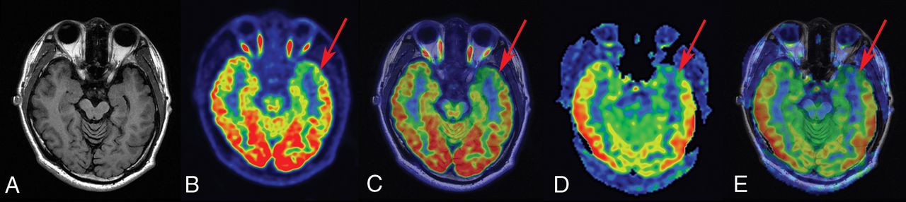
  • Fig 2.
    • Download figure
    • Open in new tab
    • Download powerpoint
    Fig 2.

    FCD type Ib in a 23-year-old patient with a history of seizures, onset at 14 years of age. A, T1-weighted axial image has normal findings. B and C, PET and PET-T1WI fused images (arrow) indicate a well-defined area of focal hypometabolism in the left temporal lobe region. D and E, ASL and ASL-T1WI fused images (arrow) show hypoperfusion in the same brain region. After a left anterior temporal lobectomy, histopathologic findings showed FCD type Ib. After a postoperative follow-up of at least 1 year, the patient was classified as having an Engel class I outcome.
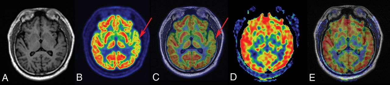
  • Fig 3.
    • Download figure
    • Open in new tab
    • Download powerpoint
    Fig 3.

    FCD type IIIa–HS in a 29-year-old patient with a history of seizures, onset at 15 years of age. A, T1-weighted axial image has normal findings. B and C, PET and PET-T1WI fused images (arrows) indicate a well-defined area of focal hypometabolism in the left temporal lobe region. However, ASL and ASL-T1WI fused images (D and E, arrows) have normal findings in the same brain region. After a left anterior temporal lobectomy, histopathologic findings of the surgical specimen were consistent with FCD type IIIa–HS. This patient with TLE had an Engel class I outcome after >1 year of follow-up.
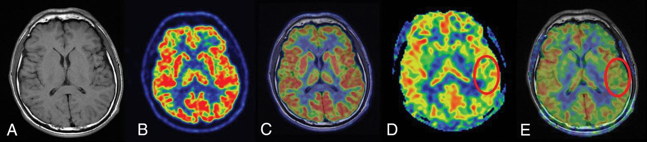
  • Fig 4.
    • Download figure
    • Open in new tab
    • Download powerpoint
    Fig 4.

    FCD type I in a 26-year-old patient with a history of seizure onset at 14 years of age. The T1-weighted axial image (A), PET image (B), and fused image (C) have normal findings. D and E, ASL and ASL-T1WI fused images (circles) show a well-defined area of focal hypoperfusion in the left temporal lobe region. After a left temporal lobe resection, pathologic findings were consistent with FCD type I. The patient had a follow-up time of >1 year, showing Engel class I outcome.
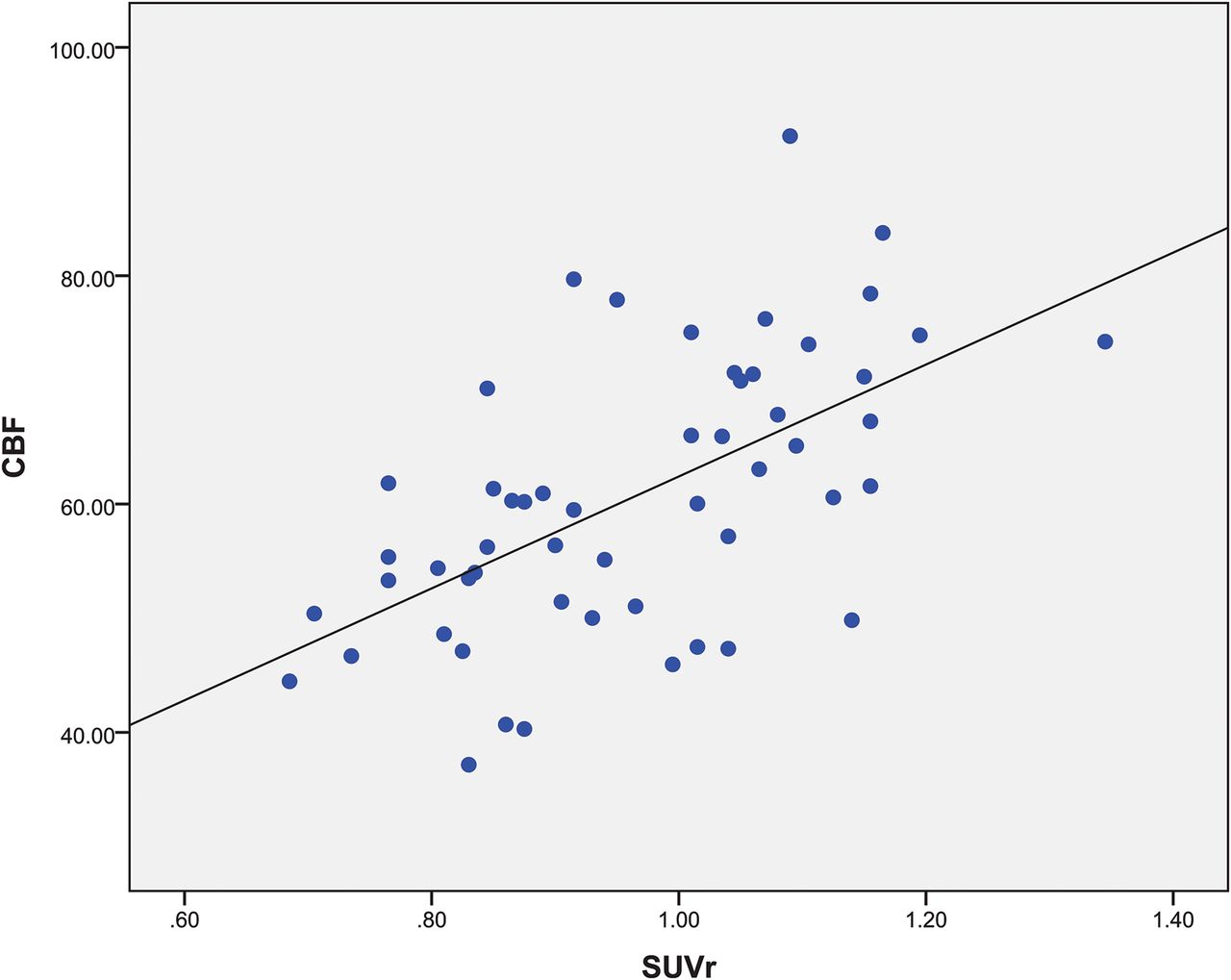
  • Fig 5.
    • Download figure
    • Open in new tab
    • Download powerpoint
    Fig 5.

    Regional comparison across the 20 patients for ROIs with hypometabolism in PET and hypoperfusion in ASL (r = 0.587, P < .001).

  • Fig 6.
    • Download figure
    • Open in new tab
    • Download powerpoint
    Fig 6.

    SPM analysis of [18F] FDG-PET images in localizing the EZ between patients and healthy controls. The hypometabolic region is mainly identified in the middle temporal gyrus. The threshold P value is set at .001.
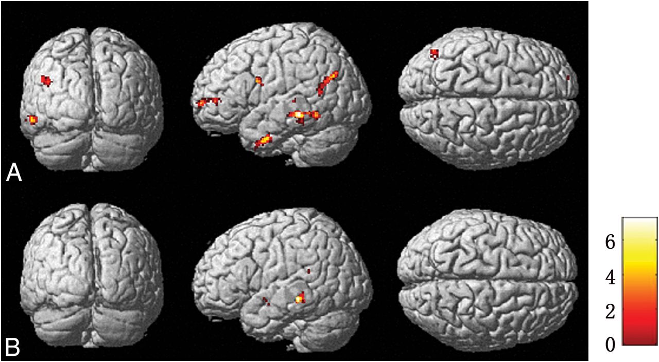
  • Fig 7.
    • Download figure
    • Open in new tab
    • Download powerpoint
    Fig 7.

    Brain regions of metabolism and perfusion asymmetry of patients compared with controls by SPM analysis in PET (A) and ASL images (B), respectively. The regions of metabolism and perfusion asymmetry are mainly identified in the middle temporal gyrus and superior temporal gyrus, respectively. The threshold P value is set at .001.

Tables

  • Figures
    • View popup
    Table 1:

    Study population demographics

    Patient No.SexAge (yr)Age at Onset (yr)Epilepsy Duration (yr)Seizure FrequencySide of OperationHistopathology
    1F2614124–5 Times/moLeftLeft temporal region (FCD type I)
    2M287211–2 Times/dayLeftLeft temporal region (FCD type I)
    3M312652–4 Times/moLeftLeft temporal region (FCD type I)
    4M231492–5 Times/moLeftLeft temporal region (FCD type Ib)
    5M2915146–8 Times/moLeftLeft temporal region (FCD type IIIa-HS)
    6F2412122–3 Times/dayLeftLeft temporal region (FCD type IIIa-HS)
    7F3817213–5 Times/moLeftLeft temporal region (FCD type IIIa-HS)
    8F272252–3 Times/moRightRight temporal region (FCD type IIIa-HS)
    9M231673–4 Times/moRightRight temporal region (FCD type I)
    10F14862–3 Times/dayLeftLeft temporal region (FCD type I)
    11M161066–9 Times/moLeftLeft temporal region (FCD type I)
    12M2614123–4 Times/moRightRight temporal region (FCD type I)
    13M295241 Time/dayRightRight temporal region (FCD type I)
    14F161331 Time/moRightRight temporal region (FCD type Ib)
    15M2411132–4 Times/moRightRight temporal region (FCD type IIIa-HS)
    16M3512233–5 Times/moRightRight temporal region (FCD type Ic)
    17M191453 Times/moRightRight temporal region (FCD type Ib)
    18M221577–8 Times/moRightRight temporal region (FCD type Ic)
    19M171161–2 Times/moLeftLeft temporal region (FCD type Ic)
    20F5212402–3 Times/moLeftLeft temporal region (FCD type Ic)
    • View popup
    Table 2:

    Summary of findings of PET and ASL in hybrid PET/MR imaging and histopathology

    Patient No.PET Findings in PET/MR ImagingASL Findings in PET/MR ImagingHistopathology
    1NormalLeft temporalLeft temporal region (FCD type I)
    2Left parietal and temporalLeft frontal, parietal, and temporalLeft temporal region (FCD type I)
    3Left temporalNegativeLeft temporal region (FCD type I)
    4Left temporalLeft temporalLeft temporal region (FCD type Ib)
    5Left temporalNegativeLeft temporal region (FCD type IIIa-HS)
    6Left temporalLeft temporalLeft temporal region (FCD type IIIa-HS)
    7Left frontal, parietal, and temporalLeft frontal, parietal, and temporalLeft temporal region (FCD type IIIa-HS)
    8Right temporalNegativeRight temporal region (FCD type IIIa-HS)
    9Right temporalRight temporalRight temporal region (FCD type I)
    10Left temporalLeft temporalLeft temporal region (FCD type I)
    11Left frontal, parietal, and temporalLeft frontal, parietal, and temporalLeft temporal region (FCD type I)
    12Right temporalRight temporalRight temporal region (FCD type I)
    13Left frontal and right temporalRight temporalRight temporal region (FCD type I)
    14Right temporalNegativeRight temporal region (FCD type Ib)
    15Right temporalRight temporalRight temporal region (FCD type IIIa-HS)
    16Left temporalLeft temporalRight temporal region (FCD type Ic)
    17Right temporalRight temporalRight temporal region (FCD type Ib)
    18Right temporalRight temporalRight temporal region (FCD type Ic)
    19Right temporalNegativeLeft temporal region (FCD type Ic)
    20Left temporalNegativeLeft temporal region (FCD type Ic)
Next
Back to top
Advertisement
Print
Download PDF
Email Article

Thank you for your interest in spreading the word on American Journal of Neuroradiology.

NOTE: We only request your email address so that the person you are recommending the page to knows that you wanted them to see it, and that it is not junk mail. We do not capture any email address.

Enter multiple addresses on separate lines or separate them with commas.
Clinical Value of Hybrid TOF-PET/MR Imaging–Based Multiparametric Imaging in Localizing Seizure Focus in Patients with MRI-Negative Temporal Lobe Epilepsy
(Your Name) has sent you a message from American Journal of Neuroradiology
(Your Name) thought you would like to see the American Journal of Neuroradiology web site.
CAPTCHA
This question is for testing whether or not you are a human visitor and to prevent automated spam submissions.
Cite this article
K. Shang, J. Wang, X. Fan, B. Cui, J. Ma, H. Yang, Y. Zhou, G. Zhao, J. Lu
Clinical Value of Hybrid TOF-PET/MR Imaging–Based Multiparametric Imaging in Localizing Seizure Focus in Patients with MRI-Negative Temporal Lobe Epilepsy
American Journal of Neuroradiology Sep 2018, DOI: 10.3174/ajnr.A5814

Citation Manager Formats

  • BibTeX
  • Bookends
  • EasyBib
  • EndNote (tagged)
  • EndNote 8 (xml)
  • Medlars
  • Mendeley
  • Papers
  • RefWorks Tagged
  • Ref Manager
  • RIS
  • Zotero
0 Responses
Respond to this article
Share
Bookmark this article
Clinical Value of Hybrid TOF-PET/MR Imaging–Based Multiparametric Imaging in Localizing Seizure Focus in Patients with MRI-Negative Temporal Lobe Epilepsy
K. Shang, J. Wang, X. Fan, B. Cui, J. Ma, H. Yang, Y. Zhou, G. Zhao, J. Lu
American Journal of Neuroradiology Sep 2018, DOI: 10.3174/ajnr.A5814
del.icio.us logo Twitter logo Facebook logo Mendeley logo
  • Tweet Widget
  • Facebook Like
  • Google Plus One
Purchase

Jump to section

  • Article
    • Abstract
    • ABBREVIATIONS:
    • Materials and Methods
    • Results
    • Discussion
    • Conclusions
    • Acknowledgments
    • Footnotes
    • References
  • Figures & Data
  • Info & Metrics
  • Responses
  • References
  • PDF

Related Articles

  • No related articles found.
  • PubMed
  • Google Scholar

Cited By...

  • Simultaneously Acquired MRI Arterial Spin-Labeling and Interictal FDG-PET Improves Diagnosis of Pediatric Temporal Lobe Epilepsy
  • Diagnostic Accuracy of Arterial Spin-Labeling MR Imaging in Detecting the Epileptogenic Zone: Systematic Review and Meta-analysis
  • Crossref
  • Google Scholar

This article has not yet been cited by articles in journals that are participating in Crossref Cited-by Linking.

More in this TOC Section

  • Diagnostic Neuroradiology of Monoclonal Antibodies
  • NCCT vs. MRI for Brain Atrophy in Acute Stroke
  • Clinical Outcomes After Chiari I Decompression
Show more Adult Brain

Similar Articles

Advertisement

Indexed Content

  • Current Issue
  • Accepted Manuscripts
  • Article Preview
  • Past Issues
  • Editorials
  • Editor's Choice
  • Fellows' Journal Club
  • Letters to the Editor
  • Video Articles

Cases

  • Case Collection
  • Archive - Case of the Week
  • Archive - Case of the Month
  • Archive - Classic Case

More from AJNR

  • Trainee Corner
  • Imaging Protocols
  • MRI Safety Corner
  • Book Reviews

Multimedia

  • AJNR Podcasts
  • AJNR Scantastics

Resources

  • Turnaround Time
  • Submit a Manuscript
  • Submit a Video Article
  • Submit an eLetter to the Editor/Response
  • Manuscript Submission Guidelines
  • Statistical Tips
  • Fast Publishing of Accepted Manuscripts
  • Graphical Abstract Preparation
  • Imaging Protocol Submission
  • Evidence-Based Medicine Level Guide
  • Publishing Checklists
  • Author Policies
  • Become a Reviewer/Academy of Reviewers
  • News and Updates

About Us

  • About AJNR
  • Editorial Board
  • Editorial Board Alumni
  • Alerts
  • Permissions
  • Not an AJNR Subscriber? Join Now
  • Advertise with Us
  • Librarian Resources
  • Feedback
  • Terms and Conditions
  • AJNR Editorial Board Alumni

American Society of Neuroradiology

  • Not an ASNR Member? Join Now

© 2025 by the American Society of Neuroradiology All rights, including for text and data mining, AI training, and similar technologies, are reserved.
Print ISSN: 0195-6108 Online ISSN: 1936-959X

Powered by HighWire