Advertisement
  • Main menu
  • User menu
  • Search
  • English ▼
    • English
    • Afrikaans
    • Albanian
    • Amharic
    • Arabic
    • Armenian
    • Azerbaijani
    • Basque
    • Belarusian
    • Bengali
    • Bosnian
    • Bulgarian
    • Catalan
    • Cebuano
    • Chichewa
    • Chinese (Simplified)
    • Chinese (Traditional)
    • Corsican
    • Croatian
    • Czech
    • Danish
    • Dutch
    • Esperanto
    • Estonian
    • Filipino
    • Finnish
    • French
    • Frisian
    • Galician
    • Georgian
    • German
    • Greek
    • Gujarati
    • Haitian Creole
    • Hausa
    • Hawaiian
    • Hebrew
    • Hindi
    • Hmong
    • Hungarian
    • Icelandic
    • Igbo
    • Indonesian
    • Irish
    • Italian
    • Japanese
    • Javanese
    • Kannada
    • Kazakh
    • Khmer
    • Korean
    • Kurdish (Kurmanji)
    • Kyrgyz
    • Lao
    • Latin
    • Latvian
    • Lithuanian
    • Luxembourgish
    • Macedonian
    • Malagasy
    • Malay
    • Malayalam
    • Maltese
    • Maori
    • Marathi
    • Mongolian
    • Myanmar (Burmese)
    • Nepali
    • Norwegian
    • Pashto
    • Persian
    • Polish
    • Portuguese
    • Punjabi
    • Romanian
    • Russian
    • Samoan
    • Scottish Gaelic
    • Serbian
    • Sesotho
    • Shona
    • Sindhi
    • Sinhala
    • Slovak
    • Slovenian
    • Somali
    • Spanish
    • Sudanese
    • Swahili
    • Swedish
    • Tajik
    • Tamil
    • Telugu
    • Thai
    • Turkish
    • Ukrainian
    • Urdu
    • Uzbek
    • Vietnamese
    • Welsh
    • Xhosa
    • Yiddish
    • Yoruba
    • Zulu

Main menu

  • Home
  • Content
    • Current Issue
    • Accepted Manuscripts
    • Article Preview
    • Past Issue Archive
    • Video Articles
    • AJNR Case Collection
    • Case of the Week Archive
    • Case of the Month Archive
    • Classic Case Archive
  • Special Collections
    • Advancing NeuroMRI with High-Relaxivity Contrast Agents
    • AJNR Awards
    • Low-Field MRI
    • Alzheimer Disease
    • ASNR Foundation Special Collection
    • Photon-Counting CT
    • View All
  • Multimedia
    • AJNR Podcasts
    • AJNR SCANtastic
    • Trainee Corner
    • MRI Safety Corner
    • Imaging Protocols
  • For Authors
    • Submit a Manuscript
    • Submit a Video Article
    • Submit an eLetter to the Editor/Response
    • Manuscript Submission Guidelines
    • Statistical Tips
    • Fast Publishing of Accepted Manuscripts
    • Graphical Abstract Preparation
    • Imaging Protocol Submission
    • Author Policies
  • About Us
    • About AJNR
    • Editorial Board
    • Editorial Board Alumni
  • More
    • Become a Reviewer/Academy of Reviewers
    • Subscribers
    • Permissions
    • Alerts
    • Feedback
    • Advertisers
    • ASNR Home

User menu

  • Alerts
  • Log in

Search

  • Advanced search
American Journal of Neuroradiology

American Journal of Neuroradiology

ASHNR American Society of Functional Neuroradiology ASHNR American Society of Pediatric Neuroradiology ASSR
  • Alerts
  • Log in

Advanced Search

English ▼
  • English
  • Afrikaans
  • Albanian
  • Amharic
  • Arabic
  • Armenian
  • Azerbaijani
  • Basque
  • Belarusian
  • Bengali
  • Bosnian
  • Bulgarian
  • Catalan
  • Cebuano
  • Chichewa
  • Chinese (Simplified)
  • Chinese (Traditional)
  • Corsican
  • Croatian
  • Czech
  • Danish
  • Dutch
  • Esperanto
  • Estonian
  • Filipino
  • Finnish
  • French
  • Frisian
  • Galician
  • Georgian
  • German
  • Greek
  • Gujarati
  • Haitian Creole
  • Hausa
  • Hawaiian
  • Hebrew
  • Hindi
  • Hmong
  • Hungarian
  • Icelandic
  • Igbo
  • Indonesian
  • Irish
  • Italian
  • Japanese
  • Javanese
  • Kannada
  • Kazakh
  • Khmer
  • Korean
  • Kurdish (Kurmanji)
  • Kyrgyz
  • Lao
  • Latin
  • Latvian
  • Lithuanian
  • Luxembourgish
  • Macedonian
  • Malagasy
  • Malay
  • Malayalam
  • Maltese
  • Maori
  • Marathi
  • Mongolian
  • Myanmar (Burmese)
  • Nepali
  • Norwegian
  • Pashto
  • Persian
  • Polish
  • Portuguese
  • Punjabi
  • Romanian
  • Russian
  • Samoan
  • Scottish Gaelic
  • Serbian
  • Sesotho
  • Shona
  • Sindhi
  • Sinhala
  • Slovak
  • Slovenian
  • Somali
  • Spanish
  • Sudanese
  • Swahili
  • Swedish
  • Tajik
  • Tamil
  • Telugu
  • Thai
  • Turkish
  • Ukrainian
  • Urdu
  • Uzbek
  • Vietnamese
  • Welsh
  • Xhosa
  • Yiddish
  • Yoruba
  • Zulu
  • Home
  • Content
    • Current Issue
    • Accepted Manuscripts
    • Article Preview
    • Past Issue Archive
    • Video Articles
    • AJNR Case Collection
    • Case of the Week Archive
    • Case of the Month Archive
    • Classic Case Archive
  • Special Collections
    • Advancing NeuroMRI with High-Relaxivity Contrast Agents
    • AJNR Awards
    • Low-Field MRI
    • Alzheimer Disease
    • ASNR Foundation Special Collection
    • Photon-Counting CT
    • View All
  • Multimedia
    • AJNR Podcasts
    • AJNR SCANtastic
    • Trainee Corner
    • MRI Safety Corner
    • Imaging Protocols
  • For Authors
    • Submit a Manuscript
    • Submit a Video Article
    • Submit an eLetter to the Editor/Response
    • Manuscript Submission Guidelines
    • Statistical Tips
    • Fast Publishing of Accepted Manuscripts
    • Graphical Abstract Preparation
    • Imaging Protocol Submission
    • Author Policies
  • About Us
    • About AJNR
    • Editorial Board
    • Editorial Board Alumni
  • More
    • Become a Reviewer/Academy of Reviewers
    • Subscribers
    • Permissions
    • Alerts
    • Feedback
    • Advertisers
    • ASNR Home
  • Follow AJNR on Twitter
  • Visit AJNR on Facebook
  • Follow AJNR on Instagram
  • Join AJNR on LinkedIn
  • RSS Feeds

AJNR Awards, New Junior Editors, and more. Read the latest AJNR updates


Improved Turnaround Times | Median time to first decision: 12 days

Research ArticlePediatric Neuroimaging

The Bone Does Not Predict the Brain in Sturge-Weber Syndrome

R.R. Warne, O.M. Carney, G. Wang, D. Bhattacharya, W.K. Chong, S.E. Aylett and K. Mankad
American Journal of Neuroradiology July 2018, DOI: https://doi.org/10.3174/ajnr.A5722
R.R. Warne
aFrom the Department of Paediatric Neuroradiology (R.R.W., O.M.C., D.B., W.K.C., K.M.), Great Ormond Street Hospital for Children, NHS Foundation Trust, London, UK
  • Find this author on Google Scholar
  • Find this author on PubMed
  • Search for this author on this site
  • ORCID record for R.R. Warne
O.M. Carney
aFrom the Department of Paediatric Neuroradiology (R.R.W., O.M.C., D.B., W.K.C., K.M.), Great Ormond Street Hospital for Children, NHS Foundation Trust, London, UK
  • Find this author on Google Scholar
  • Find this author on PubMed
  • Search for this author on this site
  • ORCID record for O.M. Carney
G. Wang
cDepartment of Biostatistics (G.W.), University of Sydney School of Public Health, Sydney, New South Wales, Australia 2006.
  • Find this author on Google Scholar
  • Find this author on PubMed
  • Search for this author on this site
  • ORCID record for G. Wang
D. Bhattacharya
aFrom the Department of Paediatric Neuroradiology (R.R.W., O.M.C., D.B., W.K.C., K.M.), Great Ormond Street Hospital for Children, NHS Foundation Trust, London, UK
  • Find this author on Google Scholar
  • Find this author on PubMed
  • Search for this author on this site
  • ORCID record for D. Bhattacharya
W.K. Chong
aFrom the Department of Paediatric Neuroradiology (R.R.W., O.M.C., D.B., W.K.C., K.M.), Great Ormond Street Hospital for Children, NHS Foundation Trust, London, UK
  • Find this author on Google Scholar
  • Find this author on PubMed
  • Search for this author on this site
  • ORCID record for W.K. Chong
S.E. Aylett
bNeurosciences (S.E.A.), Great Ormond Street Hospital for Children and Developmental Neurosciences University College London, NHS Foundation Trust, London UK
  • Find this author on Google Scholar
  • Find this author on PubMed
  • Search for this author on this site
  • ORCID record for S.E. Aylett
K. Mankad
aFrom the Department of Paediatric Neuroradiology (R.R.W., O.M.C., D.B., W.K.C., K.M.), Great Ormond Street Hospital for Children, NHS Foundation Trust, London, UK
  • Find this author on Google Scholar
  • Find this author on PubMed
  • Search for this author on this site
  • ORCID record for K. Mankad
  • Article
  • Figures & Data
  • Info & Metrics
  • Responses
  • References
  • PDF
Loading

Article Figures & Data

Figures

  • Tables
  • Fig 1.
    • Download figure
    • Open in new tab
    • Download powerpoint
    Fig 1.

    Associated findings of SWS. A 4-month-old boy with SWS. A, Postcontrast T1WI of the orbits shows a left ocular choroidal hemangioma (clinically confirmed). B, Coronal T2WI of the brain reveals volume loss of the left hemisphere with associated accelerated myelination (arrow). C, Postcontrast axial T1WI of the brain shows an enlarged and enhancing left glomus angioma (arrow) and prominent transmedullary veins (circle).

  • Fig 2.
    • Download figure
    • Open in new tab
    • Download powerpoint
    Fig 2.

    Calvarial bone marrow abnormality. A 6-month-old boy with right PWS only. A, Axial T2-weighted STIR shows high-signal abnormality in the right calvaria (arrows). B, Graphic of a child with right-sided PWS. Reproduced with permission from Scio21/Bigstock.com.

  • Fig 3.
    • Download figure
    • Open in new tab
    • Download powerpoint
    Fig 3.

    Bilateral PWS with bilateral calvarial marrow abnormality. An 8-month-old boy with SWS. Axial T2-weighted-STIR (A) and axial T1-weighted postcontrast (B) images reveal a bilateral marrow T2 high-signal calvarial abnormality and enhancement. C, Axial T1WI postcontrast shows a left-sided temporo-occipital pial angioma. Coronal T1-weighted precontrast (D) and postcontrast (E) imaging show bilateral marrow and dural enhancement.
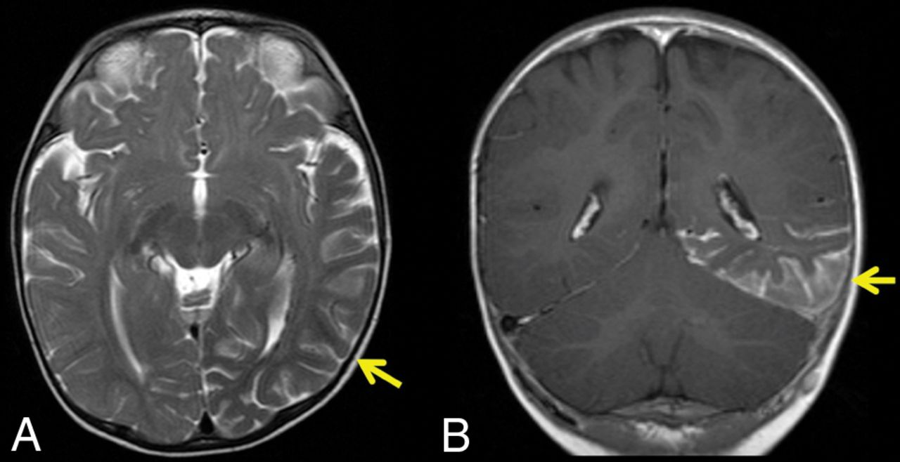
  • Fig 4.
    • Download figure
    • Open in new tab
    • Download powerpoint
    Fig 4.

    SWS with absent PWS. An 8-month-old girl with SWS. A, Axial T2-weighted STIR shows left temporo-occipital calvarial thinning with no bone marrow abnormality. B, Axial postcontrast T1WI shows a corresponding thick enhancing left hemispheric pial angioma.

  • Fig 5.
    • Download figure
    • Open in new tab
    • Download powerpoint
    Fig 5.

    The interpeduncular cistern sign. Upper panel, a 9-month-old boy with SWS. A and B, Postcontrast axial T1WI shows a left-sided unilateral pial angioma and a zoomed-in view of the interpeduncular cistern confirming the unilaterality (arrow). Lower panel, a 10-month-old girl with SWS. C and D, Bilateral pial angiomas with a zoomed-in view confirming the bilaterality (arrows), positive for the “warning sign of Warne-Mankad.”

  • Fig 6.
    • Download figure
    • Open in new tab
    • Download powerpoint
    Fig 6.

    “Warning sign of Warne-Mankad.” Bilateral interpeduncular cistern enhancement. A 10-month-old girl with SWS and bilateral PWS. A, Axial T2-weighted STIR shows bilateral hemispheric volume loss. B, Axial postcontrast T1WI shows a right-frontal and left-hemispheric pial angioma with the warning sign confirming the bilateral interpeduncular cistern enhancement (C). Coronal T2-weighted STIR (D) shows bilateral calvarial high signal and marrow enhancement (E). Coronal postcontrast FLAIR image (F) shows bilateral marrow enhancement and left-sided dural thickening.

  • Fig 7.
    • Download figure
    • Open in new tab
    • Download powerpoint
    Fig 7.

    Schematic illustration of the major calvarial diploic venous channel routes over the skull. Reproduced with permission from Springer Nature (Tsutsumi et al16). MMV indicates middle meningeal vein; OC, occipitocervical route; OFO, orbital part of the fronto-orbital route; OP, occipitoparietal route; PFO, pterional part of the fronto-orbital route; PFP, pteriofrontparietal route; PP, pterygoid plexus; SS, sigmoid sinus; SSS, superior sagittal sinus; TS, transverse sinus.

Tables

  • Figures
  • Intraosseous signal abnormality versus same-sided PWS

    PWSIntraosseous Signal Abnormality
    PresentAbsent
    Present10910
    Absent312
Next
Back to top
Advertisement
Print
Download PDF
Email Article
Cite this article
0 Responses
Respond to this article
Share
Bookmark this article
The Bone Does Not Predict the Brain in Sturge-Weber Syndrome
R.R. Warne, O.M. Carney, G. Wang, D. Bhattacharya, W.K. Chong, S.E. Aylett, K. Mankad
American Journal of Neuroradiology Jul 2018, DOI: 10.3174/ajnr.A5722
del.icio.us logo Twitter logo Facebook logo Mendeley logo
  • Tweet Widget
Purchase

Jump to section

  • Article
    • Abstract
    • ABBREVIATIONS:
    • Materials and Methods
    • Results
    • Discussion
    • Conclusions
    • Footnotes
    • References
  • Figures & Data
  • Info & Metrics
  • Responses
  • References
  • PDF

Related Articles

  • No related articles found.
  • PubMed
  • Google Scholar

Cited By...

  • Teaching NeuroImage: Sturge-Weber Syndrome in an Adult
  • Crossref
  • Google Scholar

This article has not yet been cited by articles in journals that are participating in Crossref Cited-by Linking.

More in this TOC Section

  • Pediatric Glioma:Imaging Clusters,Molecular Traits
  • Clinical SVR of Fetal Brain MRI
  • FRACTURE MR in Congenital Vertebral Anomalies
Show more PEDIATRIC NEUROIMAGING

Similar Articles

Advertisement

Indexed Content

  • Current Issue
  • Accepted Manuscripts
  • Article Preview
  • Past Issues
  • Editorials
  • Editor's Choice
  • Fellows' Journal Club
  • Letters to the Editor
  • Video Articles

Cases

  • Case Collection
  • Archive - Case of the Week
  • Archive - Case of the Month
  • Archive - Classic Case

More from AJNR

  • Trainee Corner
  • Imaging Protocols
  • MRI Safety Corner
  • Book Reviews

Multimedia

  • AJNR Podcasts
  • AJNR Scantastics

Resources

  • Turnaround Time
  • Submit a Manuscript
  • Submit a Video Article
  • Submit an eLetter to the Editor/Response
  • Manuscript Submission Guidelines
  • Statistical Tips
  • Fast Publishing of Accepted Manuscripts
  • Graphical Abstract Preparation
  • Imaging Protocol Submission
  • Evidence-Based Medicine Level Guide
  • Publishing Checklists
  • Author Policies
  • Become a Reviewer/Academy of Reviewers
  • News and Updates

About Us

  • About AJNR
  • Editorial Board
  • Editorial Board Alumni
  • Alerts
  • Permissions
  • Not an AJNR Subscriber? Join Now
  • Advertise with Us
  • Librarian Resources
  • Feedback
  • Terms and Conditions
  • AJNR Editorial Board Alumni

American Society of Neuroradiology

  • Not an ASNR Member? Join Now

© 2025 by the American Society of Neuroradiology All rights, including for text and data mining, AI training, and similar technologies, are reserved.
Print ISSN: 0195-6108 Online ISSN: 1936-959X

Powered by HighWire
Email this Article

Thank you for your interest in spreading the word on American Journal of Neuroradiology.

NOTE: We only request your email address so that the person you are recommending the page to knows that you wanted them to see it, and that it is not junk mail. We do not capture any email address.

Enter multiple addresses on separate lines or separate them with commas.
The Bone Does Not Predict the Brain in Sturge-Weber Syndrome
(Your Name) has sent you a message from American Journal of Neuroradiology
(Your Name) thought you would like to see the American Journal of Neuroradiology web site.
CAPTCHA
This question is for testing whether or not you are a human visitor and to prevent automated spam submissions.
Cite this article
R.R. Warne, O.M. Carney, G. Wang, D. Bhattacharya, W.K. Chong, S.E. Aylett, K. Mankad
The Bone Does Not Predict the Brain in Sturge-Weber Syndrome
American Journal of Neuroradiology Jul 2018, DOI: 10.3174/ajnr.A5722

Citation Manager Formats

  • BibTeX
  • Bookends
  • EasyBib
  • EndNote (tagged)
  • EndNote 8 (xml)
  • Medlars
  • Mendeley
  • Papers
  • RefWorks Tagged
  • Ref Manager
  • RIS
  • Zotero

We use cookies on this site to enhance your user experience. By clicking any link on this page you are giving your consent for us to set cookies.