Advertisement

Main menu

  • Home
  • Content
    • Current Issue
    • Accepted Manuscripts
    • Article Preview
    • Past Issue Archive
    • Video Articles
    • AJNR Case Collection
    • Case of the Week Archive
    • Case of the Month Archive
    • Classic Case Archive
  • Special Collections
    • AJNR Awards
    • Low-Field MRI
    • Alzheimer Disease
    • ASNR Foundation Special Collection
    • Photon-Counting CT
    • View All
  • Multimedia
    • AJNR Podcasts
    • AJNR SCANtastic
    • Trainee Corner
    • MRI Safety Corner
    • Imaging Protocols
  • For Authors
    • Submit a Manuscript
    • Submit a Video Article
    • Submit an eLetter to the Editor/Response
    • Manuscript Submission Guidelines
    • Statistical Tips
    • Fast Publishing of Accepted Manuscripts
    • Graphical Abstract Preparation
    • Imaging Protocol Submission
    • Author Policies
  • About Us
    • About AJNR
    • Editorial Board
    • Editorial Board Alumni
  • More
    • Become a Reviewer/Academy of Reviewers
    • Subscribers
    • Permissions
    • Alerts
    • Feedback
    • Advertisers
    • ASNR Home

User menu

  • Alerts
  • Log in

Search

  • Advanced search
American Journal of Neuroradiology

American Journal of Neuroradiology

ASHNR American Society of Functional Neuroradiology ASHNR American Society of Pediatric Neuroradiology ASSR
  • Alerts
  • Log in

Advanced Search

  • Home
  • Content
    • Current Issue
    • Accepted Manuscripts
    • Article Preview
    • Past Issue Archive
    • Video Articles
    • AJNR Case Collection
    • Case of the Week Archive
    • Case of the Month Archive
    • Classic Case Archive
  • Special Collections
    • AJNR Awards
    • Low-Field MRI
    • Alzheimer Disease
    • ASNR Foundation Special Collection
    • Photon-Counting CT
    • View All
  • Multimedia
    • AJNR Podcasts
    • AJNR SCANtastic
    • Trainee Corner
    • MRI Safety Corner
    • Imaging Protocols
  • For Authors
    • Submit a Manuscript
    • Submit a Video Article
    • Submit an eLetter to the Editor/Response
    • Manuscript Submission Guidelines
    • Statistical Tips
    • Fast Publishing of Accepted Manuscripts
    • Graphical Abstract Preparation
    • Imaging Protocol Submission
    • Author Policies
  • About Us
    • About AJNR
    • Editorial Board
    • Editorial Board Alumni
  • More
    • Become a Reviewer/Academy of Reviewers
    • Subscribers
    • Permissions
    • Alerts
    • Feedback
    • Advertisers
    • ASNR Home
  • Follow AJNR on Twitter
  • Visit AJNR on Facebook
  • Follow AJNR on Instagram
  • Join AJNR on LinkedIn
  • RSS Feeds

AJNR Awards, New Junior Editors, and more. Read the latest AJNR updates

Research ArticleHead and Neck Imaging

MSVAT-SPACE-STIR and SEMAC-STIR for Reduction of Metallic Artifacts in 3T Head and Neck MRI

T. Hilgenfeld, M. Prager, F.S. Schwindling, M. Nittka, P. Rammelsberg, M. Bendszus, S. Heiland and A. Juerchott
American Journal of Neuroradiology May 2018, DOI: https://doi.org/10.3174/ajnr.A5678
T. Hilgenfeld
aFrom the Department of Neuroradiology, (T.H., M.P., M.B., S.H., A.J.)
  • Find this author on Google Scholar
  • Find this author on PubMed
  • Search for this author on this site
  • ORCID record for T. Hilgenfeld
M. Prager
aFrom the Department of Neuroradiology, (T.H., M.P., M.B., S.H., A.J.)
bSection of Experimental Radiology (M.P., S.H.), University of Heidelberg, Heidelberg, Germany
  • Find this author on Google Scholar
  • Find this author on PubMed
  • Search for this author on this site
  • ORCID record for M. Prager
F.S. Schwindling
cDepartment of Prosthodontics (F.S.S., P.R.), Heidelberg University Hospital, Heidelberg, Germany
  • Find this author on Google Scholar
  • Find this author on PubMed
  • Search for this author on this site
  • ORCID record for F.S. Schwindling
M. Nittka
dSiemens Healthcare (M.N.), Erlangen, Germany.
  • Find this author on Google Scholar
  • Find this author on PubMed
  • Search for this author on this site
  • ORCID record for M. Nittka
P. Rammelsberg
cDepartment of Prosthodontics (F.S.S., P.R.), Heidelberg University Hospital, Heidelberg, Germany
  • Find this author on Google Scholar
  • Find this author on PubMed
  • Search for this author on this site
  • ORCID record for P. Rammelsberg
M. Bendszus
aFrom the Department of Neuroradiology, (T.H., M.P., M.B., S.H., A.J.)
  • Find this author on Google Scholar
  • Find this author on PubMed
  • Search for this author on this site
  • ORCID record for M. Bendszus
S. Heiland
aFrom the Department of Neuroradiology, (T.H., M.P., M.B., S.H., A.J.)
bSection of Experimental Radiology (M.P., S.H.), University of Heidelberg, Heidelberg, Germany
  • Find this author on Google Scholar
  • Find this author on PubMed
  • Search for this author on this site
  • ORCID record for S. Heiland
A. Juerchott
aFrom the Department of Neuroradiology, (T.H., M.P., M.B., S.H., A.J.)
  • Find this author on Google Scholar
  • Find this author on PubMed
  • Search for this author on this site
  • ORCID record for A. Juerchott
  • Article
  • Figures & Data
  • Info & Metrics
  • Responses
  • References
  • PDF
Loading

Article Figures & Data

Figures

  • Tables
  • Fig 1.
    • Download figure
    • Open in new tab
    • Download powerpoint
    Fig 1.

    Artifact volumes (signal loss and pileup artifacts) of all sequences caused by the CCT-T (A) and the Z-T (B) samples (double asterisks indicate P ≤ .001; numbers next to the bars indicate the volume of pileup and signal loss artifacts separately in milliliters). n.s. indicates not significant.

  • Fig 2.
    • Download figure
    • Open in new tab
    • Download powerpoint
    Fig 2.

    3D rendering of artifacts and source images (blue, signal loss artifacts; red, pileup artifacts) of the CCT-T (A) samples and Z-T (B) samples for all evaluated sequences.

  • Fig 3.
    • Download figure
    • Open in new tab
    • Download powerpoint
    Fig 3.

    Comparison of all 4 STIR sequences in 2 volunteers with metallic dental materials. A, A patient with artifacts caused by a retainer (signal-loss artifact within dashed lines). B, A volunteer with artifacts caused by an amalgam filling. Note the decrease of artifact sizes in MSVAT-SPACE-STIR images compared with SPACE-STIR images in both examples. Minor differences can be noted between TSE-STIR and SEMAC-STIR images, as well.
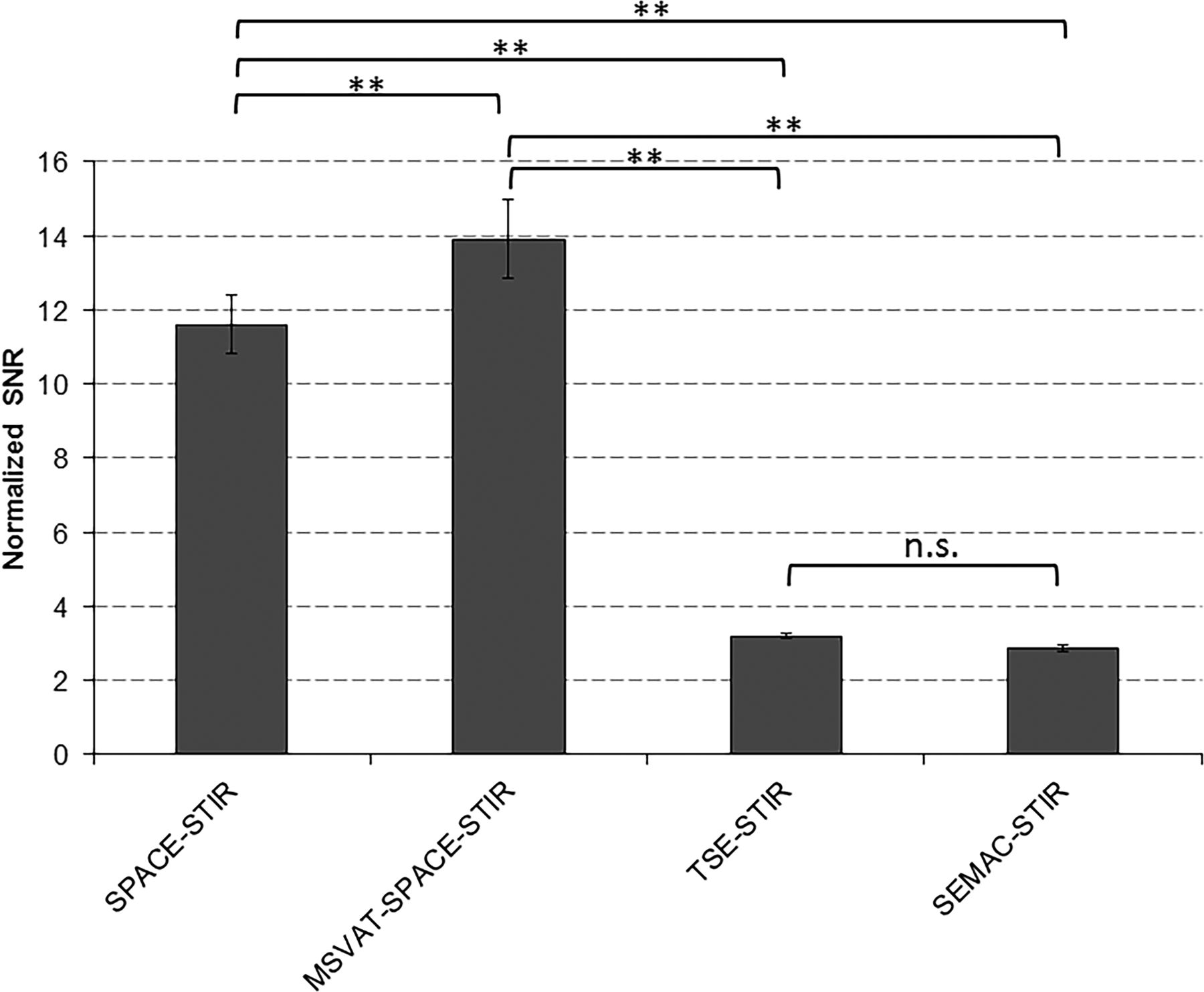
  • Fig 4.
    • Download figure
    • Open in new tab
    • Download powerpoint
    Fig 4.

    nSNR values of all used sequences. Double asterisks indicate P ≤ .001). n.s. indicates not significant.

  • Fig 5.
    • Download figure
    • Open in new tab
    • Download powerpoint
    Fig 5.

    Mean visibility scores of in vitro images of the 8 anatomic structures in all STIR sequences. The asterisk indicates P ≤ .05; double asterisks, P ≤ .001.

  • Fig 6.
    • Download figure
    • Open in new tab
    • Download powerpoint
    Fig 6.

    Two patients examined with MSVAT-SPACE-STIR. A, A 25-year-old woman with dysesthesia in the right mandible and chin after wisdom tooth extraction in the right mandible (asterisk indicates the extraction site). Increased signal intensity of the neurovascular bundle (white solid arrow) compared with the healthy side (white dashed arrow) in curved multiplane reconstructions of MSVAT-SPACE-STIR, suggesting nerve damage. Note the small amount of artifacts around the implant-supported crown (hash tag) and incomplete bone marrow conversion resulting in bright signal in STIR images on both sides (double asterisks). B, An 8-year-old child after drainage of an abscess in the right mandible with residual soft-tissue inflammation in the right lateral gingiva (white arrows in reformatted axial [left]) and coronal [right]) images).

Tables

  • Figures
  • Parameters of all sequences

    SequenceTR/TE (ms)Voxel Size (mm)FOV (mm)MatrixReadout Bandwidth (Hz/Px)SlicesSlice-Encoding Steps or Oversampling (%)VATTime (min:sec)
    SPACE-STIR2500/1310.55 × 0.55 × 0.55140 × 1242565017255.6No14:02
    MSVAT-SPACE-STIR2500/1990.55 × 0.55 × 0.55140 × 842565287255.6Yes06:04
    TSE-STIR5100/440.59 × 0.59 × 1.5150 × 15025659225NoNo03:36
    SEMAC-STIR5100/450.59 × 0.59 × 1.5150 × 150256592254Yes06:19
    • Note:—VAT indicates view angle tilting.

Next
Back to top
Advertisement
Print
Download PDF
Email Article

Thank you for your interest in spreading the word on American Journal of Neuroradiology.

NOTE: We only request your email address so that the person you are recommending the page to knows that you wanted them to see it, and that it is not junk mail. We do not capture any email address.

Enter multiple addresses on separate lines or separate them with commas.
MSVAT-SPACE-STIR and SEMAC-STIR for Reduction of Metallic Artifacts in 3T Head and Neck MRI
(Your Name) has sent you a message from American Journal of Neuroradiology
(Your Name) thought you would like to see the American Journal of Neuroradiology web site.
CAPTCHA
This question is for testing whether or not you are a human visitor and to prevent automated spam submissions.
Cite this article
T. Hilgenfeld, M. Prager, F.S. Schwindling, M. Nittka, P. Rammelsberg, M. Bendszus, S. Heiland, A. Juerchott
MSVAT-SPACE-STIR and SEMAC-STIR for Reduction of Metallic Artifacts in 3T Head and Neck MRI
American Journal of Neuroradiology May 2018, DOI: 10.3174/ajnr.A5678

Citation Manager Formats

  • BibTeX
  • Bookends
  • EasyBib
  • EndNote (tagged)
  • EndNote 8 (xml)
  • Medlars
  • Mendeley
  • Papers
  • RefWorks Tagged
  • Ref Manager
  • RIS
  • Zotero
0 Responses
Respond to this article
Share
Bookmark this article
MSVAT-SPACE-STIR and SEMAC-STIR for Reduction of Metallic Artifacts in 3T Head and Neck MRI
T. Hilgenfeld, M. Prager, F.S. Schwindling, M. Nittka, P. Rammelsberg, M. Bendszus, S. Heiland, A. Juerchott
American Journal of Neuroradiology May 2018, DOI: 10.3174/ajnr.A5678
del.icio.us logo Twitter logo Facebook logo Mendeley logo
  • Tweet Widget
  • Facebook Like
  • Google Plus One
Purchase

Jump to section

  • Article
    • Abstract
    • ABBREVIATIONS:
    • Materials and Methods
    • Results
    • Discussion
    • Conclusions
    • Acknowledgments
    • Footnotes
    • References
  • Figures & Data
  • Info & Metrics
  • Responses
  • References
  • PDF

Related Articles

  • No related articles found.
  • PubMed
  • Google Scholar

Cited By...

  • No citing articles found.
  • Crossref (16)
  • Google Scholar

This article has been cited by the following articles in journals that are participating in Crossref Cited-by Linking.

  • Recent advances in MRI of the head and neck, skull base and cranial nerves: new and evolving sequences, analyses and clinical applications
    Philip Touska, Steve E. J. Connor
    The British Journal of Radiology 2019 92 1104
  • Radiomic biomarkers for head and neck squamous cell carcinoma
    Stephanie Tanadini-Lang, Panagiotis Balermpas, Matthias Guckenberger, Matea Pavic, Oliver Riesterer, Diem Vuong, Marta Bogowicz
    Strahlentherapie und Onkologie 2020 196 10
  • In vivo accuracy of dental magnetic resonance imaging in assessing maxillary molar furcation involvement: A feasibility study in humans
    Alexander Juerchott, Meysam Sohani, Franz Sebastian Schwindling, Johann M. E. Jende, Felix T. Kurz, Peter Rammelsberg, Sabine Heiland, Martin Bendszus, Tim Hilgenfeld
    Journal of Clinical Periodontology 2020 47 7
  • Comparison of non‐contrast‐enhanced dental magnetic resonance imaging and cone‐beam computed tomography in assessing the horizontal and vertical components of furcation defects in maxillary molars: An in vivo feasibility study
    Alexander Juerchott, Meysam Sohani, Franz Sebastian Schwindling, Johann M. E. Jende, Felix T. Kurz, Peter Rammelsberg, Sabine Heiland, Martin Bendszus, Tim Hilgenfeld
    Journal of Clinical Periodontology 2020 47 12
  • MRI Metal Artifact Reduction Sequence for Auditory Implants: First Results with a Transcutaneous Bone Conduction Implant
    Wilhelm Wimmer, Arsany Hakim, Claus Kiefer, Manuela Pastore-Wapp, Lukas Anschuetz, Marco D. Caversaccio, Franca Wagner
    Audiology and Neurotology 2019 24 2
  • Fully guided implant surgery using Magnetic Resonance Imaging – An in vitro study on accuracy in human mandibles
    Tabea Flügge, Ute Ludwig, Gita Winter, Philipp Amrein, Florian Kernen, Katja Nelson
    Clinical Oral Implants Research 2020 31 8
  • Artifacts in magnetic resonance imaging caused by dental materials: a systematic review
    Lauren Bohner, Marcel Hanisch, Newton Sesma, Moritz Blanck-Lubarsch, Johannes Kleinheinz
    Dentomaxillofacial Radiology 2022 51 6
  • Basic and Advanced Metal-Artifact Reduction Techniques at Ultra-High Field 7-T Magnetic Resonance Imaging—Phantom Study Investigating Feasibility and Efficacy
    Christoph Germann, Anna L. Falkowski, Constantin von Deuster, Daniel Nanz, Reto Sutter
    Investigative Radiology 2022 57 6
  • 3D MRI of the Spine
    Meghan Sahr, Ek Tsoon Tan, Darryl B. Sneag
    Seminars in Musculoskeletal Radiology 2021 25 03
  • Magnetic resonance imaging artefacts caused by orthodontic appliances and/or implant-supported prosthesis: a systematic review
    Katrine Mølgaard Johannsen, João Marcus de Carvalho E Silva Fuglsig, Brian Hansen, Ann Wenzel, Rubens Spin-Neto
    Oral Radiology 2023 39 2

More in this TOC Section

  • ASL Sensitivity for Head and Neck Paraganglioma
  • Post SRS Peritumoral Hyperintense Signal of VSs
  • Contrast Enhanced Pituitary CISS/FIESTA
Show more Head and Neck Imaging

Similar Articles

Advertisement

Indexed Content

  • Current Issue
  • Accepted Manuscripts
  • Article Preview
  • Past Issues
  • Editorials
  • Editor's Choice
  • Fellows' Journal Club
  • Letters to the Editor
  • Video Articles

Cases

  • Case Collection
  • Archive - Case of the Week
  • Archive - Case of the Month
  • Archive - Classic Case

More from AJNR

  • Trainee Corner
  • Imaging Protocols
  • MRI Safety Corner
  • Book Reviews

Multimedia

  • AJNR Podcasts
  • AJNR Scantastics

Resources

  • Turnaround Time
  • Submit a Manuscript
  • Submit a Video Article
  • Submit an eLetter to the Editor/Response
  • Manuscript Submission Guidelines
  • Statistical Tips
  • Fast Publishing of Accepted Manuscripts
  • Graphical Abstract Preparation
  • Imaging Protocol Submission
  • Evidence-Based Medicine Level Guide
  • Publishing Checklists
  • Author Policies
  • Become a Reviewer/Academy of Reviewers
  • News and Updates

About Us

  • About AJNR
  • Editorial Board
  • Editorial Board Alumni
  • Alerts
  • Permissions
  • Not an AJNR Subscriber? Join Now
  • Advertise with Us
  • Librarian Resources
  • Feedback
  • Terms and Conditions
  • AJNR Editorial Board Alumni

American Society of Neuroradiology

  • Not an ASNR Member? Join Now

© 2025 by the American Society of Neuroradiology All rights, including for text and data mining, AI training, and similar technologies, are reserved.
Print ISSN: 0195-6108 Online ISSN: 1936-959X

Powered by HighWire