
Article Figures & Data
Figures
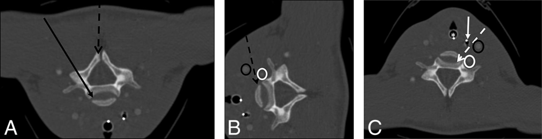
- Fig 1.
CT-guided cervical spine biopsies were performed using anterolateral, posterolateral, posterior, or lateral approaches. A, CT angiogram of the neck flipped vertically to depict prone positioning for a posterolateral- or posterior-approach cervical spine biopsy. The black arrow demonstrates a posterolateral-approach biopsy of the vertebral body or disc. The dashed black arrow demonstrates a directly posterior approach to a lesion in the spinous process. B, CT angiogram of the neck rotated to depict decubitus positioning for a lateral-approach biopsy to the vertebral body (dashed black arrow). Note that the course of the biopsy needle is between the carotid artery (black oval) and vertebral artery (white oval). C, CT angiogram of the neck with the patient in a supine position for an anterolateral-approach cervical spine biopsy. The needle (dashed white arrow) passes between the intubated trachea (white asterisk), nasogastric tube/esophagus (solid white arrow), carotid artery (black oval), and vertebral artery (white oval).
- Fig 2.
A 70-year-old woman with a history of lung cancer and a new right C5 mass extending into the adjacent soft tissues. A, Axial T1 fat-suppressed postcontrast MR image with a large right C5 mass (black asterisk) extending into the adjacent soft tissues and vertebral canal. B, Intraprocedural CT angiogram with the patient in a left lateral decubitus position shows the mass (white asterisk) and adjacent vascular structures (vertebral artery, white arrow; common carotid artery, white oval; jugular vein, black oval). C, Intraprocedural CT image with the patient in the left lateral decubitus position demonstrates the biopsy using a coaxial soft-tissue biopsy needle passing between vascular structures (vertebral artery, white arrow; common carotid artery, white oval; jugular vein, black oval) into the mass (white asterisk) using a lateral approach. The surgical pathology result was metastatic lung adenocarcinoma.
- Fig 3.
A 55-year-old woman with right neck and arm pain. A, Axial T2-weighted MR image with a large right C2 vertebral mass (black asterisk) surrounding the vertebral artery (white arrow). B, Intraprocedural CT angiogram soft-tissue-windowed image with the patient prone shows the mass (white asterisk) and adjacent vascular structures (deep cervical vein, white oval; vertebral artery, black oval). C, Intraprocedural CT bone-windowed image with the patient prone demonstrates the bone-access needle (white arrow) in the posterolateral right lamina of C2 and a soft-tissue biopsy needle (dashed white arrow) placed coaxially into the mass (white asterisk), using a posterolateral approach. The surgical pathology result was chordoma.
- Fig 4.
A 51-year-old woman with prior discectomy and interbody fusion. A, Sagittal T1 fat-suppressed postcontrast MR image with evidence of osteomyelitis at C7 (black asterisk), prevertebral inflammation, and epidural abscess (white arrow). Intraprocedural oblique axial (B) and oblique sagittal (C) CT reconstructed images during the biopsy procedure show the biopsy needle placed from a posterolateral approach, through the right C7 pedicle (white arrow) into the vertebral body (black asterisk) and subsequently the C6–C7 disc space (black arrow). Microbiology grew Staphylococcus aureus, and pathology showed osteomyelitis.


Tables
Biopsy Needle Bone Lesion Samples Bonopty 14/15 (AprioMed) 8 Arrow OnControl 11/13 (Teleflex) 12 Arrow OnControl 12/14 (Teleflex) 5 Achieve 16 (CareFusion) 5 Bard 14 (Bard Peripheral Vascular) 5 Bard 16 (Bard Peripheral Vascular) 1 Tru-Cut 18 (CareFusion) 3 Tru-Cut 20 (CareFusion) 1 ↵a Six procedures used both bone and soft-tissue biopsy needle systems for 40 total needle systems used.
Biopsy Needle Soft-Tissue Samples Bonopty 14/15 (AprioMed) 16 Arrow OnControl 11/13 (Teleflex) 20 Arrow OnControl 12/14 (Teleflex) 3 Achieve 14 (CareFusion) 1 Achieve 16 (CareFusion) 1 Achieve 18 (CareFusion) 1 Temno Evolution 18 (CareFusion) 2 Tru-Cut 16 (CareFusion) 1 Tru-Cut 20 (CareFusion) 1 ↵a Seven procedures used both bone and soft-tissue biopsy needle systems, 9 procedures used only soft-tissue needle systems, and 23 procedures used only bone biopsy needle systems for 46 total needle systems used.
- Table 3:
Histopathologic results of CT-guided cervical bone biopsies for primary tumors or metastatic disease
Malignant (n = 24 cases) Myeloma 4 NSCLC/adenocarcinoma 4 Osteoid osteoma 3 Chordoma 2 Giant cell tumor 2 Indeterminate (abnormal) 2 Metastatic carcinoma Unknown primary 2 Epithelioid hemangioendothelioma 1 Metastatic carcinoma Breast primary 1 Metastatic papillary Thyroid carcinoma 1 Aneurysmal bone cyst focal 1 Eosinophilic granuloma 1 Benign (n = 10 cases) Normala 6 Marrow fibrosis 1 Chronic inflammation 1 Micrococcus species 1 Osteomyelitis 1 Note:—NSCLC indicates non-small-cell lung carcinoma.
↵a Normal means biopsy demonstrating normal bone seen on histopathology.


Jump to section
Related Articles
Cited By...
- Diagnostic yield, accuracy, and complication rate of CT-guided biopsy for spinal lesions: a systematic review and meta-analysis
- Percutaneous CT-Guided Core Needle Biopsies of Head and Neck Masses: Technique, Histopathologic Yield, and Safety at a Single Academic Institution
- Reduction of Radiation Dose and Scanning Time While Preserving Diagnostic Yield: A Comparison of Battery-Powered and Manual Bone Biopsy Systems