
Article Figures & Data
Figures
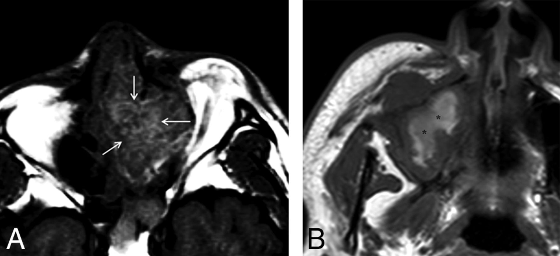
- Fig 1.
Two different types of SNM containing high signal intensity on T1WI with the presence (A) or absence (B) of a T1-SP. Although both tumors have intrinsic high signal intensity, only A demonstrates a regular pattern of the alternating hyperintense and hypointense striations, so-called T1-SP (arrows). In contrast, the high signal intensity in B appears amorphous without the alternating hyperintense and hypointense striations in a regular pattern (asterisks).
- Fig 2.
SNM in the right frontal sinus displaying a diffuse T1-SP. Precontrast T1WI demonstrates a mass with the alternating hyperintense and hypointense bands, the so-called T1-SP.
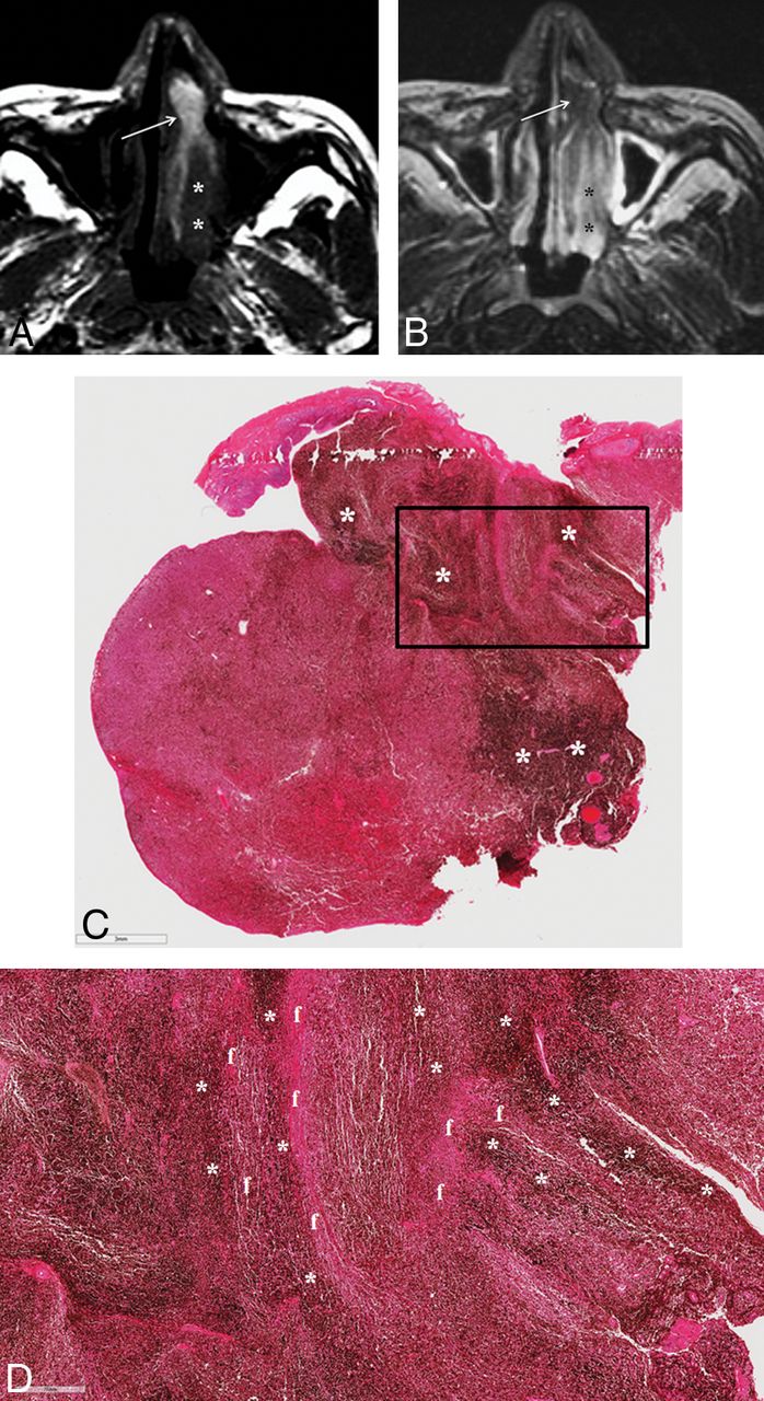
- Fig 3.
SNM displaying a partial T1-SP. A, Precontrast axial T1WI demonstrates an elongated mass with heterogeneous signal intensity in the left nasal cavity. While the anterior portion of the mass shows the alternating hyperintense and hypointense bands, the so-called T1-SP (arrow), the posterior portion is the soft-tissue component with the signal intensity isointense to the brain stem (asterisks). B, On fat-suppressed axial T2WI, the anterior portion of the mass appears isointense to the brain stem (arrow), while the posterior portion becomes hyperintense (asterisks). Insufficient facial and buccal fat suppression was caused by magnetic field inhomogeneity due to metallic dental hardware. C. Photomicrograph reveals an uneven distribution of melanin pigments (dark bands marked with asterisks). The bar on the left bottom indicates 3 mm (hematoxylin-eosin, original magnification ×7). D, Photomicrograph with higher magnification corresponding to the box in C shows the area of the alternating melanin (dark bands marked with asterisks) and fibrous (f) bands. The bar on the left bottom indicates 700 μm (hematoxylin-eosin, original magnification ×30).
- Fig 4.
Examples of nonmelanomatous sinonasal tumors displaying a T1-SP (arrows). A, Squamous cell carcinoma. B, Lymphoma.


Tables
- Table 1:
Visualization of a T1-SP on MR imaging in sinonasal melanomas and nonmelanomatous malignant sinonasal tumors based on consensus readinga
Total Hyperintense Foci on T1WI Present Absent T1-SP (+) T1-SP (−) Sinonasal melanomab 31 23 4 4 Nonmelanomatous malignant tumorb 106 3 19 84 Squamous cell carcinoma 45 2 6 37 Lymphoma 22 1 2 19 Adenoid cystic carcinoma 10 0 4 6 Rhabdomyosarcoma 5 0 0 5 Neuroendocrine carcinoma 4 0 2 2 Adenocarcinoma 4 0 2 2 Malignant fibrous histiocytoma 3 0 1 2 Poorly differentiated carcinoma 3 0 0 3 Spindle cell sarcoma 3 0 0 3 Esthesioneuroblastoma 1 0 0 1 Small round cell sarcoma 1 0 1 0 Inflammatory myofibroblastic sarcoma 1 0 0 1 Malignant peripheral nerve sheath tumor 1 0 0 1 Ewing sarcoma 1 0 1 0 Chondrosarcoma 1 0 0 1 Myoepithelial carcinoma 1 0 0 1 Presence or Absence of T1-SP Observer 1 Observer 2 Observer 3 Overallb + − + − + − + − Sinonasal melanoma 22 9 26 5 20 11 23 8 Nonmelanomatous malignant sinonasal tumors 7 99 1 105 3 103 3 103 - Table 3:
Correlation of histopathologic and MR imaging features of 23 sinonasal melanomasa
T1-SP (+) T1-SP (−) P Valueb Melanin .915 Melanotic 14 4 .043c Abundant 9 0 Moderate 5 3 Amelanotic 4 1 Hemorrhage .280 Present 12 2 Absent 6 3 Cell type .399 Epithelioid 12 3 Spindle 3 0 Mixed 3 2 Note:—+ indicates presence; −, absence.
↵a Data are presented as numbers of tumors.
↵b Comparison of the prevalence of a T1-SP according to the presence of melanin, the presence of hemorrhage, and the different cell types using the χ2 test.
↵c Comparison of the prevalence of a T1-SP between tumors with abundant melanin and the group of tumors with no and moderate melanin by the χ2 test.


