Advertisement
  • Main menu
  • User menu
  • Search
  • English ▼
    • English
    • Afrikaans
    • Albanian
    • Amharic
    • Arabic
    • Armenian
    • Azerbaijani
    • Basque
    • Belarusian
    • Bengali
    • Bosnian
    • Bulgarian
    • Catalan
    • Cebuano
    • Chichewa
    • Chinese (Simplified)
    • Chinese (Traditional)
    • Corsican
    • Croatian
    • Czech
    • Danish
    • Dutch
    • Esperanto
    • Estonian
    • Filipino
    • Finnish
    • French
    • Frisian
    • Galician
    • Georgian
    • German
    • Greek
    • Gujarati
    • Haitian Creole
    • Hausa
    • Hawaiian
    • Hebrew
    • Hindi
    • Hmong
    • Hungarian
    • Icelandic
    • Igbo
    • Indonesian
    • Irish
    • Italian
    • Japanese
    • Javanese
    • Kannada
    • Kazakh
    • Khmer
    • Korean
    • Kurdish (Kurmanji)
    • Kyrgyz
    • Lao
    • Latin
    • Latvian
    • Lithuanian
    • Luxembourgish
    • Macedonian
    • Malagasy
    • Malay
    • Malayalam
    • Maltese
    • Maori
    • Marathi
    • Mongolian
    • Myanmar (Burmese)
    • Nepali
    • Norwegian
    • Pashto
    • Persian
    • Polish
    • Portuguese
    • Punjabi
    • Romanian
    • Russian
    • Samoan
    • Scottish Gaelic
    • Serbian
    • Sesotho
    • Shona
    • Sindhi
    • Sinhala
    • Slovak
    • Slovenian
    • Somali
    • Spanish
    • Sudanese
    • Swahili
    • Swedish
    • Tajik
    • Tamil
    • Telugu
    • Thai
    • Turkish
    • Ukrainian
    • Urdu
    • Uzbek
    • Vietnamese
    • Welsh
    • Xhosa
    • Yiddish
    • Yoruba
    • Zulu

Main menu

  • Home
  • Content
    • Current Issue
    • Accepted Manuscripts
    • Article Preview
    • Past Issue Archive
    • Video Articles
    • AJNR Case Collection
    • Case of the Week Archive
    • Case of the Month Archive
    • Classic Case Archive
  • Special Collections
    • Advancing NeuroMRI with High-Relaxivity Contrast Agents
    • AJNR Awards
    • Low-Field MRI
    • Alzheimer Disease
    • ASNR Foundation Special Collection
    • Photon-Counting CT
    • View All
  • Multimedia
    • AJNR Podcasts
    • AJNR SCANtastic
    • Trainee Corner
    • MRI Safety Corner
    • Imaging Protocols
  • For Authors
    • Submit a Manuscript
    • Submit a Video Article
    • Submit an eLetter to the Editor/Response
    • Manuscript Submission Guidelines
    • Statistical Tips
    • Fast Publishing of Accepted Manuscripts
    • Graphical Abstract Preparation
    • Imaging Protocol Submission
    • Author Policies
  • About Us
    • About AJNR
    • Editorial Board
    • Editorial Board Alumni
  • More
    • Become a Reviewer/Academy of Reviewers
    • Subscribers
    • Permissions
    • Alerts
    • Feedback
    • Advertisers
    • ASNR Home

User menu

  • Alerts
  • Log in

Search

  • Advanced search
American Journal of Neuroradiology

American Journal of Neuroradiology

ASHNR American Society of Functional Neuroradiology ASHNR American Society of Pediatric Neuroradiology ASSR
  • Alerts
  • Log in

Advanced Search

English ▼
  • English
  • Afrikaans
  • Albanian
  • Amharic
  • Arabic
  • Armenian
  • Azerbaijani
  • Basque
  • Belarusian
  • Bengali
  • Bosnian
  • Bulgarian
  • Catalan
  • Cebuano
  • Chichewa
  • Chinese (Simplified)
  • Chinese (Traditional)
  • Corsican
  • Croatian
  • Czech
  • Danish
  • Dutch
  • Esperanto
  • Estonian
  • Filipino
  • Finnish
  • French
  • Frisian
  • Galician
  • Georgian
  • German
  • Greek
  • Gujarati
  • Haitian Creole
  • Hausa
  • Hawaiian
  • Hebrew
  • Hindi
  • Hmong
  • Hungarian
  • Icelandic
  • Igbo
  • Indonesian
  • Irish
  • Italian
  • Japanese
  • Javanese
  • Kannada
  • Kazakh
  • Khmer
  • Korean
  • Kurdish (Kurmanji)
  • Kyrgyz
  • Lao
  • Latin
  • Latvian
  • Lithuanian
  • Luxembourgish
  • Macedonian
  • Malagasy
  • Malay
  • Malayalam
  • Maltese
  • Maori
  • Marathi
  • Mongolian
  • Myanmar (Burmese)
  • Nepali
  • Norwegian
  • Pashto
  • Persian
  • Polish
  • Portuguese
  • Punjabi
  • Romanian
  • Russian
  • Samoan
  • Scottish Gaelic
  • Serbian
  • Sesotho
  • Shona
  • Sindhi
  • Sinhala
  • Slovak
  • Slovenian
  • Somali
  • Spanish
  • Sudanese
  • Swahili
  • Swedish
  • Tajik
  • Tamil
  • Telugu
  • Thai
  • Turkish
  • Ukrainian
  • Urdu
  • Uzbek
  • Vietnamese
  • Welsh
  • Xhosa
  • Yiddish
  • Yoruba
  • Zulu
  • Home
  • Content
    • Current Issue
    • Accepted Manuscripts
    • Article Preview
    • Past Issue Archive
    • Video Articles
    • AJNR Case Collection
    • Case of the Week Archive
    • Case of the Month Archive
    • Classic Case Archive
  • Special Collections
    • Advancing NeuroMRI with High-Relaxivity Contrast Agents
    • AJNR Awards
    • Low-Field MRI
    • Alzheimer Disease
    • ASNR Foundation Special Collection
    • Photon-Counting CT
    • View All
  • Multimedia
    • AJNR Podcasts
    • AJNR SCANtastic
    • Trainee Corner
    • MRI Safety Corner
    • Imaging Protocols
  • For Authors
    • Submit a Manuscript
    • Submit a Video Article
    • Submit an eLetter to the Editor/Response
    • Manuscript Submission Guidelines
    • Statistical Tips
    • Fast Publishing of Accepted Manuscripts
    • Graphical Abstract Preparation
    • Imaging Protocol Submission
    • Author Policies
  • About Us
    • About AJNR
    • Editorial Board
    • Editorial Board Alumni
  • More
    • Become a Reviewer/Academy of Reviewers
    • Subscribers
    • Permissions
    • Alerts
    • Feedback
    • Advertisers
    • ASNR Home
  • Follow AJNR on Twitter
  • Visit AJNR on Facebook
  • Follow AJNR on Instagram
  • Join AJNR on LinkedIn
  • RSS Feeds

AJNR Awards, New Junior Editors, and more. Read the latest AJNR updates


Improved Turnaround Times | Median time to first decision: 12 days

Research ArticleHead and Neck Imaging
Open Access

Intravoxel Incoherent Motion MR Imaging in the Differentiation of Benign and Malignant Sinonasal Lesions: Comparison with Conventional Diffusion-Weighted MR Imaging

Z. Xiao, Z. Tang, J. Qiang, S. Wang, W. Qian, Y. Zhong, R. Wang, J. Wang, L. Wu, W. Tang and Z. Zhang
American Journal of Neuroradiology January 2018, DOI: https://doi.org/10.3174/ajnr.A5532
Z. Xiao
aFrom the Departments of Radiology (Z.X., Z.T., W.Q., R.W.)
  • Find this author on Google Scholar
  • Find this author on PubMed
  • Search for this author on this site
  • ORCID record for Z. Xiao
Z. Tang
aFrom the Departments of Radiology (Z.X., Z.T., W.Q., R.W.)
  • Find this author on Google Scholar
  • Find this author on PubMed
  • Search for this author on this site
  • ORCID record for Z. Tang
J. Qiang
eDepartment of Radiology (J.Q., Y.Z.), Jinshan Hospital of Shanghai Medical School, Fudan University, Shanghai, P. R. China
  • Find this author on Google Scholar
  • Find this author on PubMed
  • Search for this author on this site
  • ORCID record for J. Qiang
S. Wang
bPathology (S.W.)
  • Find this author on Google Scholar
  • Find this author on PubMed
  • Search for this author on this site
  • ORCID record for S. Wang
W. Qian
aFrom the Departments of Radiology (Z.X., Z.T., W.Q., R.W.)
  • Find this author on Google Scholar
  • Find this author on PubMed
  • Search for this author on this site
  • ORCID record for W. Qian
Y. Zhong
eDepartment of Radiology (J.Q., Y.Z.), Jinshan Hospital of Shanghai Medical School, Fudan University, Shanghai, P. R. China
  • Find this author on Google Scholar
  • Find this author on PubMed
  • Search for this author on this site
  • ORCID record for Y. Zhong
R. Wang
aFrom the Departments of Radiology (Z.X., Z.T., W.Q., R.W.)
  • Find this author on Google Scholar
  • Find this author on PubMed
  • Search for this author on this site
  • ORCID record for R. Wang
J. Wang
cRadiotherapy (J.W.)
  • Find this author on Google Scholar
  • Find this author on PubMed
  • Search for this author on this site
  • ORCID record for J. Wang
L. Wu
dOtolaryngology (L.W.), Eye & ENT Hospital of Shanghai Medical School, Fudan University, Shanghai, P. R. China
  • Find this author on Google Scholar
  • Find this author on PubMed
  • Search for this author on this site
  • ORCID record for L. Wu
W. Tang
fSiemens Healthcare Ltd (W.T., Z.Z.), Shanghai, P. R. China.
  • Find this author on Google Scholar
  • Find this author on PubMed
  • Search for this author on this site
  • ORCID record for W. Tang
Z. Zhang
fSiemens Healthcare Ltd (W.T., Z.Z.), Shanghai, P. R. China.
  • Find this author on Google Scholar
  • Find this author on PubMed
  • Search for this author on this site
  • ORCID record for Z. Zhang
  • Article
  • Figures & Data
  • Info & Metrics
  • Responses
  • References
  • PDF
Loading

Article Figures & Data

Figures

  • Tables
  • Fig 1.
    • Download figure
    • Open in new tab
    • Download powerpoint
    Fig 1.

    Scatterplots depicting the correlations between the patients' ages and ADC values (A), the patients' ages and D values (B), the patients' ages and D* values (C), and the patients' ages and f values (D) for all sinonasal lesions.

  • Fig 2.
    • Download figure
    • Open in new tab
    • Download powerpoint
    Fig 2.

    Comparisons of the mean ADC (A), D (B), D* (C), and f (D) values between benign and malignant sinonasal lesions using the Student t test. Triple asterisks indicate P < .001; double asterisks, P < .01.
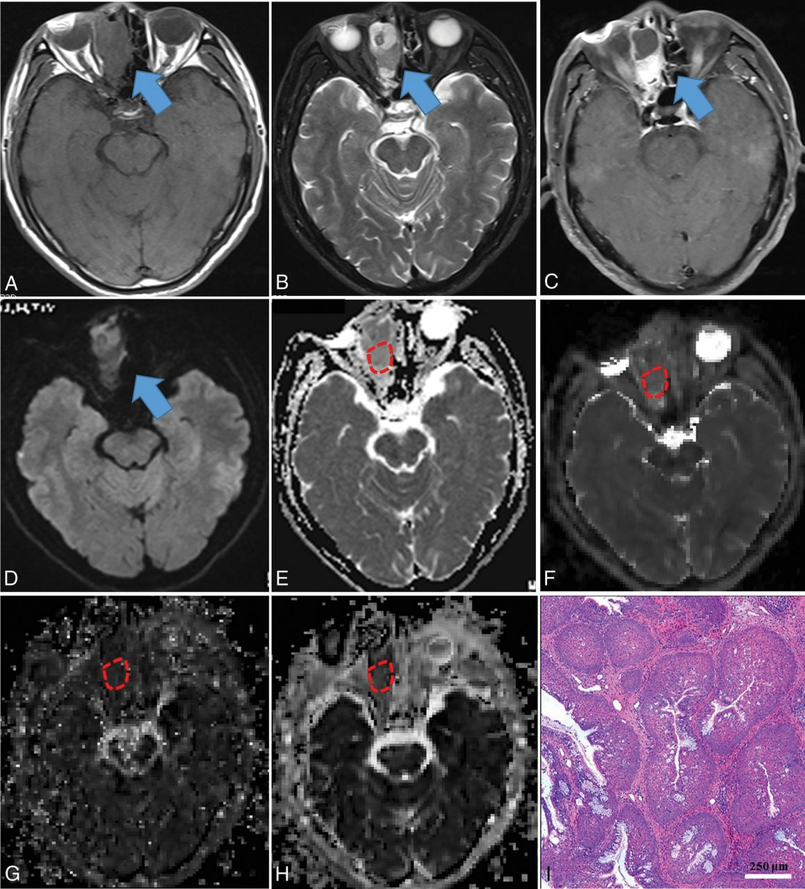
  • Fig 3.
    • Download figure
    • Open in new tab
    • Download powerpoint
    Fig 3.

    Inverted papilloma in a 71-year-old man. A mass was predominantly located in the right ethmoid sinus with involvement of the right nasal cavity (blue arrow), demonstrating heterogeneous hypo- (necrosis) to isointensity (tumor cells) on T1WI (A), iso- (tumor cells) to hyperintensity (necrosis) on T2WI (B), and heterogeneously intense enhancement on contrast-enhanced T1WI (C), compared with normal-appearing gray matter. The mass showed isointensity on the DWI (blue arrow) (D) compared with normal-appearing gray matter. On the ADC map (E), the mass showed a slightly high signal intensity with an ADC value of 1.425 × 10⁻³ mm2/s (red polygonal ROI). On IVIM images, the mass appeared obviously iso- to hyperintense on the D map (F) with a D value of 0.871 × 10⁻³ mm2/s and appeared isointense on the D* (G) and f maps (H) with D* and f values of 61.048 × 10⁻³ mm2/s and 25.651%, respectively (red polygonal ROIs). Hematoxylin-eosin staining (I) confirmed the mass as an inverted papilloma (original magnification, ×100; scale bar, 250 μm).

  • Fig 4.
    • Download figure
    • Open in new tab
    • Download powerpoint
    Fig 4.

    Squamous cell carcinoma in a 53-year-old man. A mass was predominantly located in the left ethmoid sinus with involvement of the ipsilateral orbit, nasal cavity, and sphenoid sinus (blue arrow), demonstrating heterogeneous hypo- (necrosis) to isointensity (tumor cells) on T1WI (A), iso- (tumor cells) to hyperintensity (necrosis) on T2WI (B), and heterogeneously intense enhancement on contrast-enhanced T1WI (C), compared with normal-appearing gray matter. The mass showed heterogeneously hypo- to hyperintensity on the DWI (blue arrow) (D) compared with normal-appearing gray matter. On the ADC map (E), the mass showed hypointensity with an ADC value of 0.872 × 10⁻³ mm2/s (red polygonal ROI). On IVIM images, the mass appeared dramatically hypointense on the D map (F), with a D value of 0.533 × 10⁻³ mm2/s, hypo- to isointense on the D* map (G), and iso- to hyperintense on the f map (H), with D* and f values of 77.473 × 10⁻³ mm2/s and 22.966%, respectively (red polygonal ROIs). Hematoxylin-eosin staining (I) confirmed the mass as a squamous cell carcinoma (original magnification, × 200; scale bar, 50 μm).

  • Fig 5.
    • Download figure
    • Open in new tab
    • Download powerpoint
    Fig 5.

    ROC curves with 10-fold cross-validation of single- parameter models (including ADC, D, and f) and multiparametric models (including ADC+f, ADC+D, D+f, and ADC+D+f) in the contrast-enhancing lesions for differentiating benign from malignant sinonasal lesions.

Tables

  • Figures
    • View popup
    Table 1:

    Demographics and histology of benign and malignant sinonasal lesionsa

    ParametersBenign Lesions (n = 56)Malignant Lesions (n = 75)
    Mean age (yr)43.86 ± 14.1152.27 ± 15.21
    Sex (female/male)24:3224:51
    Histologic subtypesInflammatory polyps (28)Squamous cell carcinoma (23)
    Inverted papilloma (14)Olfactory neuroblastoma (13)
    Fibroangioma (5)Malignant melanoma (12)
    Spindle cell tumor (4)Rhabdomyosarcoma (9)
    Schwannoma (2)Lymphoma (6)
    Ossifying fibroma (2)Adenoid cystic carcinoma (5)
    Enamel cell tumor (1)Undifferentiated carcinoma (2)
    Osteosarcoma (2)
    Neuroendocrine carcinoma (2)
    Malignant fibrohistiocytoma (1)
    • ↵a Data in parentheses indicate the number of corresponding patients. All inverted papillomas did not have the potential for association with/conversion to squamous cell carcinoma, proven by histopathology.

    • View popup
    Table 2:

    Inter- and intrareader reproducibility for ADC, D, D*, and f measurementsa

    ParametersICC
    InterreaderIntrareader
    ADC (×10−3 mm2/s)0.961 (0.913–0.977)0.954 (0.871–0.980)
    D (×10−3 mm2/s)0.942 (0.860–0.986)0.936 (0.884–0.975)
    D* (×10−3 mm2/s)0.840 (0.762–0.931)0.848 (0.766–0.909)
    f (%)0.908 (0.819–0.965)0.922 (0.835–0.964)
    • Note:—ICC indicates intraclass correlation coefficient.

    • ↵a Data in parentheses are 95% confidence intervals.

    • View popup
    Table 3:

    Comparisons of ADC, D, D*, and f values between benign and malignant sinonasal lesionsa

    ParametersBenign Lesions (n = 56)Malignant Lesions (n = 75)P Value
    ADC (×10−3 mm2/s)1.163 ± 0.3540.862 ± 0.258<.001
    D (× 10−3 mm2/s)1.322 ± 0.3470.677 ± 0.299<.001
    D* (×10−3 mm2/s)90.470 ± 40.75686.445 ± 22.865.474
    f (%)16.656 ± 4.27419.211 ± 4.066.001
    • ↵a Except for the P values, data are expressed as the mean ± SD.

    • View popup
    Table 4:

    Diagnostic performance of single parameters (ADC, D and f) and combined parameters (ADC+f, ADC+D, D+f, and ADC+D+f) for the differentiation of benign and malignant sinonasal lesions using receiver operating characteristic curve analysis with 10-fold cross-validation

    TVAUCCross-Validated AUCSensitivitySpecificityAccuracyPrecisionF Score
    ADC0.9190.7540.7350.8000.5470.6860.6840.738
    D0.7150.9070.8990.8410.8160.8310.8660.853
    f16.9950.6760.6560.7230.4720.6100.6270.723
    ADC+f–0.9100.9070.8570.7920.8310.8570.857
    ADC+D–0.9440.9140.8130.7780.7970.8130.813
    D+f–0.9380.9350.9140.8130.8730.8770.895
    ADC+D+f–0.9510.9440.9000.8330.8730.8870.894
    • Note:—TV indicates threshold value.

Next
Back to top
Advertisement
Print
Download PDF
Email Article
Cite this article
0 Responses
Respond to this article
Share
Bookmark this article
Intravoxel Incoherent Motion MR Imaging in the Differentiation of Benign and Malignant Sinonasal Lesions: Comparison with Conventional Diffusion-Weighted MR Imaging
Z. Xiao, Z. Tang, J. Qiang, S. Wang, W. Qian, Y. Zhong, R. Wang, J. Wang, L. Wu, W. Tang, Z. Zhang
American Journal of Neuroradiology Jan 2018, DOI: 10.3174/ajnr.A5532
del.icio.us logo Twitter logo Facebook logo Mendeley logo
  • Tweet Widget
Purchase

Jump to section

  • Article
    • Abstract
    • ABBREVIATIONS:
    • Materials and Methods
    • Results
    • Discussion
    • Conclusions
    • Footnotes
    • References
  • Figures & Data
  • Info & Metrics
  • Responses
  • References
  • PDF

Related Articles

  • No related articles found.
  • PubMed
  • Google Scholar

Cited By...

  • No citing articles found.
  • Crossref
  • Google Scholar

This article has not yet been cited by articles in journals that are participating in Crossref Cited-by Linking.

More in this TOC Section

  • Diagnostic Performance of US in Neck Node NIRADS 2
  • Hydrops Herniation into the Semicircular Canals
  • ASL Sensitivity for Head and Neck Paraganglioma
Show more HEAD AND NECK IMAGING

Similar Articles

  • ADC for Differentiation between Posttreatment Changes and Recurrence in Head and Neck Cancer: A Systematic Review and Meta-analysis
See more
Advertisement

Indexed Content

  • Current Issue
  • Accepted Manuscripts
  • Article Preview
  • Past Issues
  • Editorials
  • Editor's Choice
  • Fellows' Journal Club
  • Letters to the Editor
  • Video Articles

Cases

  • Case Collection
  • Archive - Case of the Week
  • Archive - Case of the Month
  • Archive - Classic Case

More from AJNR

  • Trainee Corner
  • Imaging Protocols
  • MRI Safety Corner
  • Book Reviews

Multimedia

  • AJNR Podcasts
  • AJNR Scantastics

Resources

  • Turnaround Time
  • Submit a Manuscript
  • Submit a Video Article
  • Submit an eLetter to the Editor/Response
  • Manuscript Submission Guidelines
  • Statistical Tips
  • Fast Publishing of Accepted Manuscripts
  • Graphical Abstract Preparation
  • Imaging Protocol Submission
  • Evidence-Based Medicine Level Guide
  • Publishing Checklists
  • Author Policies
  • Become a Reviewer/Academy of Reviewers
  • News and Updates

About Us

  • About AJNR
  • Editorial Board
  • Editorial Board Alumni
  • Alerts
  • Permissions
  • Not an AJNR Subscriber? Join Now
  • Advertise with Us
  • Librarian Resources
  • Feedback
  • Terms and Conditions
  • AJNR Editorial Board Alumni

American Society of Neuroradiology

  • Not an ASNR Member? Join Now

© 2025 by the American Society of Neuroradiology All rights, including for text and data mining, AI training, and similar technologies, are reserved.
Print ISSN: 0195-6108 Online ISSN: 1936-959X

Powered by HighWire
Email this Article

Thank you for your interest in spreading the word on American Journal of Neuroradiology.

NOTE: We only request your email address so that the person you are recommending the page to knows that you wanted them to see it, and that it is not junk mail. We do not capture any email address.

Enter multiple addresses on separate lines or separate them with commas.
Intravoxel Incoherent Motion MR Imaging in the Differentiation of Benign and Malignant Sinonasal Lesions: Comparison with Conventional Diffusion-Weighted MR Imaging
(Your Name) has sent you a message from American Journal of Neuroradiology
(Your Name) thought you would like to see the American Journal of Neuroradiology web site.
CAPTCHA
This question is for testing whether or not you are a human visitor and to prevent automated spam submissions.
Cite this article
Z. Xiao, Z. Tang, J. Qiang, S. Wang, W. Qian, Y. Zhong, R. Wang, J. Wang, L. Wu, W. Tang, Z. Zhang
Intravoxel Incoherent Motion MR Imaging in the Differentiation of Benign and Malignant Sinonasal Lesions: Comparison with Conventional Diffusion-Weighted MR Imaging
American Journal of Neuroradiology Jan 2018, DOI: 10.3174/ajnr.A5532

Citation Manager Formats

  • BibTeX
  • Bookends
  • EasyBib
  • EndNote (tagged)
  • EndNote 8 (xml)
  • Medlars
  • Mendeley
  • Papers
  • RefWorks Tagged
  • Ref Manager
  • RIS
  • Zotero

We use cookies on this site to enhance your user experience. By clicking any link on this page you are giving your consent for us to set cookies.