Advertisement

Main menu

  • Home
  • Content
    • Current Issue
    • Accepted Manuscripts
    • Article Preview
    • Past Issue Archive
    • Video Articles
    • AJNR Case Collection
    • Case of the Week Archive
    • Case of the Month Archive
    • Classic Case Archive
  • Special Collections
    • AJNR Awards
    • Low-Field MRI
    • Alzheimer Disease
    • ASNR Foundation Special Collection
    • Photon-Counting CT
    • View All
  • Multimedia
    • AJNR Podcasts
    • AJNR SCANtastic
    • Trainee Corner
    • MRI Safety Corner
    • Imaging Protocols
  • For Authors
    • Submit a Manuscript
    • Submit a Video Article
    • Submit an eLetter to the Editor/Response
    • Manuscript Submission Guidelines
    • Statistical Tips
    • Fast Publishing of Accepted Manuscripts
    • Graphical Abstract Preparation
    • Imaging Protocol Submission
    • Author Policies
  • About Us
    • About AJNR
    • Editorial Board
    • Editorial Board Alumni
  • More
    • Become a Reviewer/Academy of Reviewers
    • Subscribers
    • Permissions
    • Alerts
    • Feedback
    • Advertisers
    • ASNR Home

User menu

  • Alerts
  • Log in

Search

  • Advanced search
American Journal of Neuroradiology

American Journal of Neuroradiology

ASHNR American Society of Functional Neuroradiology ASHNR American Society of Pediatric Neuroradiology ASSR
  • Alerts
  • Log in

Advanced Search

  • Home
  • Content
    • Current Issue
    • Accepted Manuscripts
    • Article Preview
    • Past Issue Archive
    • Video Articles
    • AJNR Case Collection
    • Case of the Week Archive
    • Case of the Month Archive
    • Classic Case Archive
  • Special Collections
    • AJNR Awards
    • Low-Field MRI
    • Alzheimer Disease
    • ASNR Foundation Special Collection
    • Photon-Counting CT
    • View All
  • Multimedia
    • AJNR Podcasts
    • AJNR SCANtastic
    • Trainee Corner
    • MRI Safety Corner
    • Imaging Protocols
  • For Authors
    • Submit a Manuscript
    • Submit a Video Article
    • Submit an eLetter to the Editor/Response
    • Manuscript Submission Guidelines
    • Statistical Tips
    • Fast Publishing of Accepted Manuscripts
    • Graphical Abstract Preparation
    • Imaging Protocol Submission
    • Author Policies
  • About Us
    • About AJNR
    • Editorial Board
    • Editorial Board Alumni
  • More
    • Become a Reviewer/Academy of Reviewers
    • Subscribers
    • Permissions
    • Alerts
    • Feedback
    • Advertisers
    • ASNR Home
  • Follow AJNR on Twitter
  • Visit AJNR on Facebook
  • Follow AJNR on Instagram
  • Join AJNR on LinkedIn
  • RSS Feeds

AJNR Awards, New Junior Editors, and more. Read the latest AJNR updates

Research ArticleHead and Neck Imaging
Open Access

MR Imaging Criteria for the Detection of Nasopharyngeal Carcinoma: Discrimination of Early-Stage Primary Tumors from Benign Hyperplasia

A.D. King, L.Y.S. Wong, B.K.H. Law, K.S. Bhatia, J.K.S. Woo, Q.-Y. Ai, T.Y. Tan, J. Goh, K.L. Chuah, F.K.F. Mo, K.C.A. Chan, A.T.C. Chan and A.C. Vlantis
American Journal of Neuroradiology December 2017, DOI: https://doi.org/10.3174/ajnr.A5493
A.D. King
aFrom the Departments of Imaging and Interventional Radiology (A.D.K., L.Y.S.W., B.K.H.L., Q.-Y.A.)
  • Find this author on Google Scholar
  • Find this author on PubMed
  • Search for this author on this site
  • ORCID record for A.D. King
L.Y.S. Wong
aFrom the Departments of Imaging and Interventional Radiology (A.D.K., L.Y.S.W., B.K.H.L., Q.-Y.A.)
  • Find this author on Google Scholar
  • Find this author on PubMed
  • Search for this author on this site
  • ORCID record for L.Y.S. Wong
B.K.H. Law
aFrom the Departments of Imaging and Interventional Radiology (A.D.K., L.Y.S.W., B.K.H.L., Q.-Y.A.)
  • Find this author on Google Scholar
  • Find this author on PubMed
  • Search for this author on this site
  • ORCID record for B.K.H. Law
K.S. Bhatia
eImaging Department (K.S.B.), St Mary's Hospital, Imperial College Healthcare, National Health Service Trust, London, UK
  • Find this author on Google Scholar
  • Find this author on PubMed
  • Search for this author on this site
  • ORCID record for K.S. Bhatia
J.K.S. Woo
cOtorhinolaryngology, Head and Neck Surgery (J.K.S.W., A.C.V.)
  • Find this author on Google Scholar
  • Find this author on PubMed
  • Search for this author on this site
  • ORCID record for J.K.S. Woo
Q.-Y. Ai
aFrom the Departments of Imaging and Interventional Radiology (A.D.K., L.Y.S.W., B.K.H.L., Q.-Y.A.)
  • Find this author on Google Scholar
  • Find this author on PubMed
  • Search for this author on this site
  • ORCID record for Q.-Y. Ai
T.Y. Tan
fDepartment of Radiology (T.Y.T.), Changi General Hospital, Singapore
  • Find this author on Google Scholar
  • Find this author on PubMed
  • Search for this author on this site
  • ORCID record for T.Y. Tan
J. Goh
gDepartments of Diagnostic Radiology (J.G.)
  • Find this author on Google Scholar
  • Find this author on PubMed
  • Search for this author on this site
  • ORCID record for J. Goh
K.L. Chuah
hPathology (K.L.C.), Tan Tock Seng Hospital, Singapore.
  • Find this author on Google Scholar
  • Find this author on PubMed
  • Search for this author on this site
  • ORCID record for K.L. Chuah
F.K.F. Mo
bClinical Oncology (F.K.F.M., A.T.C.C.)
  • Find this author on Google Scholar
  • Find this author on PubMed
  • Search for this author on this site
  • ORCID record for F.K.F. Mo
K.C.A. Chan
dDepartment of Chemical Pathology (K.C.A.C.), State Key Laboratory in Oncology in South China, Li Ka Shing Institute of Health Sciences, Faculty of Medicine, The Chinese University of Hong Kong, Prince of Wales Hospital, Hong Kong, S.A.R., China
  • Find this author on Google Scholar
  • Find this author on PubMed
  • Search for this author on this site
  • ORCID record for K.C.A. Chan
A.T.C. Chan
bClinical Oncology (F.K.F.M., A.T.C.C.)
  • Find this author on Google Scholar
  • Find this author on PubMed
  • Search for this author on this site
  • ORCID record for A.T.C. Chan
A.C. Vlantis
cOtorhinolaryngology, Head and Neck Surgery (J.K.S.W., A.C.V.)
  • Find this author on Google Scholar
  • Find this author on PubMed
  • Search for this author on this site
  • ORCID record for A.C. Vlantis
  • Article
  • Figures & Data
  • Supplemental
  • Info & Metrics
  • Responses
  • References
  • PDF
Loading

Article Figures & Data

Figures

  • Tables
  • Fig 1.
    • Download figure
    • Open in new tab
    • Download powerpoint
    Fig 1.

    A, Axial T1-weighted postcontrast MR image of the nasopharynx in a 51-year-old man with NPC (open arrow). An asymmetric tumor with a lateral center at the level of the left pharyngeal recess is confined to 1 side of the nasopharynx. The tumor exhibits homogeneous low contrast enhancement with an intact deep mucosal white line (small solid arrows) along the deep margin. B, Axial T1-weighted postcontrast MR image of the nasopharynx in a 68-year-old man with NPC (open arrow). An asymmetric tumor with a lateral center at the level of the left pharyngeal recess is confined to 1 side of the nasopharynx. The tumor exhibits homogeneous moderate contrast enhancement with a focal loss of the deep mucosal white line (small solid arrows) along the deep margin. C, Axial T1-weighted postcontrast MR image of the nasopharynx in a 44-year-old woman with NPC (open arrow). An asymmetric tumor with a lateral center at the level of the right side of the roof shows heterogeneous contrast enhancement.

  • Fig 2.
    • Download figure
    • Open in new tab
    • Download powerpoint
    Fig 2.

    Axial T1-weighted postcontrast MR image of the nasopharynx in a 32-year-old woman with NPC (open arrow). An asymmetric tumor with a central center in the adenoid exhibits homogeneous contrast enhancement and loss of the normal adenoidal septa.
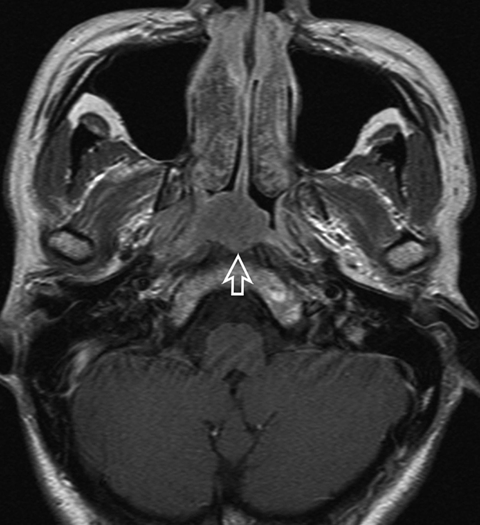
  • Fig 3.
    • Download figure
    • Open in new tab
    • Download powerpoint
    Fig 3.

    A, Axial T1-weighted postcontrast MR image of the nasopharynx in a 51-year-old man with NPC (open arrows). A symmetric tumor with diffuse involvement of all nasopharyngeal walls exhibits homogeneous contrast enhancement without a white line along the deep mucosal margin. B, Axial T1-weighted postcontrast MR image of the nasopharynx in a 59-year-old man with NPC (open arrows). A symmetric tumor with diffuse involvement of the nasopharyngeal walls exhibits homogeneous low contrast enhancement and an intact mucosal white line along the deep margin. The adenoid extends along the posterior wall from the roof, with an adenoidal “stripe” on the right (curved open arrow) but not on the left side. A small right retropharyngeal node is also indicated (solid arrow). The patient had bulky N3-stage metastatic nodes below this level.

  • Fig 4.
    • Download figure
    • Open in new tab
    • Download powerpoint
    Fig 4.

    A, Axial T1-weighted postcontrast MR image of the nasopharynx in a 53-year-old man with BH1 (open arrows). An area of diffuse symmetric mucosal thickening with homogeneous contrast enhancement is visible. B, Axial T1-weighted postcontrast MR image of the nasopharynx in a 28-year-old man with BH2 of the adenoid (open arrow). The symmetric lesion exhibits contrast-enhancing septa that run perpendicular to the nasopharyngeal wall and are separated by columns of low contrast enhancement. C, Axial T1-weighted postcontrast MR image of the nasopharynx in a 48-year-old woman with BH2 along the nasopharyngeal walls (open arrows). An area of diffuse, symmetric homogeneous low contrast enhancement and an intact deep mucosal white line along the deep margin are visible.
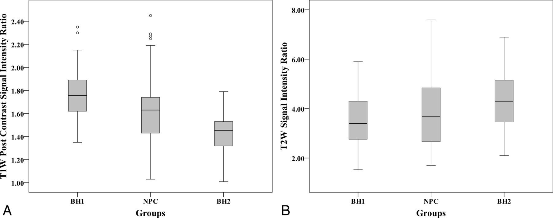
  • Fig 5.
    • Download figure
    • Open in new tab
    • Download powerpoint
    Fig 5.

    A, Boxplots showing differences in the T1 postcontrast signal intensity ratios (relative to muscle) among NPC, BH1, and BH2. B, Boxplots show differences in the T2 signal intensity ratios (relative to muscle) among NPC, BH1, and BH2.

  • Fig 6.
    • Download figure
    • Open in new tab
    • Download powerpoint
    Fig 6.

    Receiver operating characteristic curve of the percentage difference in area for NPC detection.
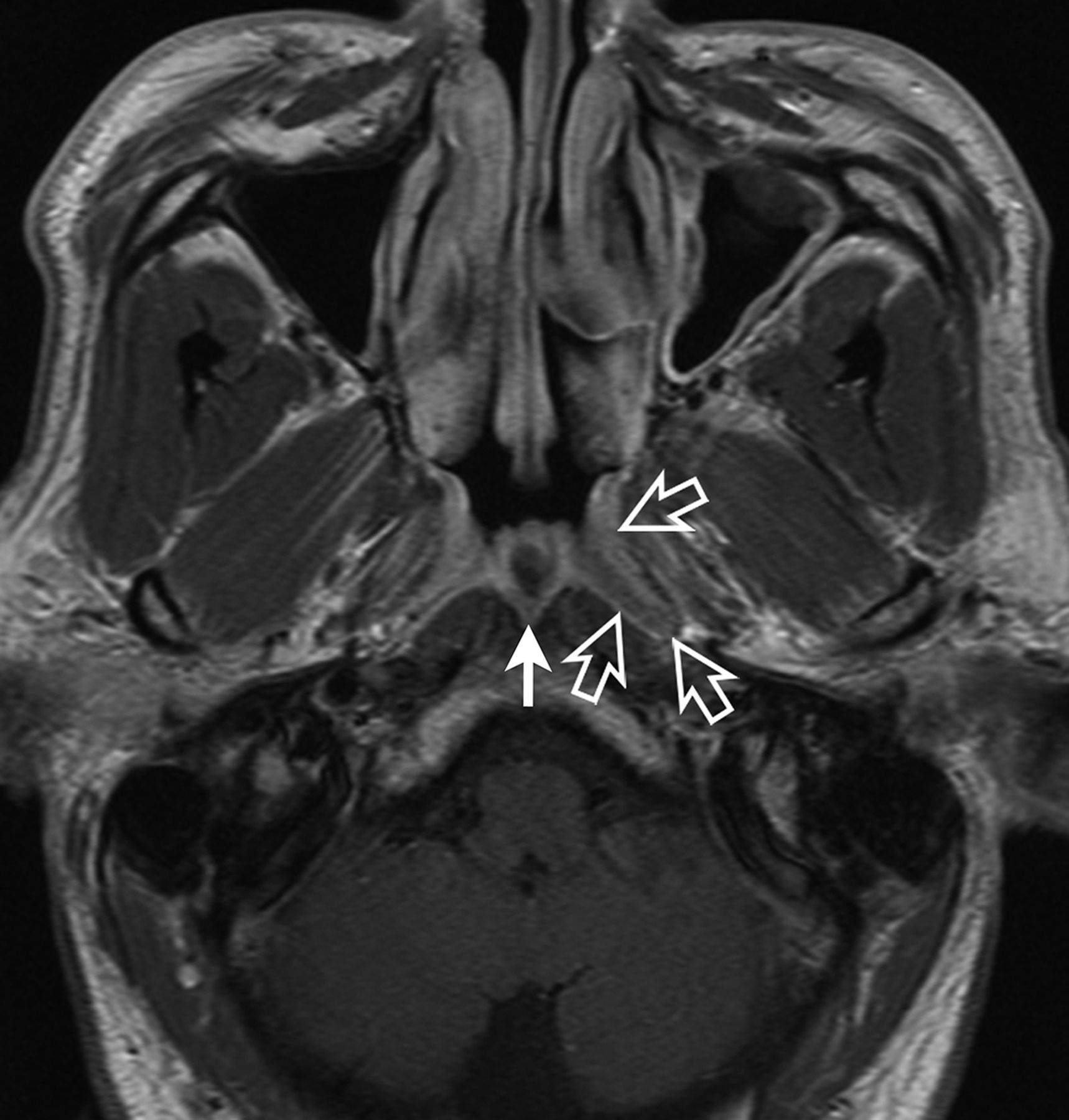
  • Fig 7.
    • Download figure
    • Open in new tab
    • Download powerpoint
    Fig 7.

    Axial T1-weighted postcontrast MR image of the nasopharynx in a 52-year-old man with asymmetric BH. Greater thickening is observed in the left side of the roof (open arrows), where a focal area of mucosal thickening comprises a superficial band of low contrast enhancement overlying an intact deep mucosal white line (BH type 2). A Tornwaldt cyst is also present (solid arrow).

Tables

  • Figures
    • View popup
    Table 1:

    MR imaging analysis of the nasopharynx

    MR Imaging Features of NPCMethod of Analysis
    SiteWalls: mucosa plus submucosa, including the lymphoid tissue layers in the mucosal space superficial to the fascia and pharyngeal muscles; wall subsites comprise the following: the pharyngeal recess, roof (above the level of the pharyngeal recess), and posterior/inferior walls (at or below the level of the pharyngeal recess)
    Adenoid: central mucosal lymphoid tissues in the roof and upper posterior wall where lymphoid tissue within the nasopharynx is abundant
    Center: lateral, central, or diffuse
    SizeVolume (area of nasopharyngeal walls and adenoid on each slice × slice thickness)
    Size asymmetryArea difference between the right and left halves of the nasopharynx on the slice with the maximum difference, expressed as the %ΔA (difference in area between the 2 sides/area on smaller side × 100)
    Signal intensityRepresentative ROI is selected on the T1-weighted postcontrast image (T2-weighted signal intensity is measured at the corresponding site); signal intensity is expressed as a ratio of signal intensity/signal intensity of the belly of the lateral pterygoid muscle
    Signal intensity asymmetrySubjective signal intensity asymmetry between the right and left halves of the nasopharynx on T1-weighted postcontrast images
    HomogeneitySubjective assessment of signal intensity on T1-weighted postcontrast images, divided into homogeneous and heterogeneous
    Deep mucosal white lineLine/band of greater contrast enhancement in the deep layer relative to the superficial layer of the nasopharyngeal wall at the site of the tumor, divided into absent, present with focal loss, and present and intact; assessment was not made at the adenoid
    Adenoidal septaContrast-enhancing septa within the adenoid, divided into absent, present and distorted (displacement or partial loss excluding that caused by adenoidal cysts), and present and not distorted (intact and symmetric)
    • View popup
    Table 2:

    Diagnostic performance of MR imaging for the discrimination of early NPC from BH

    VolumeSize AsymmetrySignal AsymmetryDeep Mucosal White LineAdenoid
    Test positive for NPCVolume ≥ 5.01 cm3%ΔA ≥ 33.7%Signal asymmetry presentFocal loss of the deep mucosal white lineSepta absent or distorted (excluding septa distorted by cysts)
    n = 333 (NPC = 189, BH = 144)n = 333 (NPC = 189, BH = 144)n = 333 (NPC = 189, BH = 144)n = 253 (NPC = 180, BH2 = 73)n = 172 (NPC = 111, BH2 = 61)
    Area under the curve (95% CI)0.676 (0.618–0.734)0.948 (0.926–0.971)
    P value<.001<.001
    True-positive (No.)119167161153111
    False-positive (No.)471326109
    True-negative (No.)971311186352
    False-negative (No.)702228270
    Sensitivity (%)63.088.485.285.0100.0
    Specificity (%)67.491.081.986.385.2
    Positive predictive value (%)71.792.886.193.992.5
    Negative predictive value (%)58.185.680.870.0100.0
    Accuracy (%)64.989.583.885.494.8
    • View popup
    Table 3:

    MR imaging criteria subgroup analysis of symmetric (%ΔA < 33.7%) NPC and BH

    VolumeSignal AsymmetryDeep Mucosal White LineAdenoid
    Test positive for NPCVolume ≥ 5.01 cm3Signal asymmetry presentFocal loss of the deep mucosal white lineSepta absent or distorted (excluding septa distorted by cysts)
    n = 153 (NPC = 22, BH = 131)n = 153 (NPC = 22, BH = 131)n = 83 (NPC = 20, BH2 = 63)n = 72 (NPC = 16, BH2 = 56)
    True-positive (No.)20111616
    False-positive (No.)421588
    True-negative (No.)891165548
    False-negative (No.)21140
    Sensitivity (%)90.150.080.0100.0
    Specificity (%)67.988.587.385.7
    Positive predictive value (%)32.342.366.766.7
    Negative predictive value (%)97.891.393.2100
    Accuracy (%)71.283.085.588.9
    • View popup
    Table 4:

    Proposed MR imaging criteria for primary NPC detection

    GradeConditionAppearance
    1NormalThin mucosa ≤3 mm; adenoid absent, vestigial or composed almost entirely of cysts
    2Probably benignDiffusely thickened >3-mm symmetric mucosa either homogeneously enhancing (benign hyperplasia type 1) or with greater contrast enhancement in the deep layer (mucosal white line) compared with the superficial layer (benign hyperplasia type 2); adenoid with intact contrast-enhancing septa (benign hyperplasia type 2)
    3IndeterminateDiffusely thickened >3-mm mucosa asymmetric in thickness; adenoid asymmetric in thickness
    4Suspicious1) Diffusely thickened >3-mm mucosa asymmetric in signal intensity; adenoid asymmetric in signal intensity
    2) Diffusely thickened mucosa >3 mm with focal loss of the mucosal white line; absence/distortion of the thin contrast-enhancing adenoidal septa (not attributed to cysts)
    3) Focal mass confined within the nasopharynx
    5Probably malignantSuperficial extension to the nasal cavity/oropharynx or deep extension to the sites bordering the nasopharynx
Next
Back to top
Advertisement
Print
Download PDF
Email Article

Thank you for your interest in spreading the word on American Journal of Neuroradiology.

NOTE: We only request your email address so that the person you are recommending the page to knows that you wanted them to see it, and that it is not junk mail. We do not capture any email address.

Enter multiple addresses on separate lines or separate them with commas.
MR Imaging Criteria for the Detection of Nasopharyngeal Carcinoma: Discrimination of Early-Stage Primary Tumors from Benign Hyperplasia
(Your Name) has sent you a message from American Journal of Neuroradiology
(Your Name) thought you would like to see the American Journal of Neuroradiology web site.
CAPTCHA
This question is for testing whether or not you are a human visitor and to prevent automated spam submissions.
Cite this article
A.D. King, L.Y.S. Wong, B.K.H. Law, K.S. Bhatia, J.K.S. Woo, Q.-Y. Ai, T.Y. Tan, J. Goh, K.L. Chuah, F.K.F. Mo, K.C.A. Chan, A.T.C. Chan, A.C. Vlantis
MR Imaging Criteria for the Detection of Nasopharyngeal Carcinoma: Discrimination of Early-Stage Primary Tumors from Benign Hyperplasia
American Journal of Neuroradiology Dec 2017, DOI: 10.3174/ajnr.A5493

Citation Manager Formats

  • BibTeX
  • Bookends
  • EasyBib
  • EndNote (tagged)
  • EndNote 8 (xml)
  • Medlars
  • Mendeley
  • Papers
  • RefWorks Tagged
  • Ref Manager
  • RIS
  • Zotero
0 Responses
Respond to this article
Share
Bookmark this article
MR Imaging Criteria for the Detection of Nasopharyngeal Carcinoma: Discrimination of Early-Stage Primary Tumors from Benign Hyperplasia
A.D. King, L.Y.S. Wong, B.K.H. Law, K.S. Bhatia, J.K.S. Woo, Q.-Y. Ai, T.Y. Tan, J. Goh, K.L. Chuah, F.K.F. Mo, K.C.A. Chan, A.T.C. Chan, A.C. Vlantis
American Journal of Neuroradiology Dec 2017, DOI: 10.3174/ajnr.A5493
del.icio.us logo Twitter logo Facebook logo Mendeley logo
  • Tweet Widget
  • Facebook Like
  • Google Plus One
Purchase

Jump to section

  • Article
    • Abstract
    • ABBREVIATIONS:
    • Materials and Methods
    • Results
    • Discussion
    • Conclusions
    • Acknowledgments
    • Footnotes
    • References
  • Figures & Data
  • Supplemental
  • Info & Metrics
  • Responses
  • References
  • PDF

Related Articles

  • PubMed
  • Google Scholar

Cited By...

  • Quantitative T1{rho} MRI of the Head and Neck Discriminates Carcinoma and Benign Hyperplasia in the Nasopharynx
  • Early Detection of Cancer: Evaluation of MR Imaging Grading Systems in Patients with Suspected Nasopharyngeal Carcinoma
  • Crossref (41)
  • Google Scholar

This article has been cited by the following articles in journals that are participating in Crossref Cited-by Linking.

  • Development of a self-constrained 3D DenseNet model in automatic detection and segmentation of nasopharyngeal carcinoma using magnetic resonance images
    Liangru Ke, Yishu Deng, Weixiong Xia, Mengyun Qiang, Xi Chen, Kuiyuan Liu, Bingzhong Jing, Caisheng He, Chuanmiao Xie, Xiang Guo, Xing Lv, Chaofeng Li
    Oral Oncology 2020 110
  • Complementary roles of MRI and endoscopic examination in the early detection of nasopharyngeal carcinoma
    A.D. King, J.K.S. Woo, Q.Y. Ai, J.S.M. Chan, W.K.J. Lam, I.O.L. Tse, K.S. Bhatia, B.C.Y. Zee, E.P. Hui, B.B.Y. Ma, R.W.K. Chiu, A.C. van Hasselt, A.T.C. Chan, Y.M.D. Lo, K.C.A. Chan
    Annals of Oncology 2019 30 6
  • Distinguishing early-stage nasopharyngeal carcinoma from benign hyperplasia using intravoxel incoherent motion diffusion-weighted MRI
    Qi-Yong Ai, Ann D. King, Janet S. M. Chan, Weitian Chen, K. C. Allen Chan, John K. S. Woo, Benny C. Y. Zee, Anthony T. C. Chan, Darren M. C. Poon, Brigette B. Y. Ma, Edwin P. Hui, Anil T. Ahuja, Alexander C. Vlantis, Jing Yuan
    European Radiology 2019 29 10
  • Convolutional neural network for discriminating nasopharyngeal carcinoma and benign hyperplasia on MRI
    Lun M. Wong, Ann D. King, Qi Yong H. Ai, W. K. Jacky Lam, Darren M. C. Poon, Brigette B. Y. Ma, K. C. Allen Chan, Frankie K. F. Mo
    European Radiology 2021 31 6
  • MR Imaging of Nasopharyngeal Carcinoma
    Ann D. King
    Magnetic Resonance Imaging Clinics of North America 2022 30 1
  • Early Detection of Cancer: Evaluation of MR Imaging Grading Systems in Patients with Suspected Nasopharyngeal Carcinoma
    A.D. King, J.K.S. Woo, Q.-Y. Ai, F.K.F. Mo, T.Y. So, W.K.J. Lam, I.O.L. Tse, A.C. Vlantis, K.W.N. Yip, E.P. Hui, B.B.Y. Ma, R.W.K. Chiu, A.T.C. Chan, Y.M.D. Lo, K.C.A. Chan
    American Journal of Neuroradiology 2020 41 3
  • MRI detection of suspected nasopharyngeal carcinoma: a systematic review and meta-analysis
    Vineet Vijay Gorolay, Naomi Natasha Niles, Ya Ruth Huo, Navid Ahmadi, Kate Hanneman, Elizabeth Thompson, Michael Vinchill Chan
    Neuroradiology 2022 64 8
  • The contrast-enhanced MRI can be substituted by unenhanced MRI in identifying and automatically segmenting primary nasopharyngeal carcinoma with the aid of deep learning models: An exploratory study in large-scale population of endemic area
    Yishu Deng, Chaofeng Li, Xing Lv, Weixiong Xia, Lujun Shen, Bingzhong Jing, Bin Li, Xiang Guo, Ying Sun, Chuanmiao Xie, Liangru Ke
    Computer Methods and Programs in Biomedicine 2022 217
  • Dual-energy CT in the differentiation of stage T1 nasopharyngeal carcinoma and lymphoid hyperplasia
    Peng Wang, Zebin Xiao, Zuohua Tang, Jie Wang
    European Journal of Radiology 2020 124
  • Quantitative Analysis of DCE-MRI and RESOLVE-DWI for Differentiating Nasopharyngeal Carcinoma from Nasopharyngeal Lymphoid Hyperplasia
    J. Y. Yu, D. Zhang, X. L. Huang, J. Ma, C. Yang, X. J. Li, H. Xiong, B. Zhou, R. K. Liao, Z. Y. Tang
    Journal of Medical Systems 2020 44 4

More in this TOC Section

  • ASL Sensitivity for Head and Neck Paraganglioma
  • Post SRS Peritumoral Hyperintense Signal of VSs
  • FDG Uptake and Thyroid Cancer Post-BRAF Therapy
Show more Head and Neck Imaging

Similar Articles

Advertisement

Indexed Content

  • Current Issue
  • Accepted Manuscripts
  • Article Preview
  • Past Issues
  • Editorials
  • Editor's Choice
  • Fellows' Journal Club
  • Letters to the Editor
  • Video Articles

Cases

  • Case Collection
  • Archive - Case of the Week
  • Archive - Case of the Month
  • Archive - Classic Case

More from AJNR

  • Trainee Corner
  • Imaging Protocols
  • MRI Safety Corner
  • Book Reviews

Multimedia

  • AJNR Podcasts
  • AJNR Scantastics

Resources

  • Turnaround Time
  • Submit a Manuscript
  • Submit a Video Article
  • Submit an eLetter to the Editor/Response
  • Manuscript Submission Guidelines
  • Statistical Tips
  • Fast Publishing of Accepted Manuscripts
  • Graphical Abstract Preparation
  • Imaging Protocol Submission
  • Evidence-Based Medicine Level Guide
  • Publishing Checklists
  • Author Policies
  • Become a Reviewer/Academy of Reviewers
  • News and Updates

About Us

  • About AJNR
  • Editorial Board
  • Editorial Board Alumni
  • Alerts
  • Permissions
  • Not an AJNR Subscriber? Join Now
  • Advertise with Us
  • Librarian Resources
  • Feedback
  • Terms and Conditions
  • AJNR Editorial Board Alumni

American Society of Neuroradiology

  • Not an ASNR Member? Join Now

© 2025 by the American Society of Neuroradiology All rights, including for text and data mining, AI training, and similar technologies, are reserved.
Print ISSN: 0195-6108 Online ISSN: 1936-959X

Powered by HighWire