Advertisement

Main menu

  • Home
  • Content
    • Current Issue
    • Accepted Manuscripts
    • Article Preview
    • Past Issue Archive
    • Video Articles
    • AJNR Case Collection
    • Case of the Week Archive
    • Case of the Month Archive
    • Classic Case Archive
  • Special Collections
    • AJNR Awards
    • Low-Field MRI
    • Alzheimer Disease
    • ASNR Foundation Special Collection
    • Photon-Counting CT
    • View All
  • Multimedia
    • AJNR Podcasts
    • AJNR SCANtastic
    • Trainee Corner
    • MRI Safety Corner
    • Imaging Protocols
  • For Authors
    • Submit a Manuscript
    • Submit a Video Article
    • Submit an eLetter to the Editor/Response
    • Manuscript Submission Guidelines
    • Statistical Tips
    • Fast Publishing of Accepted Manuscripts
    • Graphical Abstract Preparation
    • Imaging Protocol Submission
    • Author Policies
  • About Us
    • About AJNR
    • Editorial Board
    • Editorial Board Alumni
  • More
    • Become a Reviewer/Academy of Reviewers
    • Subscribers
    • Permissions
    • Alerts
    • Feedback
    • Advertisers
    • ASNR Home

User menu

  • Alerts
  • Log in

Search

  • Advanced search
American Journal of Neuroradiology

American Journal of Neuroradiology

ASHNR American Society of Functional Neuroradiology ASHNR American Society of Pediatric Neuroradiology ASSR
  • Alerts
  • Log in

Advanced Search

  • Home
  • Content
    • Current Issue
    • Accepted Manuscripts
    • Article Preview
    • Past Issue Archive
    • Video Articles
    • AJNR Case Collection
    • Case of the Week Archive
    • Case of the Month Archive
    • Classic Case Archive
  • Special Collections
    • AJNR Awards
    • Low-Field MRI
    • Alzheimer Disease
    • ASNR Foundation Special Collection
    • Photon-Counting CT
    • View All
  • Multimedia
    • AJNR Podcasts
    • AJNR SCANtastic
    • Trainee Corner
    • MRI Safety Corner
    • Imaging Protocols
  • For Authors
    • Submit a Manuscript
    • Submit a Video Article
    • Submit an eLetter to the Editor/Response
    • Manuscript Submission Guidelines
    • Statistical Tips
    • Fast Publishing of Accepted Manuscripts
    • Graphical Abstract Preparation
    • Imaging Protocol Submission
    • Author Policies
  • About Us
    • About AJNR
    • Editorial Board
    • Editorial Board Alumni
  • More
    • Become a Reviewer/Academy of Reviewers
    • Subscribers
    • Permissions
    • Alerts
    • Feedback
    • Advertisers
    • ASNR Home
  • Follow AJNR on Twitter
  • Visit AJNR on Facebook
  • Follow AJNR on Instagram
  • Join AJNR on LinkedIn
  • RSS Feeds

AJNR Awards, New Junior Editors, and more. Read the latest AJNR updates

Research ArticleHead and Neck Imaging
Open Access

Dynamic Contrast-Enhanced MRI–Derived Intracellular Water Lifetime (τi): A Prognostic Marker for Patients with Head and Neck Squamous Cell Carcinomas

S. Chawla, L.A. Loevner, S.G. Kim, W.-T. Hwang, S. Wang, G. Verma, S. Mohan, V. LiVolsi, H. Quon and H. Poptani
American Journal of Neuroradiology November 2017, DOI: https://doi.org/10.3174/ajnr.A5440
S. Chawla
aFrom the Departments of Radiology (S.C., L.A.L., S.G.K., S.W., G.V., S.M., H.P.)
  • Find this author on Google Scholar
  • Find this author on PubMed
  • Search for this author on this site
  • ORCID record for S. Chawla
L.A. Loevner
aFrom the Departments of Radiology (S.C., L.A.L., S.G.K., S.W., G.V., S.M., H.P.)
  • Find this author on Google Scholar
  • Find this author on PubMed
  • Search for this author on this site
  • ORCID record for L.A. Loevner
S.G. Kim
aFrom the Departments of Radiology (S.C., L.A.L., S.G.K., S.W., G.V., S.M., H.P.)
eDepartment of Radiology (S.G.K.), New York University, New York, New York
  • Find this author on Google Scholar
  • Find this author on PubMed
  • Search for this author on this site
  • ORCID record for S.G. Kim
W.-T. Hwang
cBiostatistics and Epidemiology (W.-T.H.)
  • Find this author on Google Scholar
  • Find this author on PubMed
  • Search for this author on this site
  • ORCID record for W.-T. Hwang
S. Wang
aFrom the Departments of Radiology (S.C., L.A.L., S.G.K., S.W., G.V., S.M., H.P.)
  • Find this author on Google Scholar
  • Find this author on PubMed
  • Search for this author on this site
  • ORCID record for S. Wang
G. Verma
aFrom the Departments of Radiology (S.C., L.A.L., S.G.K., S.W., G.V., S.M., H.P.)
  • Find this author on Google Scholar
  • Find this author on PubMed
  • Search for this author on this site
  • ORCID record for G. Verma
S. Mohan
aFrom the Departments of Radiology (S.C., L.A.L., S.G.K., S.W., G.V., S.M., H.P.)
  • Find this author on Google Scholar
  • Find this author on PubMed
  • Search for this author on this site
  • ORCID record for S. Mohan
V. LiVolsi
dPathology and Lab Medicine (V.L.), Perelman School of Medicine, University of Pennsylvania, Philadelphia, Pennsylvania
  • Find this author on Google Scholar
  • Find this author on PubMed
  • Search for this author on this site
  • ORCID record for V. LiVolsi
H. Quon
bRadiation Oncology (H.Q.)
fDepartment of Radiation Oncology and Molecular Radiation Sciences (H.Q.), Johns Hopkins University, Baltimore, Maryland
  • Find this author on Google Scholar
  • Find this author on PubMed
  • Search for this author on this site
  • ORCID record for H. Quon
H. Poptani
aFrom the Departments of Radiology (S.C., L.A.L., S.G.K., S.W., G.V., S.M., H.P.)
gDepartment of Cellular and Molecular Physiology (H.P.), University of Liverpool, Liverpool, United Kingdom.
  • Find this author on Google Scholar
  • Find this author on PubMed
  • Search for this author on this site
  • ORCID record for H. Poptani
  • Article
  • Figures & Data
  • Info & Metrics
  • Responses
  • References
  • PDF
Loading

Article Figures & Data

Figures

  • Tables
  • Fig 1.
    • Download figure
    • Open in new tab
    • Download powerpoint
    Fig 1.

    Representative images from a patient exhibiting long survival (follow-up duration of 8.19 years). Axial T2-weighted image (A) demonstrates an enlarged heterogeneous hyperintense metastatic left level IIa lymph node (arrow). This appears hypointense on a coregistered T1-weighted image (B), with heterogeneous enhancement on the corresponding postcontrast T1-weighted image (C). DCE-MRI–derived τi (0.136 seconds [D]) and Ktrans (0.882 minutes−1 [E]) maps are shown as color images overlaid on postcontrast T1-weighted images.
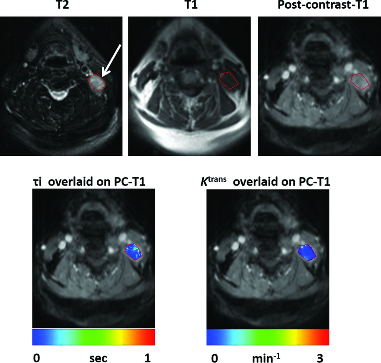
  • Fig 2.
    • Download figure
    • Open in new tab
    • Download powerpoint
    Fig 2.

    Representative images from a patient who died 2.12 years after the end of CRT. Axial T2-weighted image (A) demonstrates a heterogeneous hyperintense metastatic left level IIb lymph node (arrow). It appears hypointense on a coregistered T1-weighted image (B) with heterogeneous enhancement on postcontrast T1-weighted image (C). DCE-MRI–derived τi (0.031 seconds; [D]) and Ktrans (0.135 minutes−1 [E]) maps overlaid on postcontrast T1-weighted images demonstrating lower τi and Ktrans values from the node compared with the patient with longer survival as shown in Fig 1.

  • Fig 3.
    • Download figure
    • Open in new tab
    • Download powerpoint
    Fig 3.

    Kaplan-Meier plot for τi. Patients with higher pretreatment τi (solid curves) demonstrate longer OS compared with patients with lower τi (broken curves) for first 2-year (solid vertical line, P = .09), 5-year (dotted vertical line, P = .01), and long-term (median duration, >7 years; P = .006) follow-up periods.
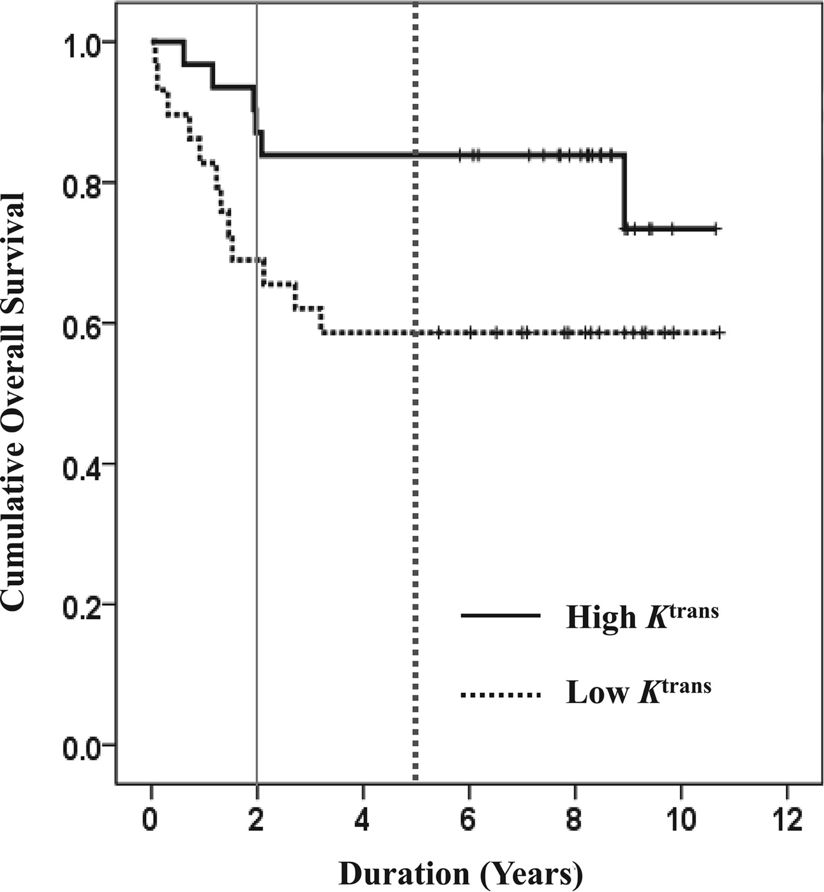
  • Fig 4.
    • Download figure
    • Open in new tab
    • Download powerpoint
    Fig 4.

    Kaplan-Meier plots for Ktrans. Patients with higher pretreatment Ktrans (solid curves) demonstrate longer OS compared with patients with lower Ktrans (broken curves) at the 2-year (solid vertical line; P = .07), 5-year (dotted vertical line; P = .028), and long-term (median duration, >7 years; P = .06) follow-up periods.

  • Fig 5.
    • Download figure
    • Open in new tab
    • Download powerpoint
    Fig 5.

    Kaplan-Meier plots for combinations of τi and Ktrans. Patients with high τi/Ktrans (thick broken curve) had the longest OS, and patients with low τi/Ktrans (thin broken curve) had the shortest OS for the first 2-year (solid vertical line; P = .02), 5-year (dotted vertical line; P < .0001), and long-term (median duration, >7 years; P < .0001) follow-up periods. In addition, patients with high τi/low Ktrans (thick solid curve) exhibited longer OS than patients with low τi/high Ktrans (thin solid curve) at all clinical end points.

  • Fig 6.
    • Download figure
    • Open in new tab
    • Download powerpoint
    Fig 6.

    Kaplan-Meier plots for p16 expression. (A) Patients with p16-positive expression (solid curve) exhibited significantly longer long-term (median duration, >7 years) OS (P < .05) than patients with p16-negative expression (broken curve) (B). Patients with positive p16 expression and high τi/high Ktrans (thick broken curve) had longer OS than p16-positive patients with low τi/low Ktrans (thin broken curve). In addition, patients with high τi/low Ktrans (gray solid curve) exhibited longer OS than patients with low τi/high Ktrans (black solid curve) at all clinical end points. However, these differences were not significant (P > .05).

Tables

  • Figures
  • Patient characteristics and treatment modalities

    Characteristics
    Number of patients60
    Mean age, yrs ± SD62.34 ± 9.18
    Sex
        Male49 (81.7%)
        Female11 (18.3%)
    Primary tumor site
        Base of tongue24 (40.0%)
        Tonsil14 (23.3%)
        Larynx7 (11.7%)
        Less common/unknown sites15 (25.0%)
    T staging
        Tx14 (23.3%)
        T02 (3.3%)
        T12 (3.3%)
        T215 (25.0%)
        T39 (15.0%)
        T418 (30.0%)
    N staging
        N12 (3.3%)
        N251 (85.0%)
        N37 (11.7%)
    M staging
        M060 (100%)
    p16 expression
        Positive21 (35.0%)
        Negative11 (18.3%)
        Unknown (insufficient specimen)28 (46.7%)
    Treatment
        Radiotherapy + concurrent chemotherapy39 (65.0%)
        Induction chemotherapy + radiotherapy + concurrent chemotherapy21 (35.0%)
Next
Back to top
Advertisement
Print
Download PDF
Email Article

Thank you for your interest in spreading the word on American Journal of Neuroradiology.

NOTE: We only request your email address so that the person you are recommending the page to knows that you wanted them to see it, and that it is not junk mail. We do not capture any email address.

Enter multiple addresses on separate lines or separate them with commas.
Dynamic Contrast-Enhanced MRI–Derived Intracellular Water Lifetime (τi): A Prognostic Marker for Patients with Head and Neck Squamous Cell Carcinomas
(Your Name) has sent you a message from American Journal of Neuroradiology
(Your Name) thought you would like to see the American Journal of Neuroradiology web site.
CAPTCHA
This question is for testing whether or not you are a human visitor and to prevent automated spam submissions.
Cite this article
S. Chawla, L.A. Loevner, S.G. Kim, W.-T. Hwang, S. Wang, G. Verma, S. Mohan, V. LiVolsi, H. Quon, H. Poptani
Dynamic Contrast-Enhanced MRI–Derived Intracellular Water Lifetime (τi): A Prognostic Marker for Patients with Head and Neck Squamous Cell Carcinomas
American Journal of Neuroradiology Nov 2017, DOI: 10.3174/ajnr.A5440

Citation Manager Formats

  • BibTeX
  • Bookends
  • EasyBib
  • EndNote (tagged)
  • EndNote 8 (xml)
  • Medlars
  • Mendeley
  • Papers
  • RefWorks Tagged
  • Ref Manager
  • RIS
  • Zotero
0 Responses
Respond to this article
Share
Bookmark this article
Dynamic Contrast-Enhanced MRI–Derived Intracellular Water Lifetime (τi): A Prognostic Marker for Patients with Head and Neck Squamous Cell Carcinomas
S. Chawla, L.A. Loevner, S.G. Kim, W.-T. Hwang, S. Wang, G. Verma, S. Mohan, V. LiVolsi, H. Quon, H. Poptani
American Journal of Neuroradiology Nov 2017, DOI: 10.3174/ajnr.A5440
del.icio.us logo Twitter logo Facebook logo Mendeley logo
  • Tweet Widget
  • Facebook Like
  • Google Plus One
Purchase

Jump to section

  • Article
    • Abstract
    • ABBREVIATIONS:
    • Materials and Methods
    • Results
    • Discussion
    • Conclusions
    • Footnotes
    • References
  • Figures & Data
  • Info & Metrics
  • Responses
  • References
  • PDF

Related Articles

  • No related articles found.
  • PubMed
  • Google Scholar

Cited By...

  • No citing articles found.
  • Crossref (27)
  • Google Scholar

This article has been cited by the following articles in journals that are participating in Crossref Cited-by Linking.

  • Using 1H2O MR to measure and map sodium pump activity in vivo
    Charles S. Springer
    Journal of Magnetic Resonance 2018 291
  • Feasibility of filter-exchange imaging (FEXI) in measuring different exchange processes in human brain
    Ruiliang Bai, Zhaoqing Li, Chaoliang Sun, Yi-Cheng Hsu, Hui Liang, Peter Basser
    NeuroImage 2020 219
  • Measurement of cellular‐interstitial water exchange time in tumors based on diffusion‐time‐dependent diffusional kurtosis imaging
    Jin Zhang, Gregory Lemberskiy, Linda Moy, Els Fieremans, Dmitry S. Novikov, Sungheon Gene Kim
    NMR in Biomedicine 2021 34 6
  • NMR shutter‐speed elucidates apparent population inversion of 1H2O signals due to active transmembrane water cycling
    Xin Li, Silvia Mangia, Jing‐Huei Lee, Ruiliang Bai, Charles S. Springer
    Magnetic Resonance in Medicine 2019 82 1
  • Improving MR cell size imaging by inclusion of transcytolemmal water exchange
    Xiaoyu Jiang, Sean P. Devan, Jingping Xie, John C. Gore, Junzhong Xu
    NMR in Biomedicine 2022 35 12
  • Shutter‐Speed DCE‐MRI Analyses of Human Glioblastoma Multiforme (GBM) Data
    Ruiliang Bai, Bao Wang, Yinhang Jia, Zejun Wang, Charles S. Springer, Zhaoqing Li, Chuanjin Lan, Yi Zhang, Peng Zhao, Yingchao Liu
    Journal of Magnetic Resonance Imaging 2020 52 3
  • The Promise of Novel Biomarkers for Head and Neck Cancer from an Imaging Perspective
    Loredana G. Marcu, Paul Reid, Eva Bezak
    International Journal of Molecular Sciences 2018 19 9
  • Time‐dependent diffusivity and kurtosis in phantoms and patients with head and neck cancer
    Eddy Solomon, Gregory Lemberskiy, Steven Baete, Kenneth Hu, Dariya Malyarenko, Scott Swanson, Amita Shukla‐Dave, Stephen E. Russek, Elcin Zan, Sungheon Gene Kim
    Magnetic Resonance in Medicine 2023 89 2
  • The direction-dependence of apparent water exchange rate in human white matter
    Zhaoqing Li, Zhenfeng Pang, Juange Cheng, Yi-Cheng Hsu, Yi Sun, Evren Özarslan, Ruiliang Bai
    NeuroImage 2022 247
  • Prediction of distant metastases in patients with squamous cell carcinoma of head and neck using DWI and DCE‐MRI
    Sanjeev Chawla, Sungheon G Kim, Laurie A Loevner, Sumei Wang, Suyash Mohan, Alexander Lin, Harish Poptani
    Head & Neck 2020 42 11

More in this TOC Section

  • ASL Sensitivity for Head and Neck Paraganglioma
  • Post SRS Peritumoral Hyperintense Signal of VSs
  • Contrast Enhanced Pituitary CISS/FIESTA
Show more Head and Neck Imaging

Similar Articles

Advertisement

Indexed Content

  • Current Issue
  • Accepted Manuscripts
  • Article Preview
  • Past Issues
  • Editorials
  • Editor's Choice
  • Fellows' Journal Club
  • Letters to the Editor
  • Video Articles

Cases

  • Case Collection
  • Archive - Case of the Week
  • Archive - Case of the Month
  • Archive - Classic Case

More from AJNR

  • Trainee Corner
  • Imaging Protocols
  • MRI Safety Corner
  • Book Reviews

Multimedia

  • AJNR Podcasts
  • AJNR Scantastics

Resources

  • Turnaround Time
  • Submit a Manuscript
  • Submit a Video Article
  • Submit an eLetter to the Editor/Response
  • Manuscript Submission Guidelines
  • Statistical Tips
  • Fast Publishing of Accepted Manuscripts
  • Graphical Abstract Preparation
  • Imaging Protocol Submission
  • Evidence-Based Medicine Level Guide
  • Publishing Checklists
  • Author Policies
  • Become a Reviewer/Academy of Reviewers
  • News and Updates

About Us

  • About AJNR
  • Editorial Board
  • Editorial Board Alumni
  • Alerts
  • Permissions
  • Not an AJNR Subscriber? Join Now
  • Advertise with Us
  • Librarian Resources
  • Feedback
  • Terms and Conditions
  • AJNR Editorial Board Alumni

American Society of Neuroradiology

  • Not an ASNR Member? Join Now

© 2025 by the American Society of Neuroradiology All rights, including for text and data mining, AI training, and similar technologies, are reserved.
Print ISSN: 0195-6108 Online ISSN: 1936-959X

Powered by HighWire