
Article Figures & Data
Figures
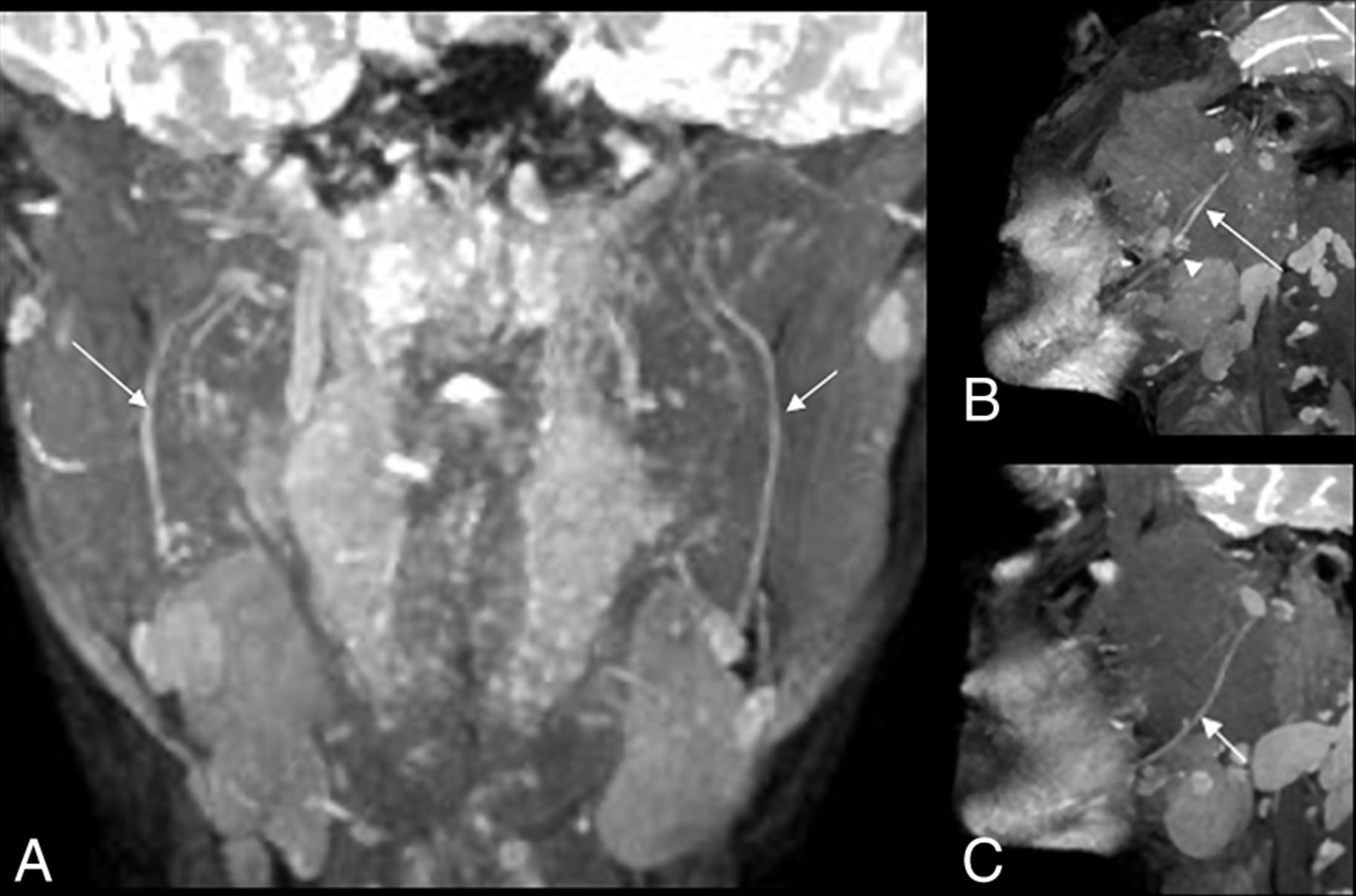
- Fig 1.
A, MIP coronal 3D PSIF image showing class II injury to the right IAN with mild increase in caliber (less than 50% of the left) and signal intensity of the right IAN (long arrow) in comparison with a normal left inferior alveolar nerve (short arrow). B, Sagittal reconstruction MIP 3D PSIF image showing increase in caliber and signal intensity of the right IAN (long arrow) proximal to injury site (arrowhead). C, Normal uniform caliber and signal intensity of the left IAN (short arrow).
- Fig 2.
A and B, MIP 3D coronal PSIF images show a hyperintense left LN (long arrow) with a 3-mm neuroma in continuity (demarcated by 3 arrowheads) compatible with class IV injury. C and D, Sagittal reconstructions show the abnormal left LN neuroma (demarcated by 3 arrowheads) compared with a normal right LN (short arrow).
- Fig 3.
Coronal 3D PSIF images showing A, localization of the site of the LN and IAN (short and long arrows, respectively) and B, signal intensity measurements on both sides.
- Fig 4.
A, MIP 3D PSIF coronal image shows class IV/V injury of the left LN with excessive granulation and possible discontinuity of its distal end (long arrow). B, On surgery, it was also called class IV/V injury (arrow) with excessive scarring and granulation tissue and was resected. The final gap was 16 mm (C) and an allograft was placed for nerve reconstruction.
- Fig 5.
κ correlations for A, MRN versus NST and B, MRN versus surgical classifications.
- Fig 6.
Differences in thickness, T2SIR, and CNR among the case and control groups.
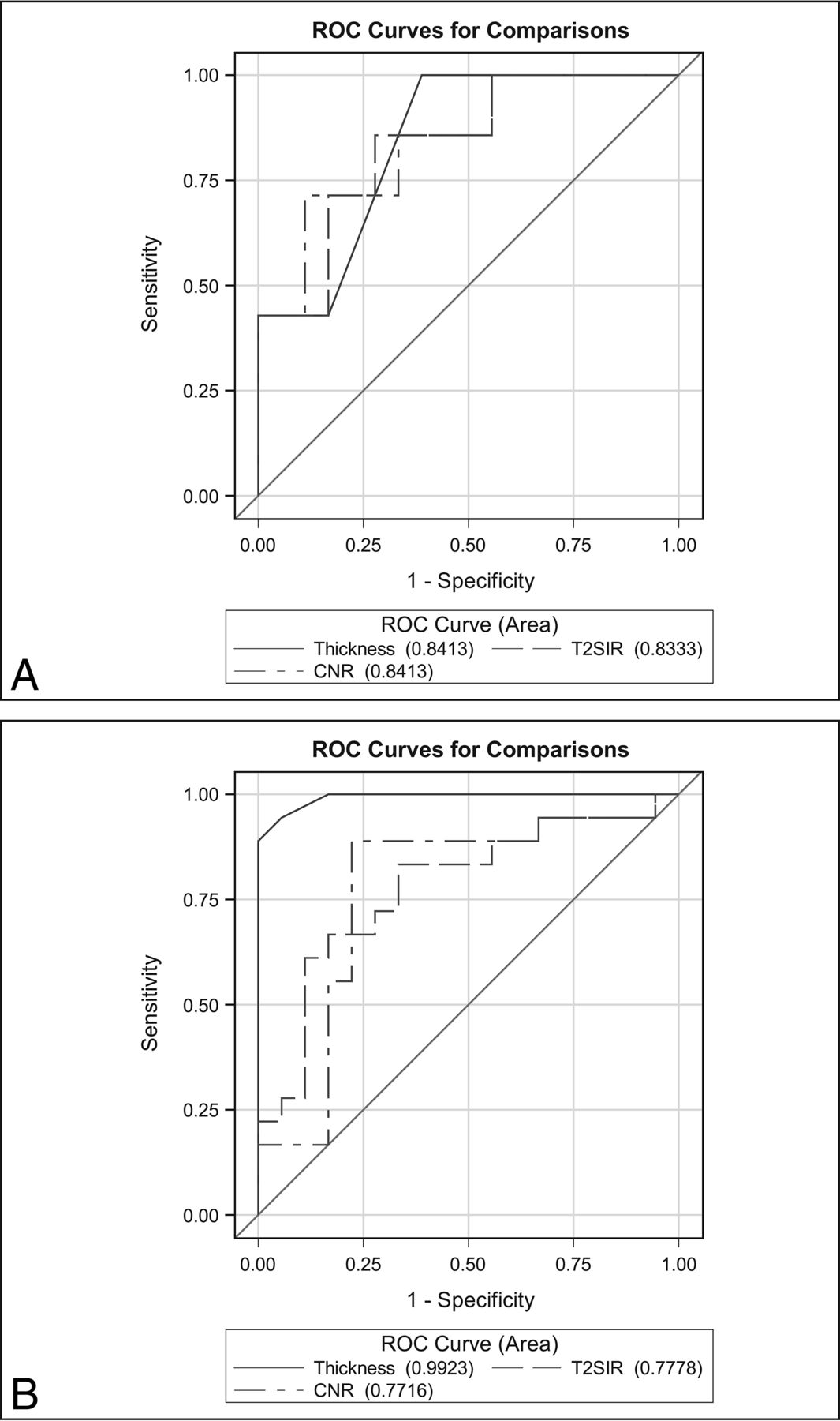
- Fig 7.
ROC curves for A, IAN and B, LN.
- Fig 8.
Correlations between differences in nerve thickness on MRN versus NST (A) and surgery (B).


Tables
Level A: Spatiotemporal Sensory Perception Direction Sensitivity <90% Static 2-Point Discrimination <18 mm Level B: Contact Detection with Monofilament <2.83 Level C: Pain Threshold and Tolerance Heat Temperature Threshold <47 Heat Temperature Tolerance <50 Pressure Pain Threshold <1.5 lb. Pressure Pain Tolerance <2.0 lb. Normal Present Present Present Mild Failed Present Present Moderate Failed Failed Present Severe Failed Failed Elevated Complete Failed Failed Absent ↵a Present: values recorded at test and control sites exhibit comparable sensitivity within published normative range. Failed: values recorded at test site sensitivity are less than that of control sites or published normative range. Elevated: values recorded at test site sensitivity are greater than that of control sites or published normative range but below maximum of test device (ie, 6 lbs.). Absent: values recorded at test site sensitivity are greater that maximum of test device (ie, 6 lbs.).
- Table 2:
Criteria for stratifying of nerve injuries on MRN and surgery based on Sunderland classification
Class MRN Surgical I Qualitative: Homogeneous increased T2 signal of nerve with no change in caliber Intact with no internal or external fibrosis, normal mobility and neuroarchitecture (visualize fascicles and Fanconi bands) Quantitative: No changes II Qualitative: Homogeneous increased T2 signal of nerve and mild nerve thickening Perineural fibrosis Intact with no internal fibrosis with external fibrosis, restricted mobility but neuroarchitecture intact (visualized fascicles and Fanconi bands once external scar removed) Quantitative: <50% larger than contralateral /normal nerve III Qualitative: Homogeneous increased T2 signal of nerve and moderate to marked nerve thickening Perineural fibrosis Intact with both internal and external fibrosis, restricted mobility and disturbance of neuroarchitecture (abnormal fascicle patterns and/or Fanconi bands not visible) Quantitative: >50% larger than contralateral/normal nerve IV Qualitative: Heterogeneous increased T2 signal of nerve and focal enlargement in otherwise continuous nerve (neuroma in continuity) Perineural and intraneural fibrosis Partial transected nerve, but some amount of distal nerve present with or without lateral neuroma Quantitative: Focal swelling with heterogeneous T2 signal or fascicular disruption V Qualitative: Discontinuous nerve with end-bulb neuroma Completely transected nerve with or without amputation (end-bulb) neuroma Quantitative: Complete disruption with gap and end-bulb neuroma Nerve Group Thickness T2SIR CNR Mean Difference SD P Value Mean Difference SD P Value Mean Difference SD P Value IAN Cases 0.60 0.33 .01 3.15 1.91 .012 6.53 4.00 .01 Controls 0.22 0.20 1.34 1.09 2.20 1.89 LN Cases 0.87 0.34 .0001 4.58 3.40 .005 6.93 4.89 .01 Controls 0.11 0.12 1.92 1.51 3.37 3.81


Jump to section
Related Articles
Cited By...
- Visualization of the Extracranial Branches of the Trigeminal Nerve Using Improved Motion-Sensitized Driven Equilibrium-Prepared 3D Inversion Recovery TSE Sequence
- Utility of MR Neurography for the Evaluation of Peripheral Trigeminal Neuropathies in the Postoperative Period
- Efficacy of MR Neurography of Peripheral Trigeminal Nerves: Correlation of Sunderland Grade versus Neurosensory Testing
- Cross-Sectional Imaging of Third Molar-Related Abnormalities