
Article Figures & Data
Figures
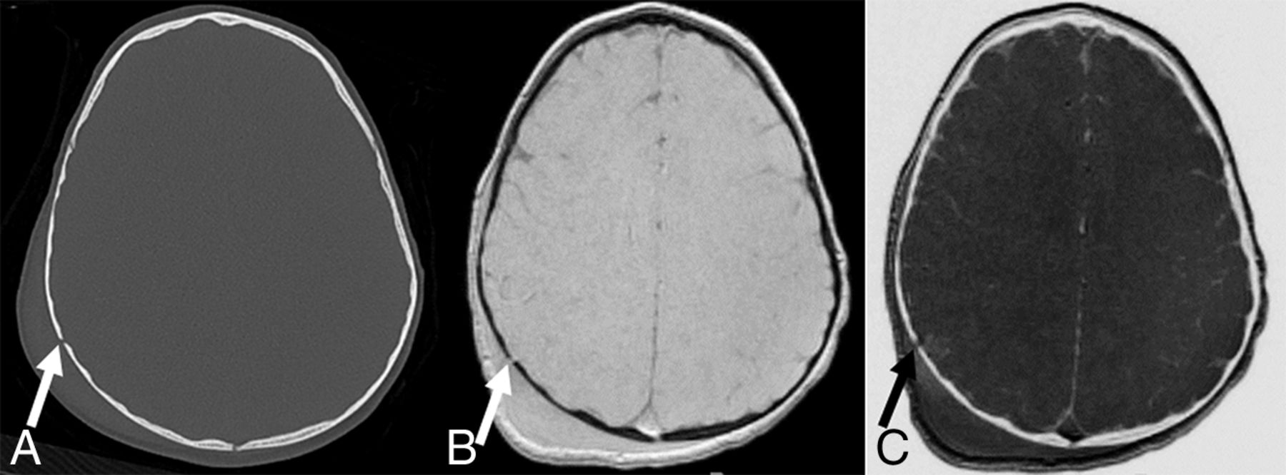
- Fig 1.
A, Axial CT image shows a nondisplaced linear fracture of the right parietal bone (arrow) with extracranial soft-tissue swelling. Black bone (B) and inverted black bone (C) MR images reveal equivalent visualization of the right parietal fracture (arrows), as well as overlying soft-tissue swelling.
- Fig 2.
A, Axial CT image shows 2 small nondisplaced linear fractures of the mastoid (arrows). On black bone (B) and inverted black bone (C) MR images, these fractures are barely visible (arrows).
- Fig 3.
A, Axial CT image does not show intracranial hemorrhage. A matching axial T2-weighted MR image (B), axial trace of diffusion (C), ADC map (D), minimal intensity projection–SWI (E), and inverted black bone MR image (F) reveal areas of T2-hyperintense signal and restricted diffusion within the temporal white matter (arrows in B–D), areas of restricted diffusion within the right frontal lobe and splenium of the corpus callosum (arrows in C and D), and foci of hypointense SWI signal within the right frontal white matter (arrows in E), suggestive of intracranial hemorrhages and diffuse axonal injury not seen on axial CT.


Tables
- Table 1:
Number and types of skull fractures and intracranial hemorrhages detected on axial 2D head CT and brain MRI including the black bone sequence in 28 children with head trauma
No. of Patients Percentage of Patients Skull fractures 12 43% Linear 11 39% Depressed 1 4% Intracranial hemorrhage 22 79% Isolated subdural 4 14% Isolated epidural 3 11% Isolated intraparenchymal 3 11% Mixed 12 43% - Table 2:
Diagnostic accuracy of axial 2D head CT compared with brain MRI including the black bone sequence for the detection of skull fractures, intracranial hemorrhages, and skull fractures and/or intracranial hemorrhages combined in 28 children with head trauma
Skull Fractures Intracranial Hemorrhages Skull Fracture and/or Intracranial Hemorrhages MRI + BB (Compared with CT) Axial CT (Compared with MRI) Axial CT MRI + BB Sensitivity 66.7% 72.7% 81.8% 100% Specificity 87.5% 83.3% 83.3% 100% PPV 80.0% 94.1% 94.7% 100% NPV 77.8% 45.5% 55.6% 100% Note:—BB indicates black bone MRI sequence.
- Table 3:
Differences in diagnostic accuracy of brain MRI including the black bone sequence for the detection of skull fractures in 28 children with head trauma, depending on the age of the child and MR imaging field strength
Age at MRI MRI Field Strength Younger Than 2 yr (n = 14) 2 yr and Older (n = 14) 1.5T (n = 12) 3T (n = 16) Sensitivity 75.0% 50.0% 83.3% 50.0% Specificity 66.7% 100% 83.3% 90.0% PPV 75.0% 100% 83.3% 75.0% NPV 66.7% 83.3% 83.3% 75.0%


Jump to section
Related Articles
Cited By...
- Comparing CT-Like Bone Images Based on Fast Field Echo Resembling a CT Using Restricted Echo Spacing (FRACTURE) MR with CT in Pediatric Congenital Vertebral Anomalies
- MR Cranial Bone Imaging: Evaluation of Both Motion-Corrected and Automated Deep Learning Pseudo-CT Estimated MR Images
- Black bone MRI morphometry for mandibular cortical bone measurement in head and neck cancer patients: Prospective method comparison with CT
- Zero TE MRI for Craniofacial Bone Imaging