Case of the Week
Section Editors: Matylda Machnowska1 and Anvita Pauranik2
1University of Toronto, Toronto, Ontario, Canada
2BC Children's Hospital, University of British Columbia, Vancouver, British Columbia, Canada
Sign up to receive an email alert when a new Case of the Week is posted.
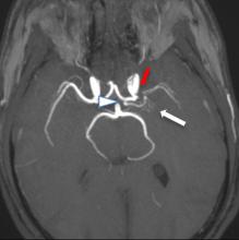
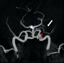
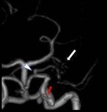
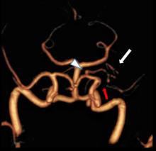
Axial FLAIR image (A) shows multiple small chronic infarcts in the frontal and parietal juxtacortical/deep white matter. Mild prominence of the overlying sulcal spaces with cortical thinning is also present. Coronal T1 postcontrast image (B) shows multiple tiny arterial collaterals in the M1 segment cistern: a "twig-like appearance" (black arrow). Axial reformatted MIP TOF (C) and 3D TOF-MRA images (D, E, and F) demonstrate proximal left MCA occlusion (red arrow) and plexiform arterial network instead of M1 (red arrow) arising from the proximal portion of ACA (arrowhead) and connecting to M2 branches. No aneurysm, stenosis, or atheromatous disease is seen within the rest of the vessels of the circle of Willis.


