Case of the Week
Section Editors: Matylda Machnowska1 and Anvita Pauranik2
1University of Toronto, Toronto, Ontario, Canada
2BC Children's Hospital, University of British Columbia, Vancouver, British Columbia, Canada
Sign up to receive an email alert when a new Case of the Week is posted.
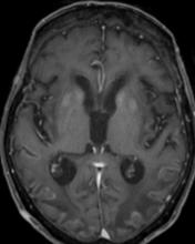
MRI 7 days after the hypoglycemic event. Axial FLAIR (A, B) demonstrates increased signal of the cortex in both hemispheres and in the basal ganglia. Axial DWI and ADC (C, D) shows restricted diffusion of the cortex and the splenium of corpus callosom. Leptomeningeal enhancement is seen in both parietal lobes (E).
Follow-up MRI done 20 days later. Axial FLAIR shows severe atrophy and increased signal in the cerebral cortex, splenium, basal ganglia, and cerebral white matter (F), without restricted diffusion (G). Axial T1 demonstrates laminar necrosis of parietal cortex and basal ganglia (H).


