Advertisement

Main menu

  • Home
  • Content
    • Current Issue
    • Accepted Manuscripts
    • Article Preview
    • Past Issue Archive
    • Video Articles
    • AJNR Case Collection
    • Case of the Week Archive
    • Case of the Month Archive
    • Classic Case Archive
  • Special Collections
    • AJNR Awards
    • Low-Field MRI
    • Alzheimer Disease
    • ASNR Foundation Special Collection
    • Photon-Counting CT
    • View All
  • Multimedia
    • AJNR Podcasts
    • AJNR SCANtastic
    • Trainee Corner
    • MRI Safety Corner
    • Imaging Protocols
  • For Authors
    • Submit a Manuscript
    • Submit a Video Article
    • Submit an eLetter to the Editor/Response
    • Manuscript Submission Guidelines
    • Statistical Tips
    • Fast Publishing of Accepted Manuscripts
    • Graphical Abstract Preparation
    • Imaging Protocol Submission
    • Author Policies
  • About Us
    • About AJNR
    • Editorial Board
    • Editorial Board Alumni
  • More
    • Become a Reviewer/Academy of Reviewers
    • Subscribers
    • Permissions
    • Alerts
    • Feedback
    • Advertisers
    • ASNR Home

User menu

  • Alerts
  • Log in
  • Log out

Search

  • Advanced search
American Journal of Neuroradiology

American Journal of Neuroradiology

ASHNR American Society of Functional Neuroradiology ASHNR American Society of Pediatric Neuroradiology ASSR
  • Alerts
  • Log in
  • Log out

Advanced Search

  • Home
  • Content
    • Current Issue
    • Accepted Manuscripts
    • Article Preview
    • Past Issue Archive
    • Video Articles
    • AJNR Case Collection
    • Case of the Week Archive
    • Case of the Month Archive
    • Classic Case Archive
  • Special Collections
    • AJNR Awards
    • Low-Field MRI
    • Alzheimer Disease
    • ASNR Foundation Special Collection
    • Photon-Counting CT
    • View All
  • Multimedia
    • AJNR Podcasts
    • AJNR SCANtastic
    • Trainee Corner
    • MRI Safety Corner
    • Imaging Protocols
  • For Authors
    • Submit a Manuscript
    • Submit a Video Article
    • Submit an eLetter to the Editor/Response
    • Manuscript Submission Guidelines
    • Statistical Tips
    • Fast Publishing of Accepted Manuscripts
    • Graphical Abstract Preparation
    • Imaging Protocol Submission
    • Author Policies
  • About Us
    • About AJNR
    • Editorial Board
    • Editorial Board Alumni
  • More
    • Become a Reviewer/Academy of Reviewers
    • Subscribers
    • Permissions
    • Alerts
    • Feedback
    • Advertisers
    • ASNR Home
  • Follow AJNR on Twitter
  • Visit AJNR on Facebook
  • Follow AJNR on Instagram
  • Join AJNR on LinkedIn
  • RSS Feeds

AJNR Awards, New Junior Editors, and more. Read the latest AJNR updates

Research ArticleHead and Neck Imaging

Inner Ear Signal Abnormalities of Adjacent Intracranial Lipochoristoma

John C. Benson, Matthew L. Carlson, Karl R. Khandalavala, Girish Bathla, Paul J. Farnsworth, Jamie J. Van Gompel, John T. Wald and John I. Lane
American Journal of Neuroradiology May 2025, 46 (5) 1016-1021; DOI: https://doi.org/10.3174/ajnr.A8603
John C. Benson
aFrom the Department of Radiology (J.C.B., G.B., P.J.F., J.T.W., J.I.L.), Mayo Clinic, Rochester, Minnesota
  • Find this author on Google Scholar
  • Find this author on PubMed
  • Search for this author on this site
  • ORCID record for John C. Benson
Matthew L. Carlson
bDepartment of Otorhinolaryngology (M.L.C., K.R.K.), Mayo Clinic, Rochester, Minnesota
  • Find this author on Google Scholar
  • Find this author on PubMed
  • Search for this author on this site
  • ORCID record for Matthew L. Carlson
Karl R. Khandalavala
bDepartment of Otorhinolaryngology (M.L.C., K.R.K.), Mayo Clinic, Rochester, Minnesota
  • Find this author on Google Scholar
  • Find this author on PubMed
  • Search for this author on this site
Girish Bathla
aFrom the Department of Radiology (J.C.B., G.B., P.J.F., J.T.W., J.I.L.), Mayo Clinic, Rochester, Minnesota
  • Find this author on Google Scholar
  • Find this author on PubMed
  • Search for this author on this site
  • ORCID record for Girish Bathla
Paul J. Farnsworth
aFrom the Department of Radiology (J.C.B., G.B., P.J.F., J.T.W., J.I.L.), Mayo Clinic, Rochester, Minnesota
  • Find this author on Google Scholar
  • Find this author on PubMed
  • Search for this author on this site
  • ORCID record for Paul J. Farnsworth
Jamie J. Van Gompel
cDepartment of Neurologic Surgery (J.J.V.G.), Mayo Clinic, Rochester, Minnesota
  • Find this author on Google Scholar
  • Find this author on PubMed
  • Search for this author on this site
  • ORCID record for Jamie J. Van Gompel
John T. Wald
aFrom the Department of Radiology (J.C.B., G.B., P.J.F., J.T.W., J.I.L.), Mayo Clinic, Rochester, Minnesota
  • Find this author on Google Scholar
  • Find this author on PubMed
  • Search for this author on this site
  • ORCID record for John T. Wald
John I. Lane
aFrom the Department of Radiology (J.C.B., G.B., P.J.F., J.T.W., J.I.L.), Mayo Clinic, Rochester, Minnesota
  • Find this author on Google Scholar
  • Find this author on PubMed
  • Search for this author on this site
  • ORCID record for John I. Lane
  • Article
  • Figures & Data
  • Info & Metrics
  • Responses
  • References
  • PDF
Loading

This article requires a subscription to view the full text. If you have a subscription you may use the login form below to view the article. Access to this article can also be purchased.

Abstract

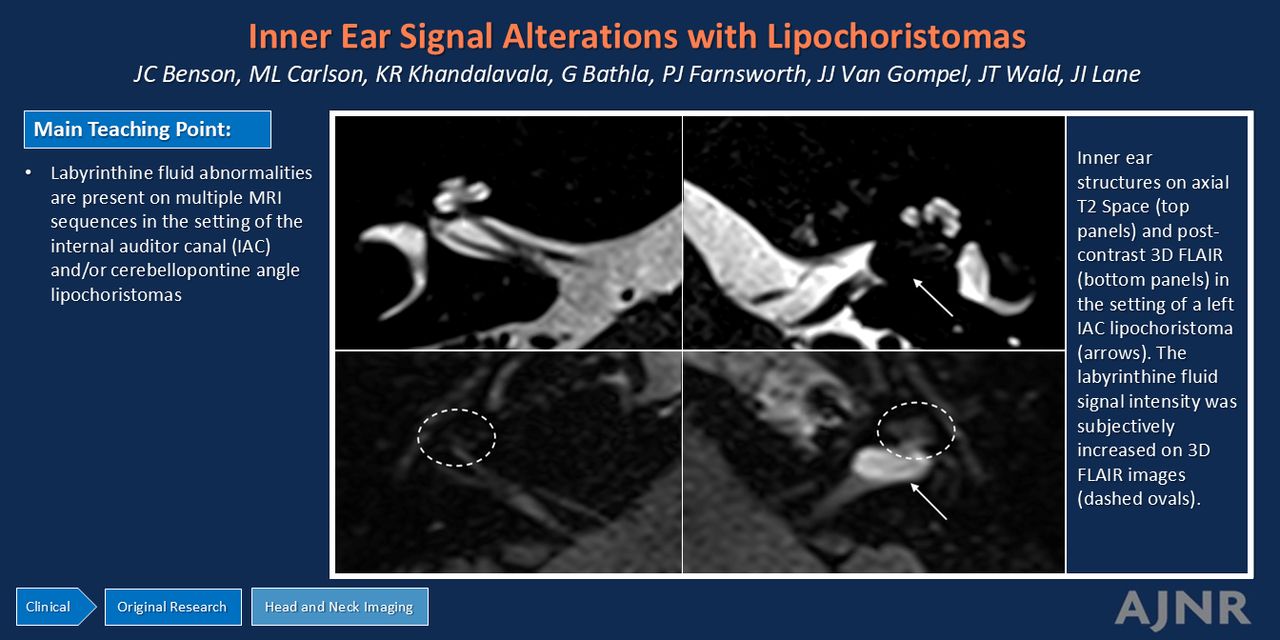
BACKGROUND AND PURPOSE: Inner ear signal abnormalities commonly develop in cases of vestibular schwannoma and are associated with hearing loss. Whether such signal alterations occur in other masses of the internal auditory canal (IAC), however, remains unknown. Here, we assessed inner ear signal abnormalities of lipochoristomas, historically termed “lipomas,” involving the IAC and cerebellopontine angle (CPA).

MATERIALS AND METHODS: A retrospective review was completed of patients with an MRI of an IAC and/or CPA intracranial lipochoristoma. The signal intensity of the ipsilateral labyrinthine structures was both subjectively and objectively compared with the contralateral side on FLAIR, FSE, T2 sampling perfection with application-optimized contrasts by using different flip angle evolution (SPACE sequence) and gradient recalled-echo (CISS) images by 2 neuroradiologists. Any initial disagreements were resolved by joint review to establish a consensus.

RESULTS: Fourteen patients were included. The average age was 53.1 (SD, 11.7) years, and 6 patients (42.9%) were women. Twelve of 14 (86%) of the lipochoristomas were in the IAC; the remaining masses were in the CPA. Regarding subjective assessment of abnormal labyrinthine signal, there was perfect interobserver agreement using FLAIR and T2 SPACE images; the Fleiss κ for CISS images was 0.7379. After consensus review, abnormal signal was noted in the adjacent labyrinthine structures in most cases on FLAIR (75%) and T2 CISS (73%); only 8% of patients had abnormal signal on T2 SPACE. Objective measurements of the cochlear signal similarly demonstrated relatively increased ipsilateral signal on FLAIR (P = .011) and relatively decreased signal on T2 CISS (P = .046). No significant difference was noted between ipsilateral and contralateral measurements on T2 SPACE (P = .093).

CONCLUSIONS: Abnormally increased FLAIR signal and decreased T2 CISS signal are present in most ipsilateral labyrinthine structures in patients with IAC and/or CPA lipochoristomas. Thus, these labyrinthine signal alterations are not exclusively restricted to vestibular schwannomas.

ABBREVIATIONS:

CPA
cerebellopontine angle
IAC
internal auditory canal
PTA
pure tone average
VS
vestibular schwannoma
WRS
word recognition score
  • © 2025 by American Journal of Neuroradiology
View Full Text

Log in using your username and password

Forgot your user name or password?

Log in through your institution

You may be able to gain access using your login credentials for your institution. Contact your library if you do not have a username and password.
PreviousNext
Back to top

In this issue

American Journal of Neuroradiology: 46 (5)
American Journal of Neuroradiology
Vol. 46, Issue 5
1 May 2025
  • Table of Contents
  • Index by author
  • Complete Issue (PDF)
Advertisement
Print
Download PDF
Email Article

Thank you for your interest in spreading the word on American Journal of Neuroradiology.

NOTE: We only request your email address so that the person you are recommending the page to knows that you wanted them to see it, and that it is not junk mail. We do not capture any email address.

Enter multiple addresses on separate lines or separate them with commas.
Inner Ear Signal Abnormalities of Adjacent Intracranial Lipochoristoma
(Your Name) has sent you a message from American Journal of Neuroradiology
(Your Name) thought you would like to see the American Journal of Neuroradiology web site.
CAPTCHA
This question is for testing whether or not you are a human visitor and to prevent automated spam submissions.
Cite this article
John C. Benson, Matthew L. Carlson, Karl R. Khandalavala, Girish Bathla, Paul J. Farnsworth, Jamie J. Van Gompel, John T. Wald, John I. Lane
Inner Ear Signal Abnormalities of Adjacent Intracranial Lipochoristoma
American Journal of Neuroradiology May 2025, 46 (5) 1016-1021; DOI: 10.3174/ajnr.A8603

Citation Manager Formats

  • BibTeX
  • Bookends
  • EasyBib
  • EndNote (tagged)
  • EndNote 8 (xml)
  • Medlars
  • Mendeley
  • Papers
  • RefWorks Tagged
  • Ref Manager
  • RIS
  • Zotero
0 Responses
Respond to this article
Share
Bookmark this article
Lipochoristomas and inner ear signal
John C. Benson, Matthew L. Carlson, Karl R. Khandalavala, Girish Bathla, Paul J. Farnsworth, Jamie J. Van Gompel, John T. Wald, John I. Lane
American Journal of Neuroradiology May 2025, 46 (5) 1016-1021; DOI: 10.3174/ajnr.A8603
del.icio.us logo Twitter logo Facebook logo Mendeley logo
  • Tweet Widget
  • Facebook Like
  • Google Plus One
Purchase

Jump to section

  • Article
    • Abstract
    • ABBREVIATIONS:
    • MATERIALS AND METHODS
    • RESULTS
    • DISCUSSION
    • CONCLUSIONS
    • Footnotes
    • References
  • Figures & Data
  • Info & Metrics
  • Responses
  • References
  • PDF

Related Articles

  • No related articles found.
  • PubMed
  • Google Scholar

Cited By...

  • No citing articles found.
  • Crossref
  • Google Scholar

This article has not yet been cited by articles in journals that are participating in Crossref Cited-by Linking.

More in this TOC Section

  • FDG Uptake and Thyroid Cancer Post-BRAF Therapy
  • Normal Facial Nerve Enhancement on VIBE Sequence
  • Comprehensive Review of Inner Ear Anatomy on Photon-Counting CT
Show more Head and Neck Imaging

Similar Articles

Advertisement

Indexed Content

  • Current Issue
  • Accepted Manuscripts
  • Article Preview
  • Past Issues
  • Editorials
  • Editor's Choice
  • Fellows' Journal Club
  • Letters to the Editor
  • Video Articles

Cases

  • Case Collection
  • Archive - Case of the Week
  • Archive - Case of the Month
  • Archive - Classic Case

More from AJNR

  • Trainee Corner
  • Imaging Protocols
  • MRI Safety Corner

Multimedia

  • AJNR Podcasts
  • AJNR Scantastics

Resources

  • Turnaround Time
  • Submit a Manuscript
  • Submit a Video Article
  • Submit an eLetter to the Editor/Response
  • Manuscript Submission Guidelines
  • Statistical Tips
  • Fast Publishing of Accepted Manuscripts
  • Graphical Abstract Preparation
  • Imaging Protocol Submission
  • Evidence-Based Medicine Level Guide
  • Publishing Checklists
  • Author Policies
  • Become a Reviewer/Academy of Reviewers
  • News and Updates

About Us

  • About AJNR
  • Editorial Board
  • Editorial Board Alumni
  • Alerts
  • Permissions
  • Not an AJNR Subscriber? Join Now
  • Advertise with Us
  • Librarian Resources
  • Feedback
  • Terms and Conditions
  • AJNR Editorial Board Alumni

American Society of Neuroradiology

  • Not an ASNR Member? Join Now

© 2025 by the American Society of Neuroradiology All rights, including for text and data mining, AI training, and similar technologies, are reserved.
Print ISSN: 0195-6108 Online ISSN: 1936-959X

Powered by HighWire