
Article Figures & Data
Figures
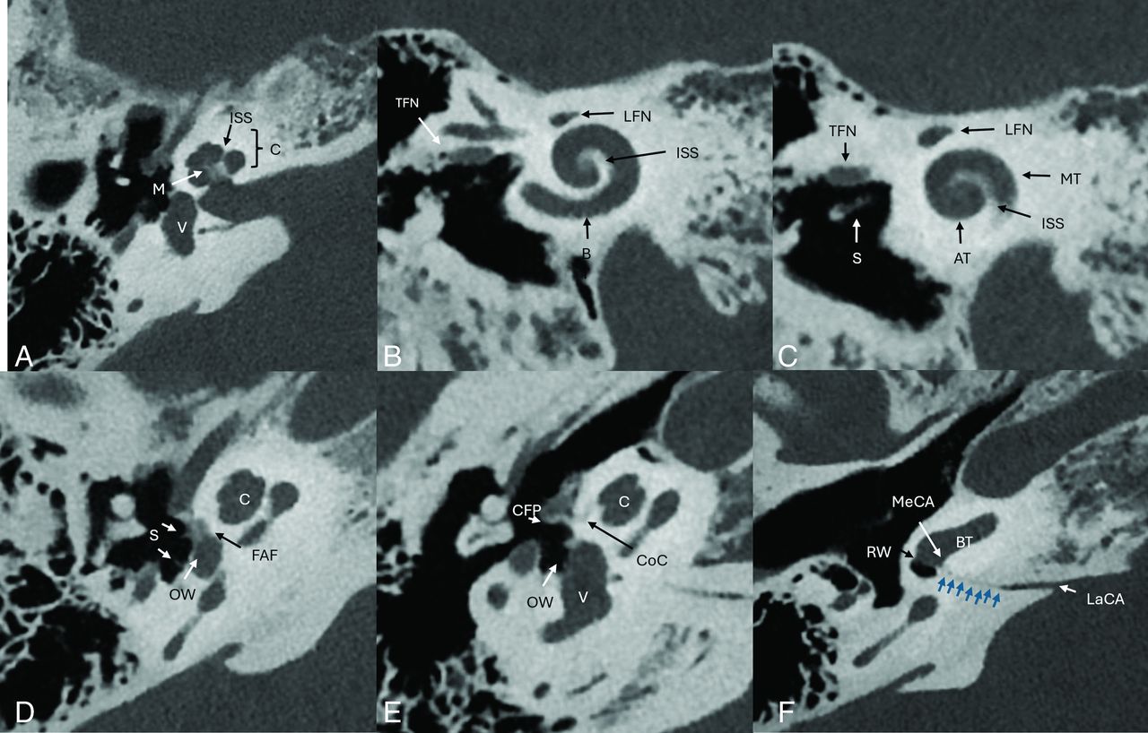
- FIG 1.
A, Axial image of the cochlea and vestibule. B and C, Stenver projection reconstructed images. D, Axial CT image demonstrating demineralized appearance of the FAF anterior to the oval window (OW). The FAF is only visible in patients with otosclerosis. Note the expansile appearance and relative posterior position to the cochlear cleft (CoC) in figure E. Note the anterior and posterior crura of the stapes. E, Axial image of the cochlea and vestibule. F, Axial image of the cochlear aqueduct. The cochlear aqueduct (blue arrows) is visible extending from the medial petrous ridge to the basal turn of the cochlea near the round window (RW). LaCA indicates lateral cochlear aqueduct orifice; MeCA, medial cochlear aqueduct orifice; M, modiolus; V, vestibule; S, stapes; C. cochlea; CFP, cochleariform process; LFN, labyrinthine segment facial nerve; BT, basal turn of the cochlea; ISS, interscalar septum; AT, apical turn of the cochlea MT, middle turn of the cochlea; TFN, tympanic segment facial nerve.
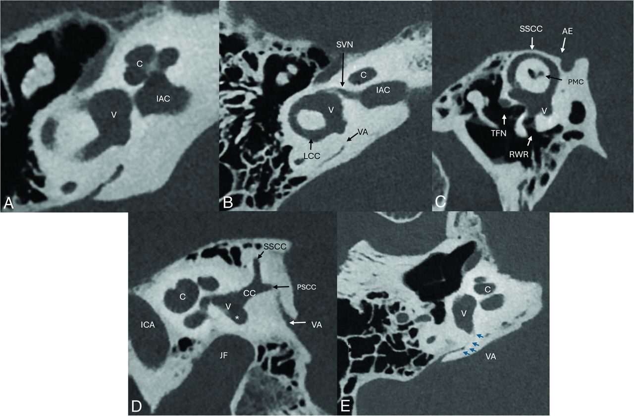
- FIG 2.
A, Axial image of the cochlea and vestibule. B, Axial image of the vestibular structures. C, Poschl reconstructed image of the superior semicircular canal (SSCC). D, Sagittal image of the vestibular structures. E, Axial image of the VA (blue arrows). PCT can demonstrate the anterior orifice in the vestibule. CC indicates common crus of the superior and posterior semicircular canal; PMC, petromastoid canal; RWR, round window recess; TFN, tympanic segment facial nerve; LCC, lateral semicircular canal; VA, vestibular aqueduct; JF, jugular fossa; PSCC, posterior semicircular canal; asterisk, the PSC ampulla; SVN, superior vestibular nerve; C, cochlea; V, vestibule; AE, arcuate eminence.
- FIG 3.
A, Axial image of the facial nerve (FN) including the labyrinthine segment of the facial nerve (LFN), facial nerve geniculate segment (GFN), which contains the geniculate ganglion. Note the superior vestibular nerve (SVN), which exits the posterior IAC to innervate the vestibular structures (V). B, Axial image of the greater superficial petrosal nerve (GSPN). C, Axial oblique image of the tympanic segment of the facial nerve (TFN). D, Sagittal oblique image demonstrates the relationship of the TFN and mastoid segments of the facial nerve (MFN) with adjacent structures. Note the nerve to the stapedius muscle (St-N) and the TFN position superior to the stapes (S). E, Sagittal oblique image demonstrating the relationship of the MFN, the auricular branch of the vagus nerve (Arnold’s nerve, [ABV]), and the stapedial nerve (St-N) and muscle (St-M). F, Axial oblique image demonstrates the St-N exiting the mastoid segment of the facial nerve (MFN) and entering the St-M. MTC indicates the middle turn of the cochlea; SMF, stylomastoid foramen.
- FIG 4.
A, Coronal oblique image of the chorda tympani origin (CT) from the mastoid segment of the facial nerve (FN). B, Sagittal oblique image of the posterior canaliculus of the CT (CT-PC) extending through the mastoid bone and entering the middle ear cavity. Note the course adjacent to the malleus (M) and incus (I). C, Axial oblique image of the CT tympanic segment (CT-T) medial to the tympanic membrane (TM). The nerve passes between the manubrium of the malleus (Ma) and long process of the incus (LoP). D, The anterior aspect of the CT exits the middle ear cavity through the medial aspect of the petrotympanic fissure (PTF), traveling through the anterior canaliculus (CT-AC), and then exits the skull base at the anterior tympanic aperture (ATA) to join the lingual nerve.
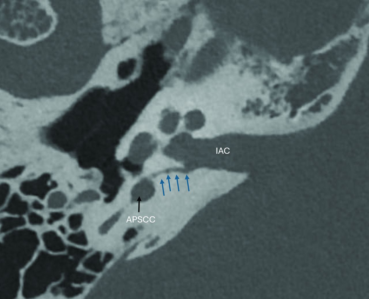
- FIG 5.
Axial image demonstrating the singular nerve (blue arrows) arising from the inferior IAC and extending through the otic capsule into the ampulla of the posterior semicircular canal (APSCC).
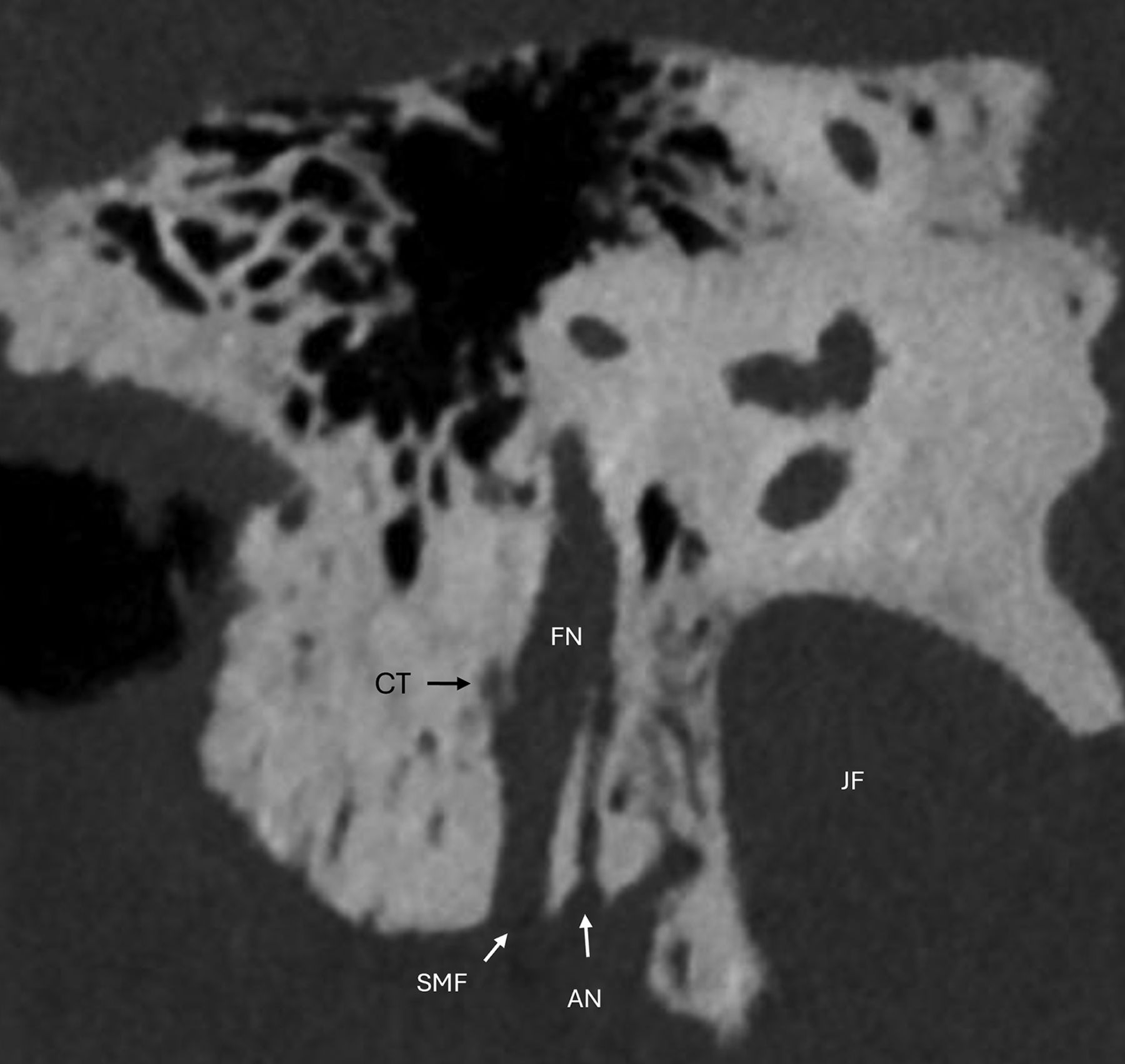
- FIG 6.
Coronal oblique image demonstrating the Arnold’s nerve (AN) as it originates from the lateral aspect of the jugular fossa (JF). AN enters the facial nerve canal (FN) mastoid segment. Note the origin of the posterior canaliculus of the chorda tympani (CT) and the stylomastoid foramen (SMF).
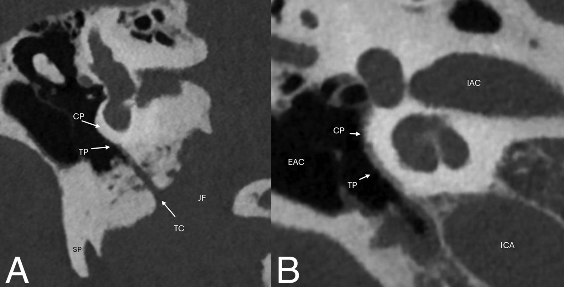
- FIG 7.
A, Coronal oblique image of the course of Jacobson’s nerve through the inferior tympanic canaliculus (TC) along the undersurface of the cochlear promontory (CP), where it forms the tympanic plexus (TP). B, Coronal oblique image demonstrates the TP along the CP. Note the position of the external auditory canal (EAC), IAC, and ICA for orientation. JF indicates jugular foramen, SP, styloid process.
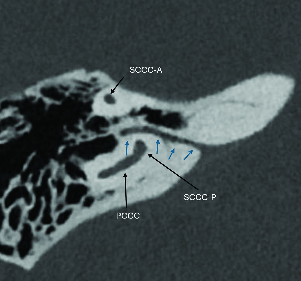
- FIG 8.
Axial image demonstrating the petromastoid canal (blue arrows), which contains the subarcuate artery and vein. The canal passes between the superior semicircular canal anterior (SCCA-A) and posterior (SCCA-P) crura. Note the position to the posterior semicircular canal (PCCC).


Jump to section
Related Articles
Cited By...
- No citing articles found.