
Article Figures & Data
Figures
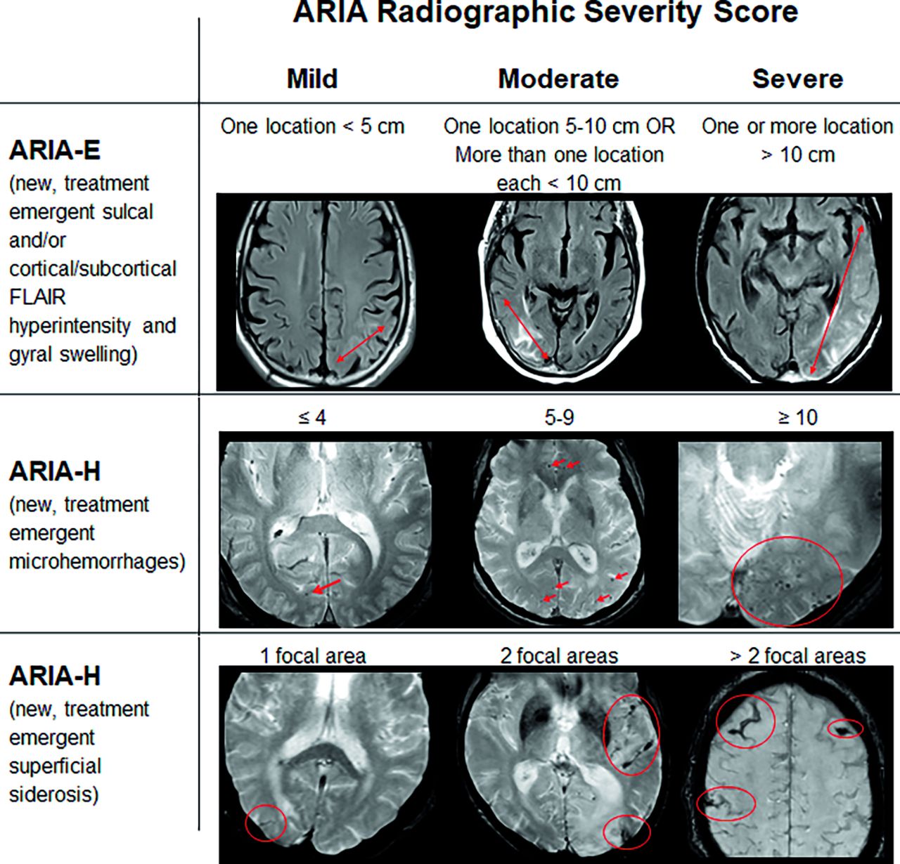
- FIG 1.
ARIA radiographic severity score. ARIA-E, ARIA-H microhemorrhages, and ARIAI-H superficial siderosis are graded separately on the basis of treatment-emergent imaging findings. Any new, transient edema/sulcal effusion and new microhemorrhages or siderosis that occur while on treatment constitute ARIA. For ARIA-E, the size indicates the greatest extent of contiguous signal abnormality/gyral swelling measured in any dimension. Mild ARIA-E: new postdosing left parietal parenchymal edema measuring less than 5 cm (line); Moderate ARIA-E: new edema and gyral swelling in the right temporal-occipital lobes measuring 5–10 cm (line); and Severe ARIA-E: new edema and gyral swelling involving the left temporal-occipital lobes measuring greater 10 cm in greatest dimension. Mild ARIA-H microhemorrhages: one right occipital treatment-emergent microhemorrhage (arrow); Moderate ARIA-H: 6 scattered treatment-emergent microhemorrhages (arrows); and Severe ARIA-H: more than 10 treatment-emergent microhemorrhages clustered in the left occipital lobe (oval). Mild ARIA-H superficial siderosis: one right occipital region of treatment-emergent siderosis (oval); Moderate ARIA-H: 2 regions of siderosis (left sylvian fissure and left occipital, ovals); and Severe ARIA-H: 3 regions of siderosis (bilateral frontal and central sulcus, ovals). Some images courtesy of Dominantly Inherited Alzheimer’s Network (DIAN).
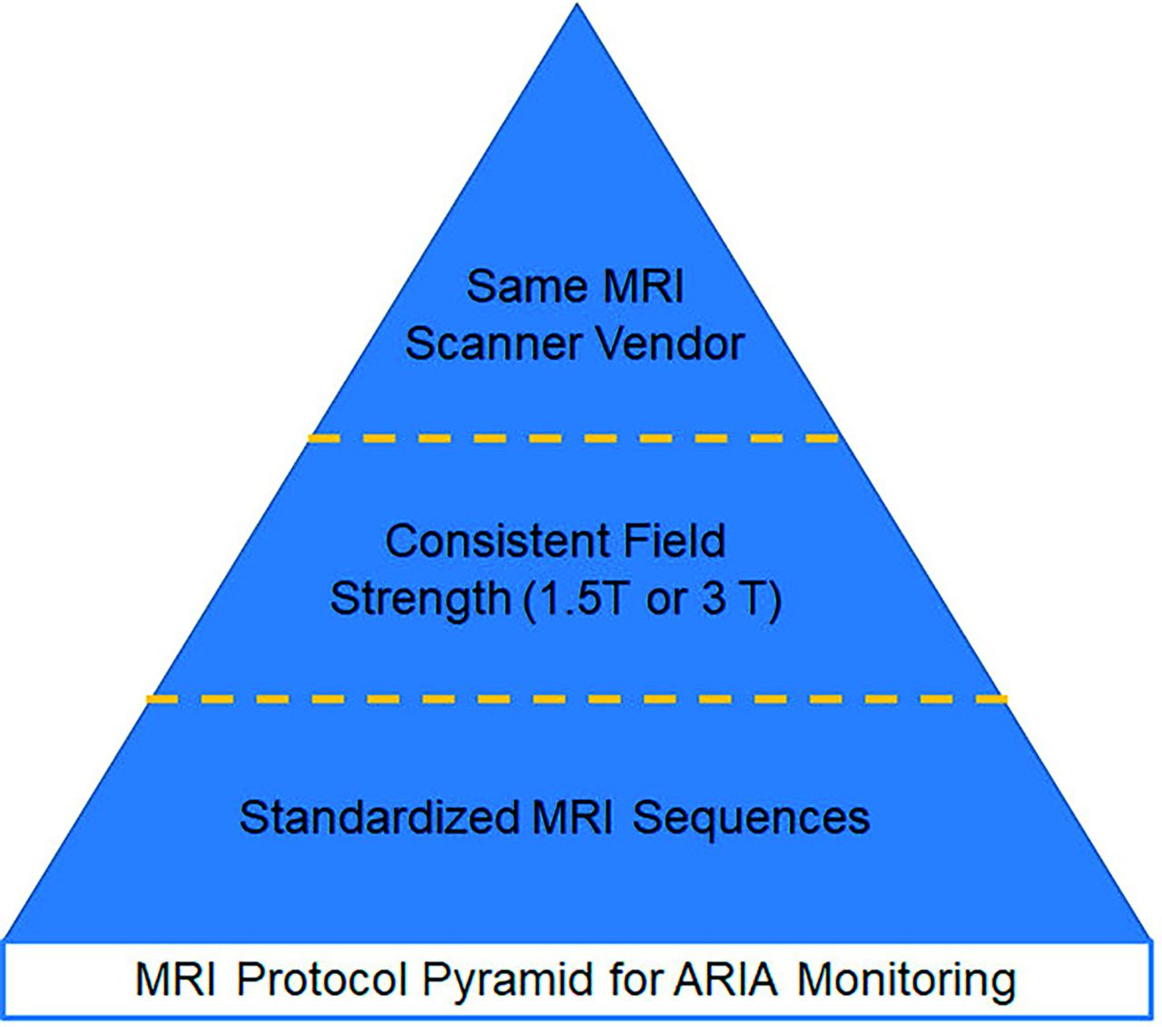
- FIG 2.
Pyramid of MRI protocol standardization. Ideally, patients are imaged using the same sequences and sequence parameters across serial examinations, using the same field strength, vendor, and scanner model. If a patient cannot be imaged on the same scanner across serial examinations, radiologists must be aware of differences that may affect the ARIA evaluation. Most important, at the pyramid base, a standardized set of MRI sequences should be used. Next in importance is the field strength, because sensitivity for heme products varies proportionally. Third, a patient would ideally be imaged using the same scanner vendor to prevent ARIA mimics on the basis of the differential appearance of white matter hyperintensities among vendors.
- FIG 3.
Temporal evolution of ARIA and reporting. AD therapy enrollment (A and D). Axial T2 FLAIR shows mild white matter hyperintensities (incompletely imaged in slice shown) and no infarcts (A). GRE shows one left occipital microhemorrhage (arrow) and no superficial siderosis (D). AD therapy monitoring (B and E). T2 FLAIR shows new T2 hyperintense signal and edema in the left-greater-than-right occipital white matter, measuring up to 3.6 cm in the greatest linear dimension. On the basis of 2 regions of signal abnormality, this finding is moderate ARIA-E (arrows, B). GRE shows a total of 13 new microhemorrhages (only some shown on this slice), severe ARIA-H microhemorrhages, and no ARIA-H siderosis (oval, E). The patient was followed with monthly MRIs until the resolution of ARIA-E and stabilization of ARIA-H, which occurred after 3 months (C and F). T2 FLAIR shows resolution of occipital T2 signal abnormality and no new FLAIR signal abnormality. No ARIA-E (C). GRE shows no new microhemorrhages. There is a total of 14 microhemorrhages, one unchanged from baseline, and 13 treatment-emergent. Unchanged severe ARIA-H (oval, F). See the Online Supplemental Data for sample reports of these findings. Images courtesy of Dominantly Inherited Alzheimer’s Network (DIAN).
- FIG 4.
Selected results from the 2023 ASNR survey gauging practice readiness for AD therapeutics imaging. A, How confident are you in your ability to identify ARIA on a brain MRI? B, Do you (your practice) have specific imaging protocols in place for AD therapeutics imaging? C, At what field strengths will your practice perform AD therapeutics imaging? D, What sequences are included in your AD therapeutics imaging protocol?


Tables
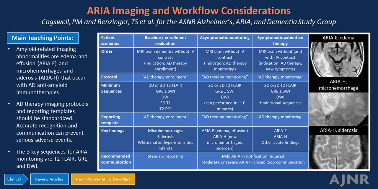
Summary of recommendations based on 3 patient scenarios
Baseline/Enrollment Evaluation Asymptomatic Monitoring Symptomatic Patient on Therapy Order MRI brain dementia without IV contrast (indication: AD therapy enrollment) MRI brain without IV contrast(indication: AD therapy monitoring) MRI brain without (and with) IV contrast (indication: AD therapy, new symptoms) Protocol AD therapy enrollment AD therapy monitoring AD therapy monitoring Minimum sequences 2D or 3D T2 FLAIRGREa ± SWIDWI3D T1T2 FSE 2D or 3D T2 FLAIRGREa ± SWIDWI 2D or 3D T2 FLAIRGREa ± SWIDWI ± additional sequences Reporting template AD therapy enrollment AD therapy monitoring AD therapy monitoring Key findings MicrohemorrhagesSiderosisWhite matter hyperintensitiesInfarcts ARIA-E (edema, effusion)ARIA-H (new microhemorrhages, siderosis) ARIA-EARIA-HOther acute findings Recommended communication Standard reporting Mild ARIA: notification requiredModerate or severe ARIA: closed-loop communication ↵a GRE must be performed with an appropriate T: 3T TE = 15–20 ms, 1.5T TE = 25–35 ms.


