
Article Figures & Data
Figures
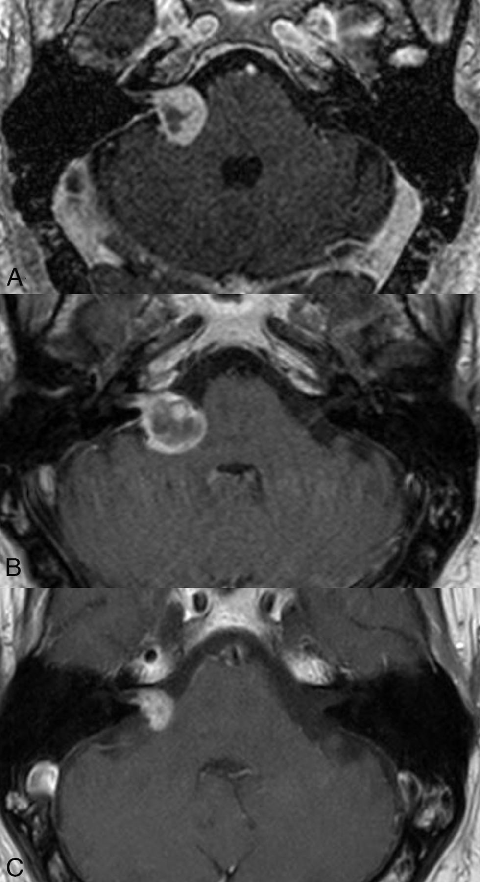
- FIG 1.
Pseudoprogression in a VS treated with SRS. The tumor volume on the planning scan (A) was 3.9 mL, which increased to 4.4 mL 6 months posttreatment. A follow-up study at 4 years (C) showed tumor regression with a volume of 0.6 mL.
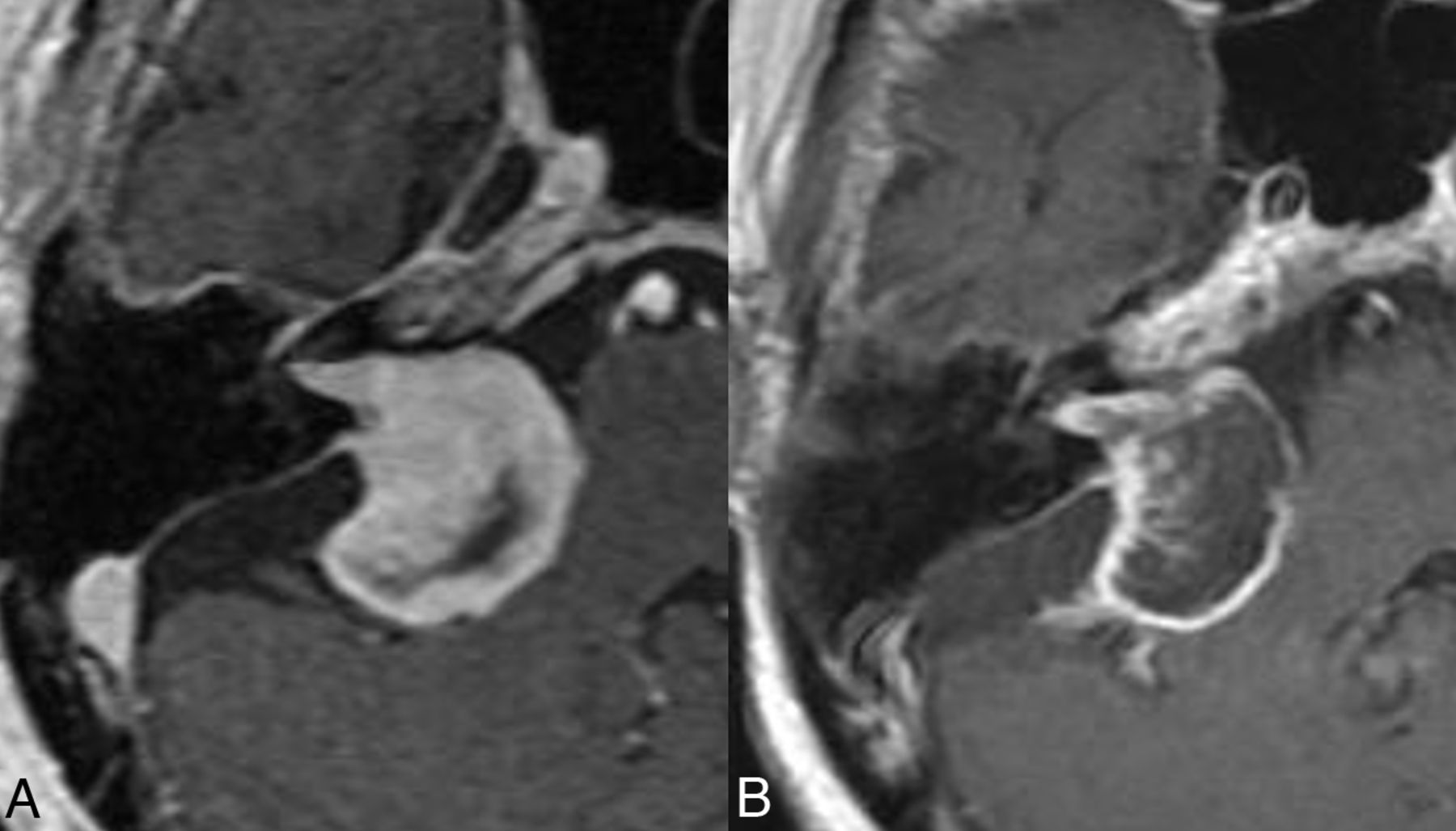
- FIG 2.
Loss of central enhancement. Postcontrast images obtained pre- (A) and 6 months post-SRS (B) show near-complete loss of central enhancement in the right VS.
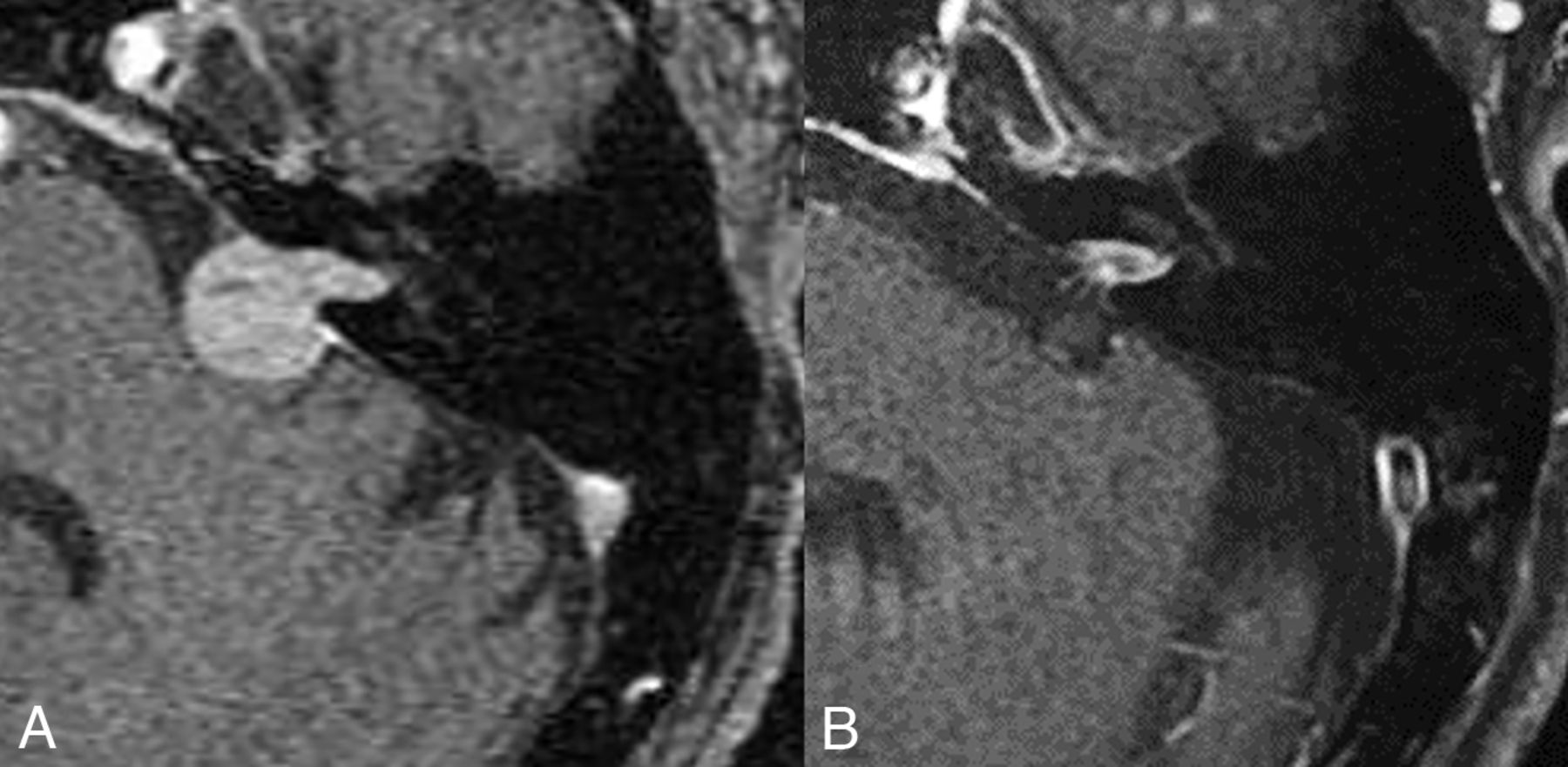
- FIG 3.
Favorable treatment response post-SRS. Axial postcontrast images obtained pre-SRS (A) and at 8 years post-SRS (B) show considerable lesion regression, consistent with a response.
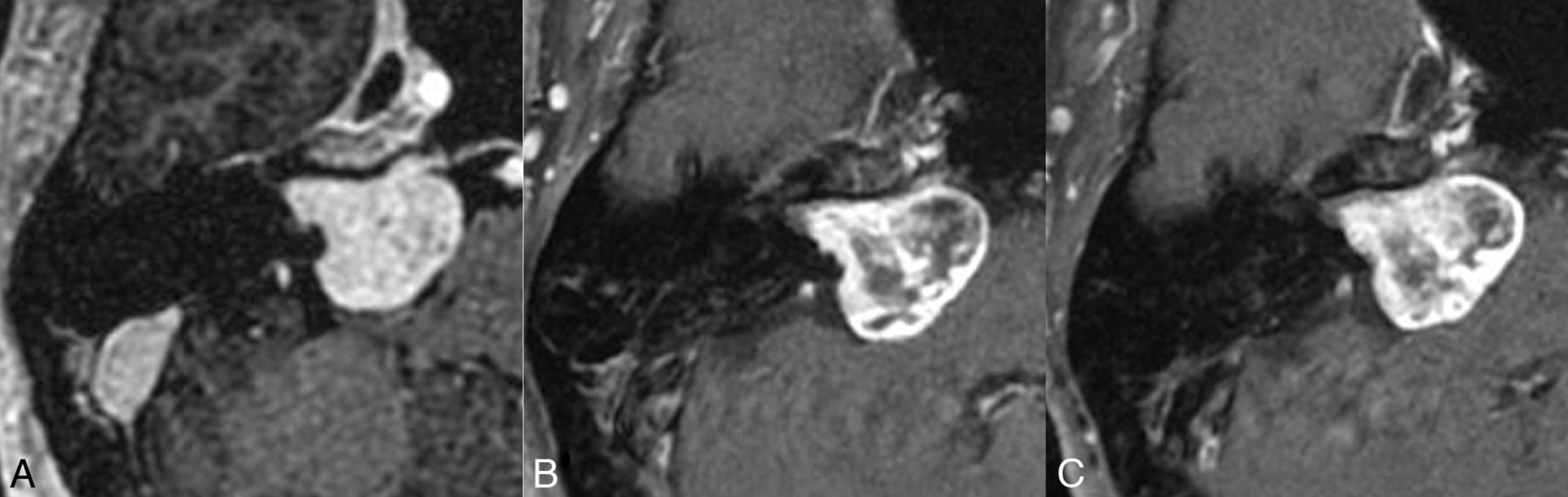
- FIG 4.
Tumor control post-SRS. Axial postcontrast images obtained pre-SRS (A) and at 1 (B) and 3 years (C) post-SRS show stable tumor size, despite considerable loss of central enhancement in the post-SRS period.
- FIG 5.
Treatment failure post-SRS. Axial postcontrast images obtained pre-SRS (A), and at 1 (B), and 2 (C) years posttherapy show a progressive increase in tumor volume from 1.9 mL at baseline to 3.2 mL at 2 years, accompanied by worsening disequilibrium clinically.
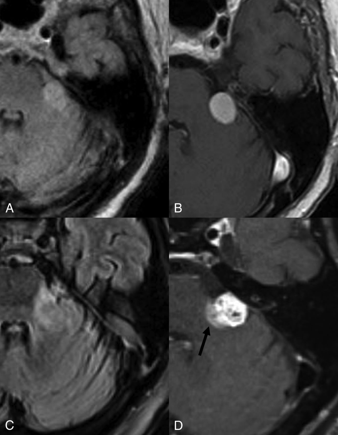
- FIG 6.
Parenchymal edema and enhancement post-SRS. Axial FLAIR (A) and postcontrast (B) images show a left-sided vestibular schwannoma abutting the left brachium pontis without edema. Post-SRS, axial FLAIR (C), and postcontrast T1WI (D) reveal parenchymal edema (C) and enhancement (arrow, D).
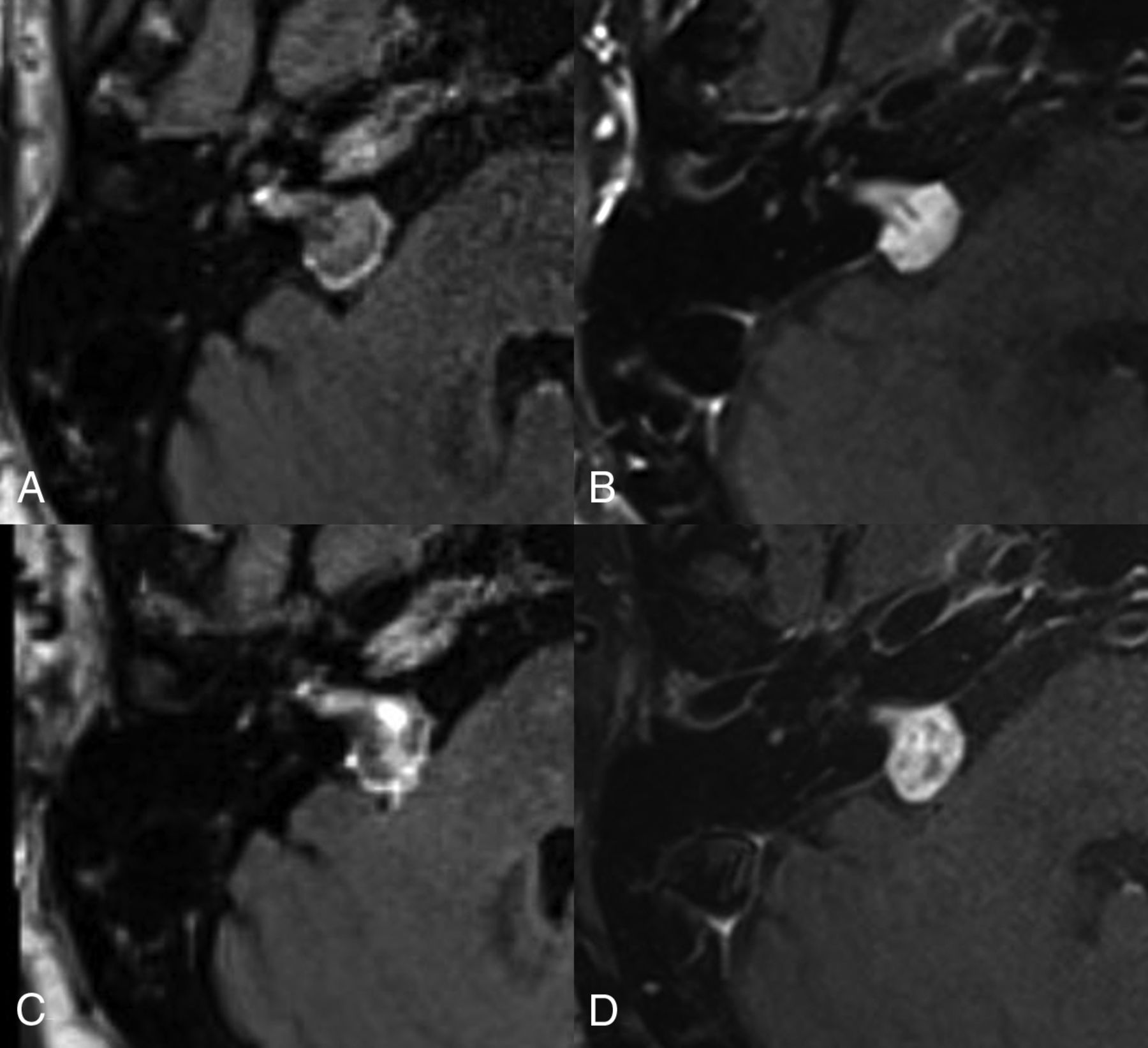
- FIG 7.
Contrast leakage post-SRS. Axial postcontrast FLAIR (A and C) and T1WI (B and D) obtained pre- (A and B), and 1 year post-SRS (C and D). On the pre-SRS images, there is a thin peritumoral halo on the FLAIR imaging (A) without any contrast leakage centrally. Post-SRS FLAIR (C) shows contrast leakage within the VS more centrally.
- FIG 8.
Hydrocephalus post-SRS. Axial postcontrast images at the level of third ventricle (A–C) and the VS (D–F) obtained at baseline (A and D), at 1 (B and E) and 2 years (C and F) post-SRS show progressive enlargement in ventricular dimensions and disproportionate enlargement of left Sylvian fissure. The underlying VS (D–F) remained stable in size and showed loss of central enhancement. The patient was diagnosed with normal pressure hydrocephalus and underwent ventricular shunting.
- FIG 9.
Glioblastoma post-SRS. Axial postcontrast, pretreatment images reveal a left VS (A). The post-SRS left temporal lobe at 2 years (B) is without any lesions. The patient subsequently presented with seizures 3 years post-SRS with a new left temporal intra-axial mass on imaging (C), which was subsequently resected and diagnosed as a glioblastoma.
- FIG 10.
Malignant transformation of a VS post-SRS. Pre-SRS (A) contrast-enhanced image shows a left-sided VS. Post-SRS image after 5 years (B) shows a mild overall increase in tumor size. However, the tumor showed increased growth at 7 years post-SRS (C). A subtotal resection was performed (D), and pathology revealed a malignant peripheral nerve sheath tumor. Follow-up imaging after 4 months (E) shows considerable recurrent tumor burden with involvement of adjacent structures.


Tables
Entity Criteria Reported Incidence New Clinical Symptoms Approximate Timeline Tumor control Lesion regression or stability >90% No NA Pseudoprogression Transient increase in tumor volume over baseline 5%–74% No 5–18 mo Delayed pseudoprogression Transient increase in tumor volume over baseline 6%–17% No 36–48 mo Tumor growth Progressive increase in tumor size/volume for 3 consecutive scans, or 40% over baseline by some authors <10% Yes Generally, not considered until 3 years post-SRS unless new symptoms Note:—NA indicates not applicable.
Post-SRS Complication/Adverse Effects Reported Incidence Risk/Prognostic Factors Vertigo and disequilibrium 1%–2% Marginal dose <13 Gy; larger tumors; female sex associated with worse outcomes Facial nerve dysfunction <1% Younger patients, smaller tumors <1.5 cm3, and radiation dose <13 Gy associated with better outcomes Trigeminal nerve dysfunction 3% at 5 years Total dose >13 Gy; brainstem dose >10 Gy; larger tumor volume associated with worse outcomes Worsening hearing loss 21%–59% at 5 years Cochlear dose >4 Gy; marginal dose >13 Gy associated with worse outcomes Hydrocephalus 2%–3% Older than 60 years of age; female sex; larger tumors associated with worse outcomes Malignant transformation <0.04% at 15 years Underlying neurofibromatosis associated with increased incidence


