Advertisement

Main menu

  • Home
  • Content
    • Current Issue
    • Accepted Manuscripts
    • Article Preview
    • Past Issue Archive
    • Video Articles
    • AJNR Case Collection
    • Case of the Week Archive
    • Case of the Month Archive
    • Classic Case Archive
  • Special Collections
    • AJNR Awards
    • Low-Field MRI
    • Alzheimer Disease
    • ASNR Foundation Special Collection
    • Photon-Counting CT
    • View All
  • Multimedia
    • AJNR Podcasts
    • AJNR SCANtastic
    • Trainee Corner
    • MRI Safety Corner
    • Imaging Protocols
  • For Authors
    • Submit a Manuscript
    • Submit a Video Article
    • Submit an eLetter to the Editor/Response
    • Manuscript Submission Guidelines
    • Statistical Tips
    • Fast Publishing of Accepted Manuscripts
    • Graphical Abstract Preparation
    • Imaging Protocol Submission
    • Author Policies
  • About Us
    • About AJNR
    • Editorial Board
    • Editorial Board Alumni
  • More
    • Become a Reviewer/Academy of Reviewers
    • Subscribers
    • Permissions
    • Alerts
    • Feedback
    • Advertisers
    • ASNR Home

User menu

  • Alerts
  • Log in

Search

  • Advanced search
American Journal of Neuroradiology

American Journal of Neuroradiology

ASHNR American Society of Functional Neuroradiology ASHNR American Society of Pediatric Neuroradiology ASSR
  • Alerts
  • Log in

Advanced Search

  • Home
  • Content
    • Current Issue
    • Accepted Manuscripts
    • Article Preview
    • Past Issue Archive
    • Video Articles
    • AJNR Case Collection
    • Case of the Week Archive
    • Case of the Month Archive
    • Classic Case Archive
  • Special Collections
    • AJNR Awards
    • Low-Field MRI
    • Alzheimer Disease
    • ASNR Foundation Special Collection
    • Photon-Counting CT
    • View All
  • Multimedia
    • AJNR Podcasts
    • AJNR SCANtastic
    • Trainee Corner
    • MRI Safety Corner
    • Imaging Protocols
  • For Authors
    • Submit a Manuscript
    • Submit a Video Article
    • Submit an eLetter to the Editor/Response
    • Manuscript Submission Guidelines
    • Statistical Tips
    • Fast Publishing of Accepted Manuscripts
    • Graphical Abstract Preparation
    • Imaging Protocol Submission
    • Author Policies
  • About Us
    • About AJNR
    • Editorial Board
    • Editorial Board Alumni
  • More
    • Become a Reviewer/Academy of Reviewers
    • Subscribers
    • Permissions
    • Alerts
    • Feedback
    • Advertisers
    • ASNR Home
  • Follow AJNR on Twitter
  • Visit AJNR on Facebook
  • Follow AJNR on Instagram
  • Join AJNR on LinkedIn
  • RSS Feeds

AJNR Awards, New Junior Editors, and more. Read the latest AJNR updates

Brief ReportPediatric Neuroimaging
Open Access

Low-Field Portable MR Imaging to Evaluate Ventricular Volumes: A Single-Center Retrospective Study

Vivek Velagapudi, Nathan S. Artz, Johnston K. Fite, Sherwin S. Chan and Adam G. Rouse
American Journal of Neuroradiology August 2024, 45 (8) 1076-1080; DOI: https://doi.org/10.3174/ajnr.A8269
Vivek Velagapudi
aFrom the School of Medicine (V.V.), University of Kansas Medical Center, Kansas City, Kansas
  • Find this author on Google Scholar
  • Find this author on PubMed
  • Search for this author on this site
  • ORCID record for Vivek Velagapudi
Nathan S. Artz
bDepartment of Radiology (N.S.A., J.K.F., S.S.C.), University of Missouri at Kansas City School of Medicine, Kansas City, Missouri
cDepartment of Radiology (N.S.A., J.K.F., S.S.C.), Children’s Mercy Kansas City, Kansas City, Missouri
  • Find this author on Google Scholar
  • Find this author on PubMed
  • Search for this author on this site
Johnston K. Fite
bDepartment of Radiology (N.S.A., J.K.F., S.S.C.), University of Missouri at Kansas City School of Medicine, Kansas City, Missouri
cDepartment of Radiology (N.S.A., J.K.F., S.S.C.), Children’s Mercy Kansas City, Kansas City, Missouri
  • Find this author on Google Scholar
  • Find this author on PubMed
  • Search for this author on this site
  • ORCID record for Johnston K. Fite
Sherwin S. Chan
bDepartment of Radiology (N.S.A., J.K.F., S.S.C.), University of Missouri at Kansas City School of Medicine, Kansas City, Missouri
cDepartment of Radiology (N.S.A., J.K.F., S.S.C.), Children’s Mercy Kansas City, Kansas City, Missouri
  • Find this author on Google Scholar
  • Find this author on PubMed
  • Search for this author on this site
  • ORCID record for Sherwin S. Chan
Adam G. Rouse
dDepartment of Neurosurgery (A.R.), Department of Neurosurgery (A.G.R.), University of Kansas Health System, Kansas City, Kansas
  • Find this author on Google Scholar
  • Find this author on PubMed
  • Search for this author on this site
  • ORCID record for Adam G. Rouse
  • Article
  • Figures & Data
  • Info & Metrics
  • Responses
  • References
  • PDF
Loading

Article Figures & Data

Figures

  • Tables
  • FIG 1.
    • Download figure
    • Open in new tab
    • Download powerpoint
    FIG 1.

    Comparison of SOC and portable clinical MR imaging as well as examples of manual segmentation in 1 participant, a 4-day-old boy without intracranial abnormalities such as ischemia or hemorrhage. Segmentation at 1 level of the lateral ventricles is shown here; SOC (A), SOC with segmentation (B), portable (C), portable with segmentation (D). Hyperfine scans were acquired with software, Version 8.1.0.1. TRA indicates transaxial; AXI, axial; R, right; L, left; P, posterior; A, anterior.

  • FIG 2.
    • Download figure
    • Open in new tab
    • Download powerpoint
    FIG 2.

    Portable MR imaging versus SOC MR imaging–estimated ventricular volumes. The ventricular volumes using Hyperfine and SOC MR images are plotted. The ventricular volumes of each set of images for the 17 patients were estimated using ITK-SNAP.
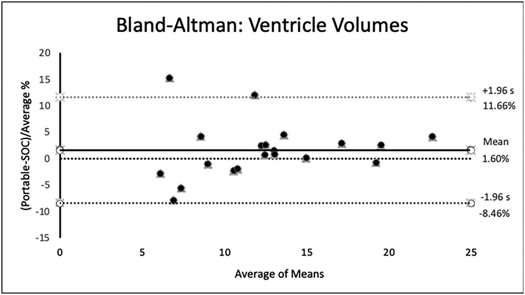
  • FIG 3.
    • Download figure
    • Open in new tab
    • Download powerpoint
    FIG 3.

    Results of the Bland-Altman analysis. Plot of differences between method A (portable) and method B (SOC), expressed as percentages of the values on the axis [(portable – SOC) / mean%)], versus the mean of the 2 measurements. The bias (mean difference) of 2.06% is constant, with greater variation for lower ventricular volumes. s indicates standard deviation.

Tables

  • Figures
    • View popup
    Table 1:

    General demographics of the study population, including indications for MR imaging that may influence ventricular volumes

    Participants
    No.%
    Biologic sex (at birth)
     Female742
     Male1058
    Ethnicity
     White1165
     African American16
     Asian212
     Other318
    Indication
     Trauma424
     Altered mental status318
     Hemorrhage212
     Ischemic stroke212
     Infection16
     Hydrocephalus00
     Lesion/mass00
    Mean time to acquire images (min)
     SOC23 (SD, 14)
     Low-field32 (SD, 13)
    Age (days)22 (SD, 7)
    • View popup
    Table 2:

    Scan parameters for SOC and Hyperfine scans used for ventricle volume quantification

    ScanNo.Scan Time (min:sec)In-Plane Resolution (mm)Section Thickness (mm)Section Spacing (mm)TR (ms)TE (ms)TI (ms)
    SOC T2-weighted TSE160:56–5:090.4–0.82–320.43250–598092–104NA
    SOC T2-weighted HASTE10:530.7–0.944.4150084NA
    Hyperfine T2-weighted TSE172:37–9:591.5–22–5NA2000195–261NA
    • Note:—NA indicates not applicable.

PreviousNext
Back to top

In this issue

American Journal of Neuroradiology: 45 (8)
American Journal of Neuroradiology
Vol. 45, Issue 8
1 Aug 2024
  • Table of Contents
  • Index by author
  • Complete Issue (PDF)
Advertisement
Print
Download PDF
Email Article

Thank you for your interest in spreading the word on American Journal of Neuroradiology.

NOTE: We only request your email address so that the person you are recommending the page to knows that you wanted them to see it, and that it is not junk mail. We do not capture any email address.

Enter multiple addresses on separate lines or separate them with commas.
Low-Field Portable MR Imaging to Evaluate Ventricular Volumes: A Single-Center Retrospective Study
(Your Name) has sent you a message from American Journal of Neuroradiology
(Your Name) thought you would like to see the American Journal of Neuroradiology web site.
CAPTCHA
This question is for testing whether or not you are a human visitor and to prevent automated spam submissions.
Cite this article
Vivek Velagapudi, Nathan S. Artz, Johnston K. Fite, Sherwin S. Chan, Adam G. Rouse
Low-Field Portable MR Imaging to Evaluate Ventricular Volumes: A Single-Center Retrospective Study
American Journal of Neuroradiology Aug 2024, 45 (8) 1076-1080; DOI: 10.3174/ajnr.A8269

Citation Manager Formats

  • BibTeX
  • Bookends
  • EasyBib
  • EndNote (tagged)
  • EndNote 8 (xml)
  • Medlars
  • Mendeley
  • Papers
  • RefWorks Tagged
  • Ref Manager
  • RIS
  • Zotero
0 Responses
Respond to this article
Share
Bookmark this article
Low-Field MR Imaging for Ventricular Volumes Study
Vivek Velagapudi, Nathan S. Artz, Johnston K. Fite, Sherwin S. Chan, Adam G. Rouse
American Journal of Neuroradiology Aug 2024, 45 (8) 1076-1080; DOI: 10.3174/ajnr.A8269
del.icio.us logo Twitter logo Facebook logo Mendeley logo
  • Tweet Widget
  • Facebook Like
  • Google Plus One
Purchase

Jump to section

  • Article
    • SUMMARY:
    • ABBREVIATIONS:
    • MATERIALS AND METHODS
    • RESULTS
    • DISCUSSION
    • CONCLUSIONS
    • Acknowledgments
    • Footnotes
    • References
  • Figures & Data
  • Info & Metrics
  • Responses
  • References
  • PDF

Related Articles

  • No related articles found.
  • PubMed
  • Google Scholar

Cited By...

  • No citing articles found.
  • Crossref
  • Google Scholar

This article has not yet been cited by articles in journals that are participating in Crossref Cited-by Linking.

More in this TOC Section

  • fetal brain development of 10 weeks gestation
  • CHARGE fetal MRI clival cleft
  • Neuroimaging Delineation and Progression of SLSMD
Show more Pediatric Neuroimaging

Similar Articles

Advertisement

Indexed Content

  • Current Issue
  • Accepted Manuscripts
  • Article Preview
  • Past Issues
  • Editorials
  • Editor's Choice
  • Fellows' Journal Club
  • Letters to the Editor
  • Video Articles

Cases

  • Case Collection
  • Archive - Case of the Week
  • Archive - Case of the Month
  • Archive - Classic Case

More from AJNR

  • Trainee Corner
  • Imaging Protocols
  • MRI Safety Corner

Multimedia

  • AJNR Podcasts
  • AJNR Scantastics

Resources

  • Turnaround Time
  • Submit a Manuscript
  • Submit a Video Article
  • Submit an eLetter to the Editor/Response
  • Manuscript Submission Guidelines
  • Statistical Tips
  • Fast Publishing of Accepted Manuscripts
  • Graphical Abstract Preparation
  • Imaging Protocol Submission
  • Evidence-Based Medicine Level Guide
  • Publishing Checklists
  • Author Policies
  • Become a Reviewer/Academy of Reviewers
  • News and Updates

About Us

  • About AJNR
  • Editorial Board
  • Editorial Board Alumni
  • Alerts
  • Permissions
  • Not an AJNR Subscriber? Join Now
  • Advertise with Us
  • Librarian Resources
  • Feedback
  • Terms and Conditions
  • AJNR Editorial Board Alumni

American Society of Neuroradiology

  • Not an ASNR Member? Join Now

© 2025 by the American Society of Neuroradiology All rights, including for text and data mining, AI training, and similar technologies, are reserved.
Print ISSN: 0195-6108 Online ISSN: 1936-959X

Powered by HighWire