
Article Figures & Data
Figures
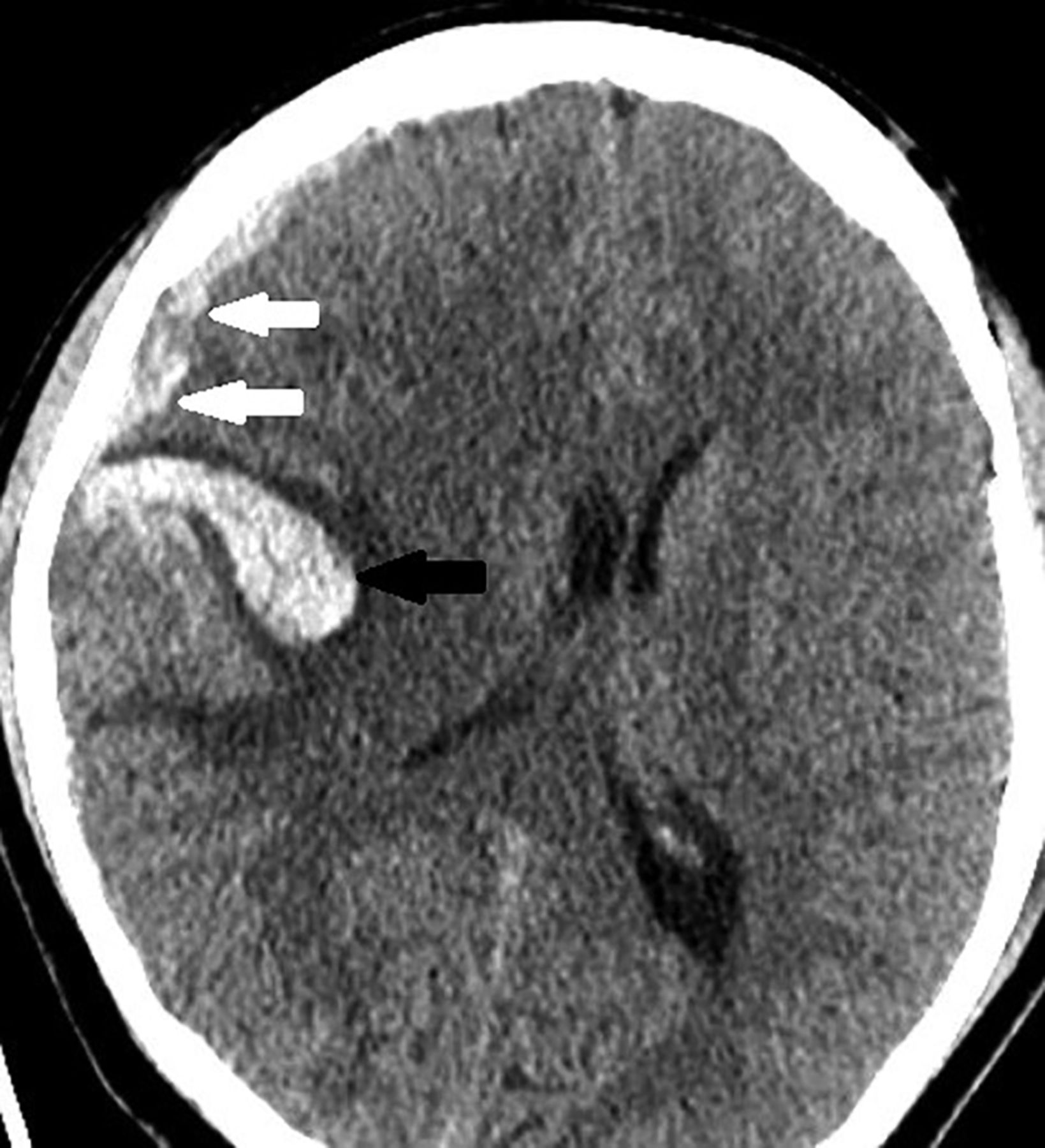
- FIG 1.
A 28-year-old patient presenting acutely with headaches over a 1-day period. Axial NCCT demonstrates acute hemorrhage within the right frontal lobe lesion (black arrow) with overlying right frontal convexity subdural hematoma (white arrows). CT also shows secondary leftward midline shift and regional mass effect.
- FIG 2.
A 10-year-old patient presenting with headaches for 14 days. A, Axial precontrast T1 3D MPRAGE demonstrates right superior parietal lobe cortical/subcortical lesion with intrinsic T1 hyperintensity reflective of internal blood products along its anterior and lateral aspects (arrows). B, SWI sequence demonstrates loss of signal within the lesion, reflective of lesion blood products (arrow). C and D, DWI and ADC sequences demonstrate diffusion restriction within the lesion that is corresponding to both the solid enhancing component and blood products noted on postcontrast and SWI sequences. E, Axial postcontrast T1 MPRAGE demonstrates homogeneous enhancement of the non hemorrhagic component (arrow). F, Single voxel short TE (35 ms) 1H-MRS demonstrates an elevated choline peak (white arrow), decreased N-acetyl aspartate peak (blue arrow), and elevated lactate peak (green arrow).
- FIG 3.
A 33-year-old patient presenting with 5-day history of worsening headaches with a rapid decrease in mentation. A, NCCT at the time of presentation demonstrates a large parenchymal hemorrhage involving the right frontal and temporal lobes with associated regional mass effect, leftward midline shift, and right ventricle effacement. B, Axial pre contrast T1 MPRAGE demonstrates areas of intrinsic T1 hyperintensity within the right temporal/insular lesion reflective of internal blood products (arrow). C, Axial postcontrast T1 MPRAGE demonstrates the homogeneous enhancement of the solid component (arrow). D, Dynamic susceptibility contrast relative cerebral blood volume color map demonstrating elevated relative cerebral blood volume within the enhancing component (arrow).
- FIG 4.
A 5-year-old presenting with headaches for 14 days. A, Sagittal precontrast T1 3D MPRAGE demonstrates a left frontal cortical/subcortical T1 intrinsically hyperintense mass that is reflective of internal blood products within the lesion (black arrow). Patient also has a left hemispheric thin subdural hematoma (white arrows). B, SWI sequence demonstrates a loss of signal within the lesion that is reflective of blood products (black arrow).
- FIG 5.
An 8-year-old patient with hypothalamic primary intracranial sarcoma, DICER1-mutant. A and B, DWI and ADC map demonstrating diffusion restriction of the left hypothalamic lesion nonenhancing component. This is partially explained by the blood products noted on SWI and the cellularity of the nonenhancing component. C, SWI sequence demonstrates a loss of signal within the lesion that is suggestive of blood products (arrow). D, Axial postcontrast T1 MPRAGE demonstrates a mixed enhancing and nonenhancing lesion with homogeneous enhancement of the solid component (arrow). E, Coronal postcontrast T1 MPRAGE demonstrates a mixed enhancing and nonenhancing lesion with homogeneous enhancement of the solid component (arrow).


Tables
- Table 1:
Demographic characteristics of patients with primary intracranial sarcoma, DICER1-mutant, included in this case series
Demographic Characteristic (Number Assessed) Finding Age (8) Mean: 16 years Standard deviation: 11.57 years Range: 4–33 Presenting symptoms (7) Headaches: 5 Extremity weakness: 2 Nausea: 2 Numbness: 2 Language deficit: 1 Blurry vision: 1 Unsteady gait: 1 Duration of symptoms (5) Average: 7 days Range: 1–14 days DICER1 mutation (8) Yes Additional mutations ATRX 5/5 patients TP53 7/7 patients Other DICER1-related tumors (7) None - Table 2:
MR imaging characteristics of patients with primary intracranial sarcoma, DICER1-mutant
MR Imaging Characteristic Finding Size (cm) Anteroposterior dimension, 4.25 cm (SD, 2.76) Transverse dimension, 3.79 cm (SD, 2.08) Craniocaudal dimension, 4.37 cm (SD, 2.5) Location Side 4 right, 3 left Cortical/deep 7 cortical/subcortical, 1 hypothalamic Cortical lobe 4 parietal, 2 frontal, 1 temporal/insula Hemorrhage Tumor 7/7 hemorrhage on SWI 8/8 hemorrhage of T1WI Extra-axial 2 SDH 2 SAH Diffusion restriction 3 patients Enhancement Presence 8/8 present Pattern 5 solid homogeneous, 2 heterogeneous, 1 peripheral Extent 5 > 75% enhancement; 2, 25%–75%; 1 < 25%


Jump to section
Related Articles
Cited By...
- No citing articles found.