
Article Figures & Data
Figures
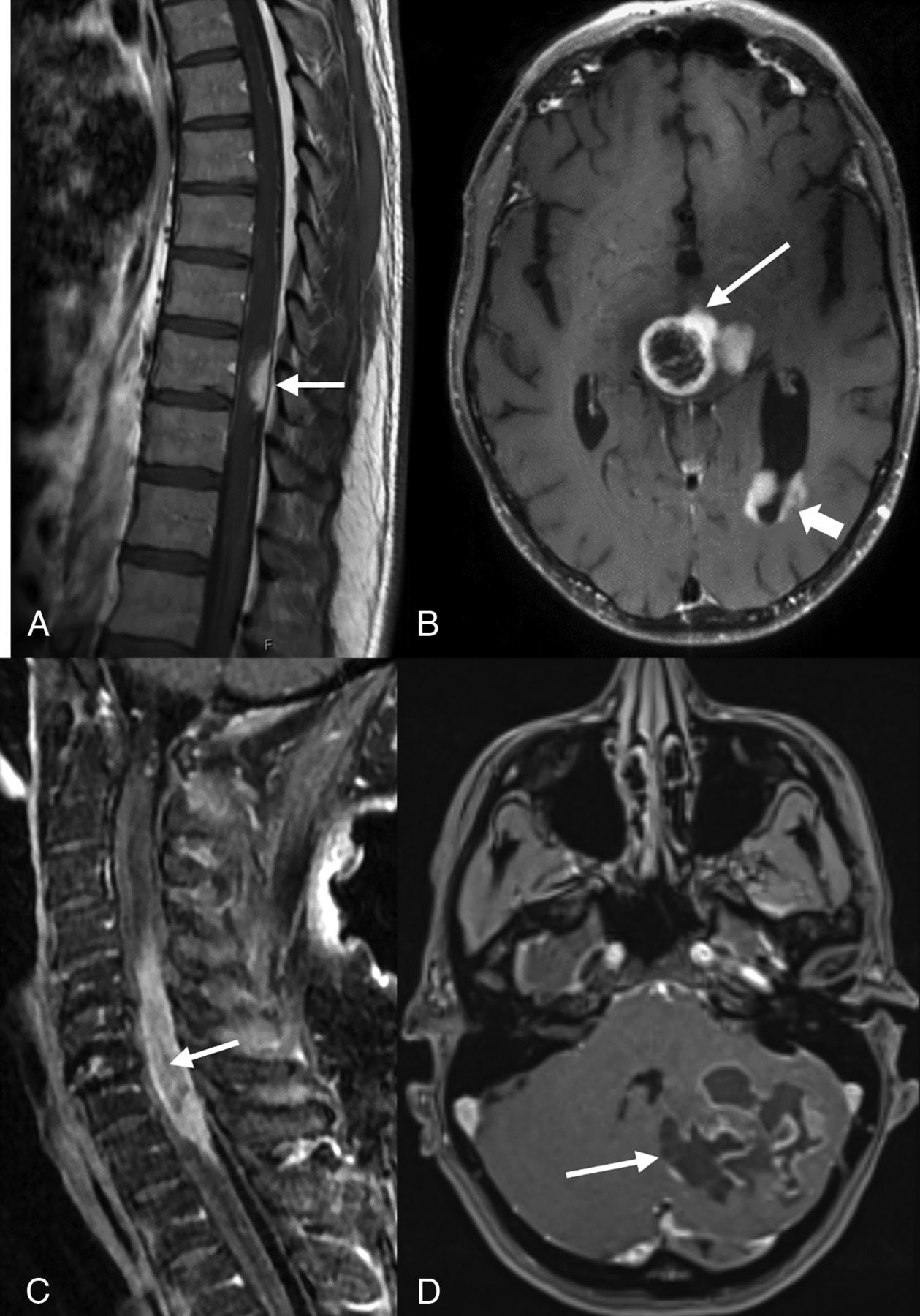
- FIG 1.
Postcontrast-T1-weighted sagittal (A) image of a 44-year-old man with midback pain shows a well-defined enhancing lesion involving the dorsal aspect of thoracic cord at the T9–T10 level (arrow). Postcontrast T1-weighted axial image in a 42-year-old man (B) shows tumor with peripheral irregular enhancement at the level of thalamus (arrow) with infratentorial pontomesencephalic and cerebellar involvement (not shown) along with diffuse intraventricular tumor seeding (arrowhead). Postcontrast T1-weighted sagittal (C) image of a 74-year-old-man with weakness and difficulty walking shows an intramedullary heterogeneously enhancing tumor involving nearly the entire cord C5–T1 (arrow) along with cord expansion. Postcontrast T1-weighted-axial (D) image in a 38-year-old man demonstrates a large multicystic lesion with peripheral enhancement in the left cerebellum (arrow).
- FIG 2.
Postcontrast FLAIR axial MR image in a 44-year-old man (A) with dyspnea and vocal cord paralysis shows an expansile hyperintense lesion in the pontomedullary region with patchy ill-defined enhancement (not shown). Postcontrast T1-weighted axial MR images (not images). B, A 32-year-old woman presenting with hearing loss demonstrates a large, right pontine tumor extending into the right cerebellopontine angle, internal auditory canal, and prepontine cistern. Postcontrast T1-weighted axial MR images (C and D) demonstrate a midline, diencephalic/thalamic region tumor with rim enhancement in a 19-year-old man (C) and a 71-year-old woman (D).
- FIG 3.
Morphologic and immunohistochemical profile of HGAP. A, H&E, 200× stained section shows a moderately cellular tumor with abundant Rosenthal fibers and eosinophilic globular bodies (arrows). B, H&E (400×) stained section highlights cells with hairlike processes imparting a piloid appearance (arrow denotes a mitotic figure). C, H&E-stained section (100×) demonstrates glomeruloid vasculature often associated with HGAP. D, H&E-stained section (400×) shows bizarre atypia seen in scattered cells. E, Immunohistochemical (200×) stains. Glial fibrillary acidic protein shows diffuse positivity. F, IDH1-R132H with absence of staining, G, ATRX stain demonstrates loss. H, Ki-67.


Tables
Demographic and clinical features of the patient population with HGAP
Patient No. Sex Age at Presentation (yr) Location Clinical Symptoms Clinical/Imaging NF1 Features 1 M 44 Intramedullary T9-T10 Midback pain None 2 M 42 Midline pontomesencephalic and thalamic tumor, intraventricular tumor seeding; drop mets on follow-up MR imaging spine (C5-C6) Confusion, fatigue, and nausea, lower backache None 3 M 74 Intramedullary C5–T1 Generalized weakness, difficulty walking, lower limb tingling and numbness Scattered cutaneous/subcutaneous neurofibromas; multiple neurofibromas along cervicodorsal spine MR imaging; postsurgical resection of sciatic nerve plexiform NF1 4 F 38 Left cerebellum Gradually progressive headache and dizziness None 5 M 44 Pontomedullary Nausea, dyspnea, vocal cord paralysis Suboccipital neurofibroma 6 F 32 Right pontine lesion extending into the right cerebellopontine angle, internal auditory canal, prepontine cistern Bilateral hearing loss Hyperpigmented macules and papules on chest; multiple plexiform neurofibromas 7 M 19 Midline diencephalic/thalamic region Severe headache None 8 F 71 Midline diencephalic/thalamic region Syncope, loss of consciousness, urinary incontinence None Note:—M indicates male; F, female; drop mets, leptomeningeal mets in spine.


Jump to section
Related Articles
Cited By...
- No citing articles found.