
Article Figures & Data
Figures
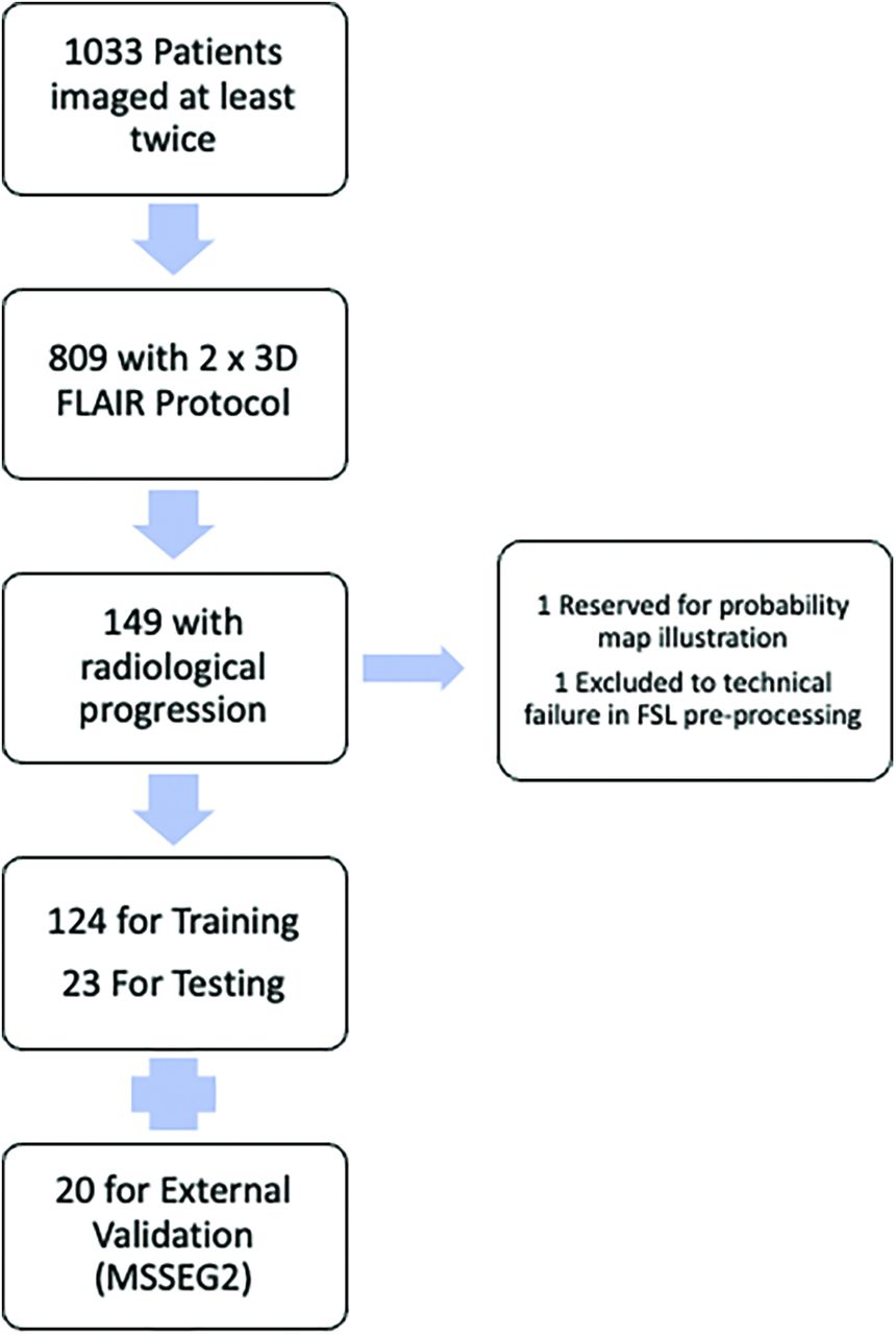
- FIG 1.
Flow chart of included patients.
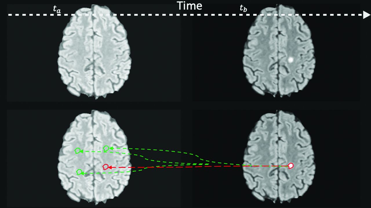
- FIG 2.
Prelesion and control masks. Expert segmentation mask at time b (tb) is projected backward to time a (ta) to the location where a lesion will occur (prelesion, red) and the other random areas in the NAWM (control, green). Note that this 2D representation is for illustrative purposes only, and for the experiments, the random translation was in 3D.
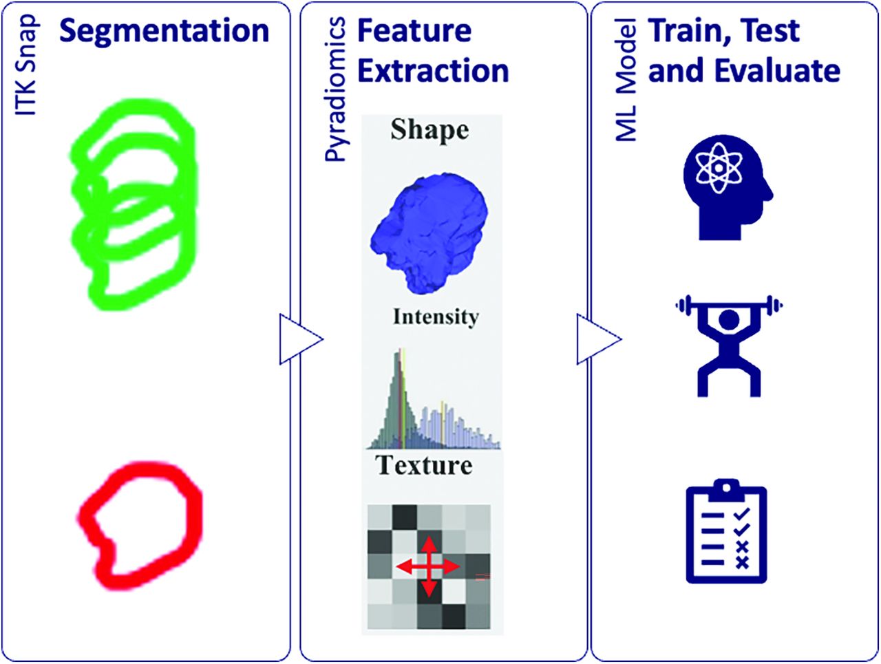
- FIG 3.
Radiomics workflow in which features are extracted from the segmented regions and passed to the machine learning models.
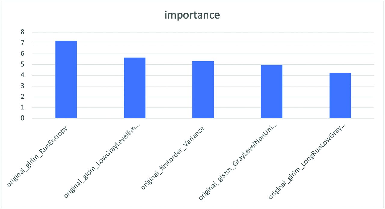
- FIG 4.
Top 5 radiomic features identified by the top performing XGBoost model.
- FIG 5.
Illustrative probability maps showing the absolute (A, Upper row) and relative (B, Lower row) probability of a new lesion occurring in each patch.
- FIG 6.
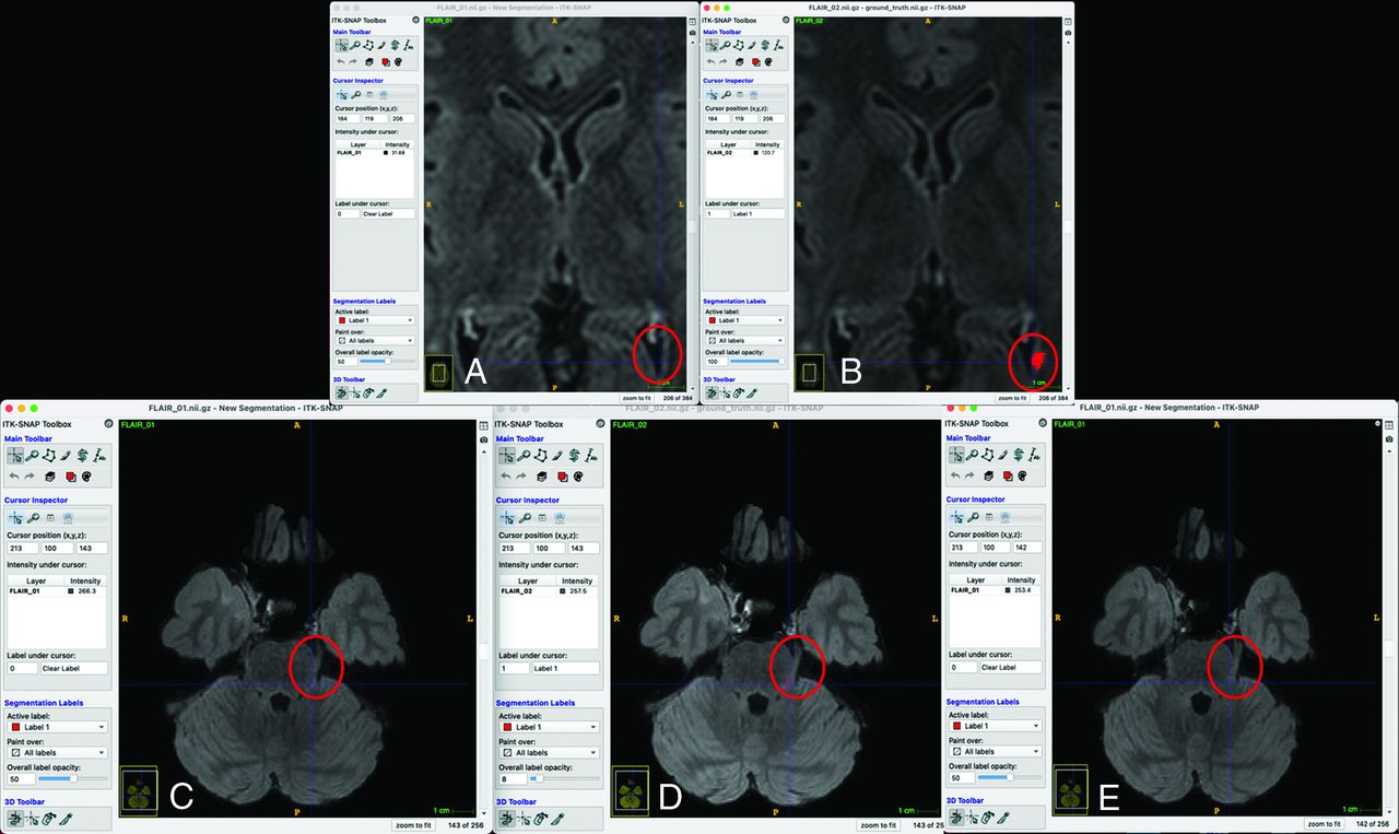
False-negative analysis (A and B, Upper row, C–E, Lower row). Two of the 3 false-negative cases in the external validation set are shown. A and B, The new lesion map falls within the ventricle on the prior image, making a negative prediction more likely because the low gray levels were associated with negative predictions (Fig 4). C, D, and E, The orientation of the proximal left trigeminal nerve is different so that the segmentation is cast onto the normal brainstem instead.


Tables
Demographics Total participants 147 Average age (yr) 42.19 Min 21 Max 74 Sex Male 45 Female 102 Note:—Max indicates maximum; Min, minimum.
Table 2: Internal test cohort results
Model Precision Recall F1-Score Accuracy Best Parameters XGBoost 0.91 0.91 0.91 0.91 {'classifier__colsample_bylevel': 0.8, 'classifier__gamma': 0, 'classifier__learning_rate': 0.2, 'classifier__max_depth': 4, 'classifier__min_child_weight': 1, 'classifier__n_estimators': 100, 'classifier__subsample': 0.5} SVC 0.90 0.89 0.89 0.89 {'classifier__C': 10, 'classifier__kernel': 'rbf'} Logistic regression 0.81 0.78 0.78 0.78 {'classifier__C': 1, 'classifier__penalty': 'l1', 'classifier__solver': 'liblinear'} KNN 0.83 0.78 0.78 0.78 {'classifier__n_neighbors': 7} Intensity baseline 0.76 0.77 0.75 0.77 NA Note:—SVC indicates support vector classifier; KNN, K nearest neighbor; NA, not applicable.
Model Precision Recall F1-Score Accuracy Best Parameters XGBoost 0.74 0.74 0.70 0.74 {'classifier__colsample_bylevel': 0.8, 'classifier__gamma': 0, 'classifier__learning_rate': 0.2, 'classifier__max_depth': 4, 'classifier__min_child_weight': 1, 'classifier__n_estimators': 100, 'classifier__subsample': 0.5} SVC 0.69 0.71 0.68 0.71 {'classifier__C': 10, 'classifier__kernel': 'rbf'} Logistic regression 0.62 0.55 0.56 0.55 {'classifier__C': 1, 'classifier__penalty': 'l1', 'classifier__solver': 'liblinear'} KNN 0.51 0.42 0.43 0.42 {'classifier__n_neighbors': 7} Intensity baseline 0.25 0.50 0.33 0.50 NA Note:—SVC indicates support vector classifier; KNN, K nearest neighbor; NA, not applicable.


Jump to section
Related Articles
Cited By...
- No citing articles found.