
Article Figures & Data
Figures
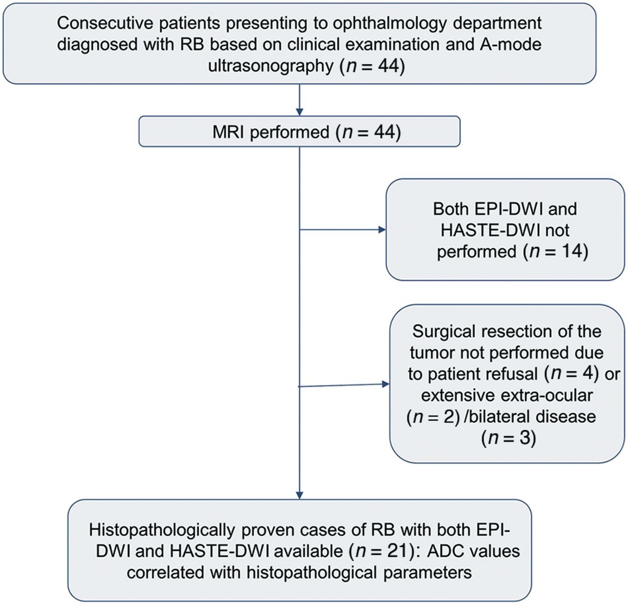
- FIG 1.
Flow chart showing patient inclusion and exclusion in the study.
- FIG 2.
Comparison of EPI-DWI and HASTE-DWI quality. Axial postcontrast T1-weighted image (A) shows heterogeneously enhancing mass (asterisk) in the posterior segment of the right eye globe. EPI (B) and HASTE (D) axial DWI (b-value, 1000 s/mm2) at the same level show high signal intensity in the mass (asterisk) with corresponding hypointensity on ADC maps (C and E, respectively, for EPI-DWI and HASTE-DWI), suggestive of diffusion restriction. There is mild elongation (arrow in C) of eye globe in EPI ADC map in contrast to maintained shape of globe in HASTE ADC map. However, there is a decreased SNR in case of HASTE-DWI and its ADC map.
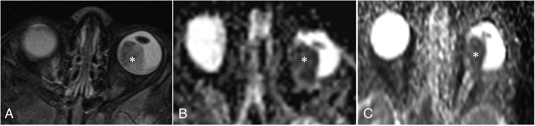
- FIG 3.
Poorly differentiated RB on histopathology. Axial T2-weighted image (A) shows a heterogeneously hypointense mass (asterisk) occupying the posterior segment of left eye globe. EPI (B) and HASTE (C) ADC maps show corresponding hypointense signal in the mass, suggestive of diffusion restriction. Mass showed low ADC values on both EPI-DWI and HASTE-DWI (mean ADC value being 0.48 × 10⁻³ mm2/s and 0.70 × 10⁻³ mm2/s for EPI-DWI and HASTE-DWI, respectively).
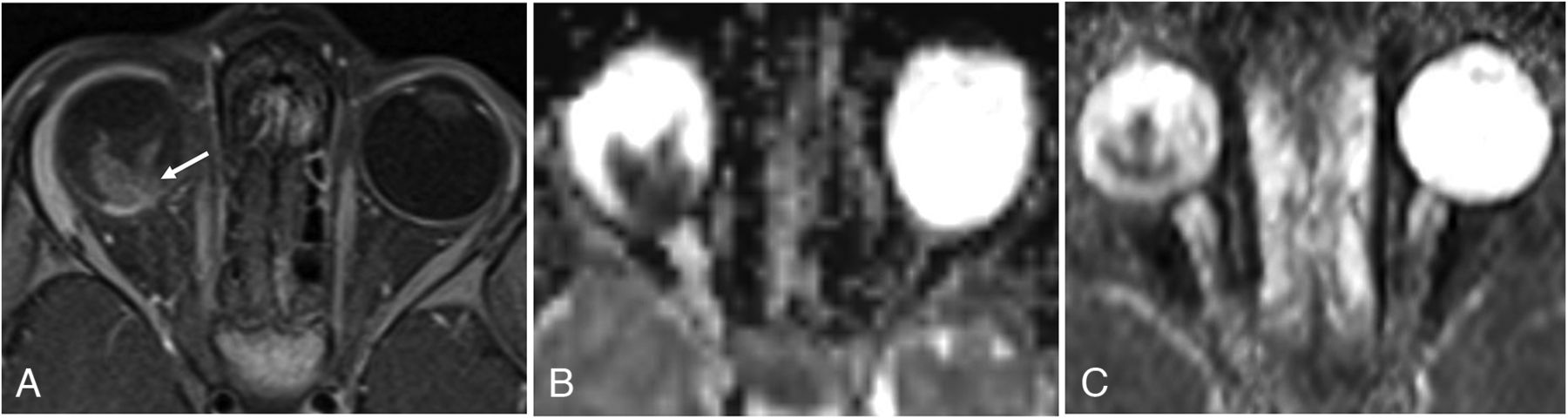
- FIG 4.
Histopathologically proved optic nerve invasion. Axial postcontrast T1-weighted image (A) at same level shows homogeneously enhancing mass with abnormal thickening and enhancement in the optic disc extending in postlaminar part of optic nerve (arrow), suggestive of postlaminar optic nerve invasion. EPI (B) and HASTE (C) ADC maps at the same level show corresponding hypointensity suggestive of diffusion restriction. Mass showed low ADC values on both EPI-DWI and HASTE-DWI (mean ADC value being 0.52 × 10⁻³ mm2/s and 0.67 × 10⁻³ mm2/s for EPI-DWI and HASTE-DWI, respectively).
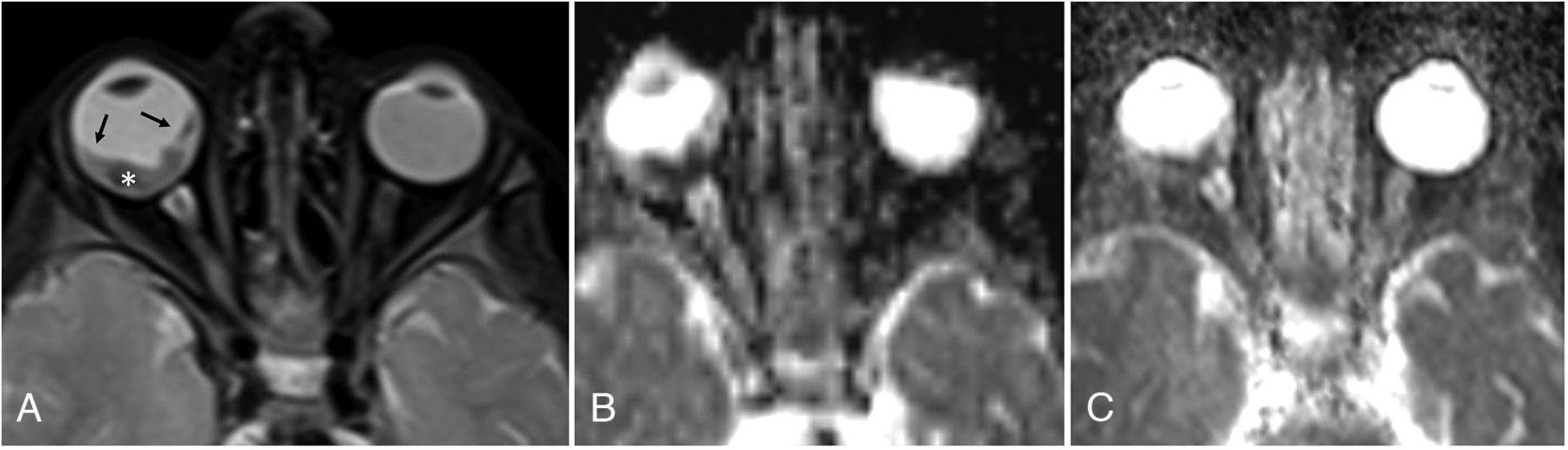
- FIG 5.
Histopathologically proved choroid invasion. Axial T2-weighted image (A) shows a hypointense small mass in the posterior segment of right eye with associated retinal detachment (arrows). EPI (B) and HASTE (C) ADC maps show corresponding hypointensity on ADC maps suggestive of diffusion restriction. Mass showed low ADC values on both EPI and HASTE-DWI (mean ADC value being 0.51 × 10⁻³ mm2/s and 0.87 × 10⁻³ mm2/s for EPI-DWI and HASTE-DWI, respectively).


Tables
Score Overall Image Quality Artifacts Tumor Sharpness Tumor Conspicuity 1 Nondiagnostic Nondiagnostic Nondiagnostic Mass unidentifiable 2 Substantial deficiency in image quality Marked impact on diagnosis Not sharp No differentiation between mass and vitreous 3 Moderate image quality Moderate impact on diagnosis Little sharp Subtle mass lesion 4 Good image quality Minimal impact on diagnosis Moderately sharp Well-seen mass lesion 5 Excellent image quality No artifact Good sharpness Very well-seen mass lesion Characteristic Value Median age at enucleation 36 months (range, 9–72 months) Sex Male: 15 (71.4%) Female: 6 (29.6%) Tumor laterality Unilateral: 21 (100%) Bilateral: 0 Right eye: 13 (61.9%) Left eye: 8 (38.1%) Median tumor size 15.3 mm (range, 10.2–36 mm) Mean follow-up period 265.4 days (range, 155–410 days) Parameter EPI-DWI HASTE-DWI P Value Qualitative parameters (n = 21): 5-point Likert scale Overall image quality 3.33 4.19 <.001 Artifacts 3.67 4.38 <.001 Tumor sharpness 3.24 4.14 <.001 Tumor conspicuity 3.52 4.33 <.001 Quantitative parameters (n = 21) Geometric distortion Transverse ocular diameter deviation (mean ± SD) 0.610 ± 0.301 mm 0.648 ± 0.339 mm .568 Anteroposterior ocular diameter deviation (mean ± SD) 6.105 ± 1.82 mm 0.643 ± 0.43 mm <.001 SNR b = 0 10.616 ± 1.844 8.283 ± 1.691 <.001 b = 500 10.268 ± 1.843 7.875 ± 1.124 <.001 b = 1000 9.578 ± 1.48 7.461 ± 1.1 <.001 CNR b = 0 10.529 ± 1.553 8.3 ± 1.527 <.001 b = 500 10.12 ± 1.254 7.553 ± 1.33 <.001 b = 1000 9.4 ± 1.789 7.295 ± 1.368 <.001 - Table 4:
Correlation of tumor ADC values on EPI-DWI and HASTE-DWI with prognostic parametersb
Prognostic Parameter Mean ADC Value for EPI-DWI (× 10−3 mm2/s) P Value for EPI-DWI Mean ADC Value for HASTE-DWI (× 10−3 mm2/s) P Value for HASTE-DWI Tumor grade Poorly differentiated (n = 13) 0.59 ± 0.16 .37 0.79 ± 0.16 .22 Moderately differentiated (n = 8) 0.65 ± 0.09 0.89 ± 0.18 Tumor size <15 mm (n = 9) 0.66 ± 0.16 .2 0.89 ± 0.19 .14 >15 mm (n = 12) 0.58 ± 0.11 0.78 ± 0.14 Optic nerve invasion Absent (n = 11) 0.65 ± 0.17 .35b 0.88 ± 0.20 .14b Prelaminar (n = 5) 0.64 ± 0.07 0.86 ± 0.13 Postlaminar (n = 5) 0.52 ± 0.04 0.69 ± 0.05 Choroid invasion Present (n = 12) 0.57 ± 0.11 .086 0.81 ± 0.18 .66 Absent (n = 9) 0.68 ± 0.15 0.85 ± 0.16 Anterior eye segment enhancement on postcontrast MRI Present (n = 6) 0.70 ± 0.18 .056 0.91 ± 0.23 .15 Absent (n = 15) 0.58 ± 0.10 0.80 ± 0.13


Jump to section
Related Articles
Cited By...
- No citing articles found.