Advertisement

Main menu

  • Home
  • Content
    • Current Issue
    • Accepted Manuscripts
    • Article Preview
    • Past Issue Archive
    • Video Articles
    • AJNR Case Collection
    • Case of the Week Archive
    • Case of the Month Archive
    • Classic Case Archive
  • Special Collections
    • AJNR Awards
    • Low-Field MRI
    • Alzheimer Disease
    • ASNR Foundation Special Collection
    • Photon-Counting CT
    • View All
  • Multimedia
    • AJNR Podcasts
    • AJNR SCANtastic
    • Trainee Corner
    • MRI Safety Corner
    • Imaging Protocols
  • For Authors
    • Submit a Manuscript
    • Submit a Video Article
    • Submit an eLetter to the Editor/Response
    • Manuscript Submission Guidelines
    • Statistical Tips
    • Fast Publishing of Accepted Manuscripts
    • Graphical Abstract Preparation
    • Imaging Protocol Submission
    • Author Policies
  • About Us
    • About AJNR
    • Editorial Board
    • Editorial Board Alumni
  • More
    • Become a Reviewer/Academy of Reviewers
    • Subscribers
    • Permissions
    • Alerts
    • Feedback
    • Advertisers
    • ASNR Home

User menu

  • Alerts
  • Log in

Search

  • Advanced search
American Journal of Neuroradiology

American Journal of Neuroradiology

ASHNR American Society of Functional Neuroradiology ASHNR American Society of Pediatric Neuroradiology ASSR
  • Alerts
  • Log in

Advanced Search

  • Home
  • Content
    • Current Issue
    • Accepted Manuscripts
    • Article Preview
    • Past Issue Archive
    • Video Articles
    • AJNR Case Collection
    • Case of the Week Archive
    • Case of the Month Archive
    • Classic Case Archive
  • Special Collections
    • AJNR Awards
    • Low-Field MRI
    • Alzheimer Disease
    • ASNR Foundation Special Collection
    • Photon-Counting CT
    • View All
  • Multimedia
    • AJNR Podcasts
    • AJNR SCANtastic
    • Trainee Corner
    • MRI Safety Corner
    • Imaging Protocols
  • For Authors
    • Submit a Manuscript
    • Submit a Video Article
    • Submit an eLetter to the Editor/Response
    • Manuscript Submission Guidelines
    • Statistical Tips
    • Fast Publishing of Accepted Manuscripts
    • Graphical Abstract Preparation
    • Imaging Protocol Submission
    • Author Policies
  • About Us
    • About AJNR
    • Editorial Board
    • Editorial Board Alumni
  • More
    • Become a Reviewer/Academy of Reviewers
    • Subscribers
    • Permissions
    • Alerts
    • Feedback
    • Advertisers
    • ASNR Home
  • Follow AJNR on Twitter
  • Visit AJNR on Facebook
  • Follow AJNR on Instagram
  • Join AJNR on LinkedIn
  • RSS Feeds

AJNR Awards, New Junior Editors, and more. Read the latest AJNR updates

Research ArticleHead and Neck Imaging
Open Access

Reliable Initial Trauma CT Findings of Supraclavicular Brachial Plexus Injury in Patients Sustaining Blunt Injuries

M.R. Povlow, J.R. Davis, A.M. Betts, S.M. Clayton, F.J. Cloran, J.K. Aden and J.L. Ritter
American Journal of Neuroradiology August 2023, 44 (8) 951-958; DOI: https://doi.org/10.3174/ajnr.A7919
M.R. Povlow
aFrom the Department of Radiology (M.R.P., J.R.D., A.M.B., S.M.C., F.J.C., J.L.R.)
  • Find this author on Google Scholar
  • Find this author on PubMed
  • Search for this author on this site
  • ORCID record for M.R. Povlow
J.R. Davis
aFrom the Department of Radiology (M.R.P., J.R.D., A.M.B., S.M.C., F.J.C., J.L.R.)
  • Find this author on Google Scholar
  • Find this author on PubMed
  • Search for this author on this site
  • ORCID record for J.R. Davis
A.M. Betts
aFrom the Department of Radiology (M.R.P., J.R.D., A.M.B., S.M.C., F.J.C., J.L.R.)
  • Find this author on Google Scholar
  • Find this author on PubMed
  • Search for this author on this site
  • ORCID record for A.M. Betts
S.M. Clayton
aFrom the Department of Radiology (M.R.P., J.R.D., A.M.B., S.M.C., F.J.C., J.L.R.)
  • Find this author on Google Scholar
  • Find this author on PubMed
  • Search for this author on this site
  • ORCID record for S.M. Clayton
F.J. Cloran
aFrom the Department of Radiology (M.R.P., J.R.D., A.M.B., S.M.C., F.J.C., J.L.R.)
  • Find this author on Google Scholar
  • Find this author on PubMed
  • Search for this author on this site
  • ORCID record for F.J. Cloran
J.K. Aden
bUnited States Army Graduate Medical Education (J.K.A.), Joint Base San Antonio-Fort Sam Houston, San Antonio, Texas
  • Find this author on Google Scholar
  • Find this author on PubMed
  • Search for this author on this site
  • ORCID record for J.K. Aden
J.L. Ritter
aFrom the Department of Radiology (M.R.P., J.R.D., A.M.B., S.M.C., F.J.C., J.L.R.)
  • Find this author on Google Scholar
  • Find this author on PubMed
  • Search for this author on this site
  • ORCID record for J.L. Ritter
  • Article
  • Figures & Data
  • Supplemental
  • Info & Metrics
  • Responses
  • References
  • PDF
Loading

Article Figures & Data

Figures

  • Tables
  • FIG 1.
    • Download figure
    • Open in new tab
    • Download powerpoint
    FIG 1.

    Anatomy of the brachial plexus. A frontal illustration (A) shows the anatomy of the brachial plexus as it arises from the neural foramina from the C4 to T1 levels, coursing between the anterior and middle scalene muscles and giving rise to many nerve branches. The cervical nerve roots of the brachial plexus are numbered 1 higher than the level where they originate (eg, the C5 nerve root arises from the C4 level, the C6 root from the C5 level, and so forth) because the C1 nerve root arises from above the C1 vertebral body at the skull base, and subsequently, there is a C8 nerve root but not a C8 vertebral body. The brachial plexus is divided into 5 roots, 3 trunks, 6 divisions, 3 cords, and terminal branches. A cross-sectional illustration (B) shows the anatomy of the proximal portions of the brachial plexus and nerve rootlets as they exit the spinal cord. Fibers from the dorsal rootlet complex with first-order sensory nerves in the dorsal root ganglion and fibers from both ventral and nerve rootlets blend before separating into ventral and dorsal nerve rami outside the neural foramina. The ventral rami go on to become the roots of the brachial plexus.

  • FIG 2.
    • Download figure
    • Open in new tab
    • Download powerpoint
    FIG 2.

    Inclusion and exclusion criteria. A flowchart delineates the inclusion and exclusion criteria used in this study, resulting in a case group of 36 and a control group of 50.

  • FIG 3.
    • Download figure
    • Open in new tab
    • Download powerpoint
    FIG 3.

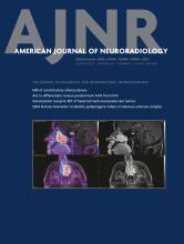
    Normal and abnormal scalene muscles in different patients. An axial CT image in a 48-year-old man (A) without BPI shows normal anterior (black arrow), middle (white arrow), and posterior (arrowhead) scalene muscles without enlargement or edema. An axial CT image in a 52-year-old man (B) with BPI shows enlargement of the right scalene muscles (arrows) with extensive surrounding edema (arrowhead). A coronal CT image in the same patient (C) shows the extent of the asymmetric edema (arrows). A coronal T2 STIR MR imaging performed a day later in the 52-year-old patient (D) again shows scalene enlargement and edema (black arrow). Thickening and signal hyperintensity within the right C7 root (white arrow) are consistent with a stretch injury; similar findings were present at other levels.

  • FIG 4.
    • Download figure
    • Open in new tab
    • Download powerpoint
    FIG 4.

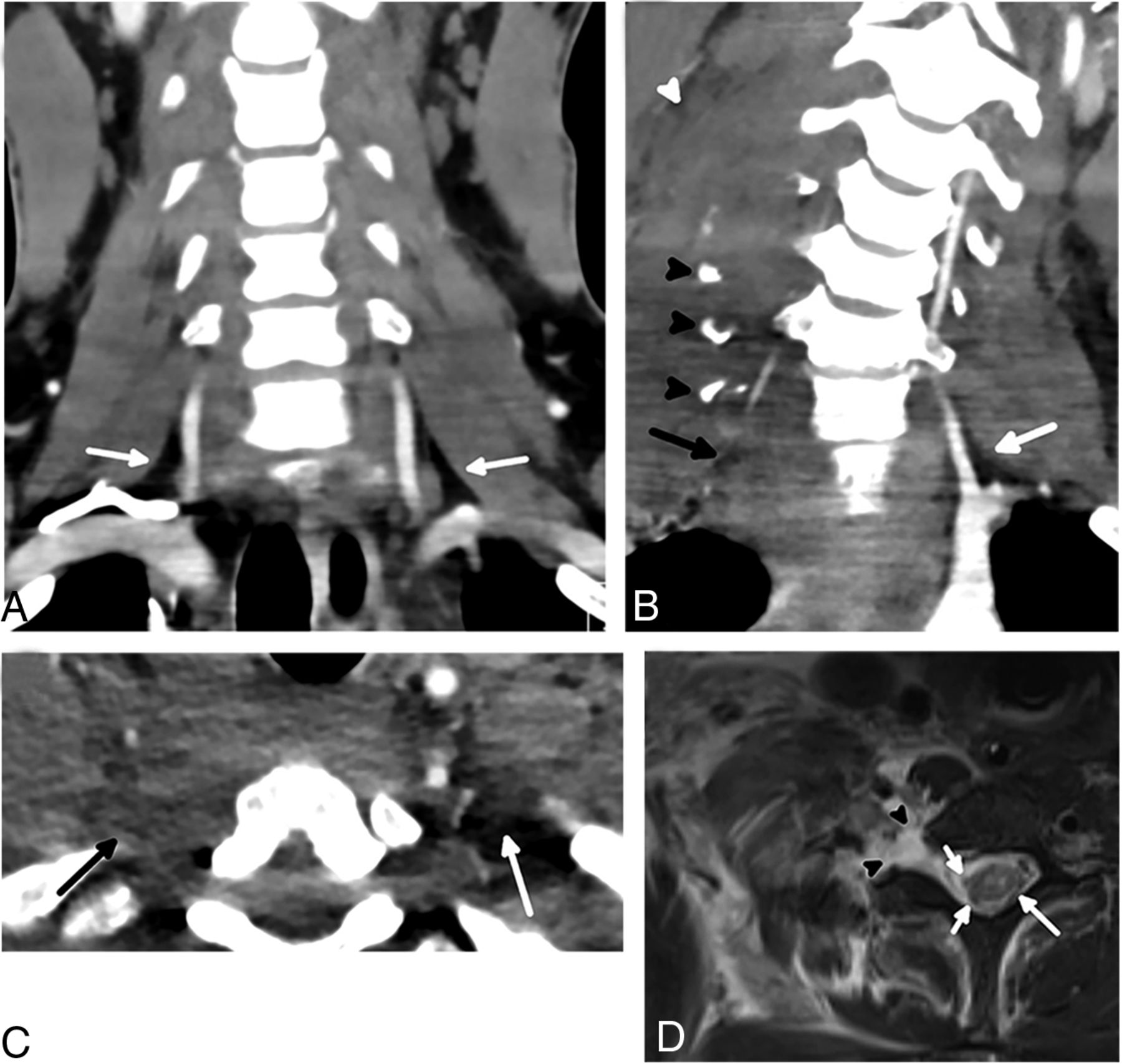
    Normal and abnormal interscalene fat pads in different patients. A coronal CT scan in a 26-year-old man (A) without BPI demonstrates smoothly marginated scalene muscles with normal interscalene fat pad (arrows). The coronal plane is a good place to assess the interscalene fat pad quickly by looking for this triangle of fat in the region of the proximal vertebral arteries, which should not have any stranding or hematoma. A coronal CT scan in a 25-year-old man (B) with BPI shows complete effacement of the right interscalene fat pad (black arrow) compared with the normal left interscalene fat pad (white arrow). Multiple displaced cervical transverse process fractures (black arrowheads) with scalene enlargement and edema (white arrowhead) are also present. An axial CT in the 25-year-old patient (C) shows the interscalene fat pad effacement (black arrow) in a different plane, compared with the normal left interscalene fat pad (white arrow). An axial T2-weighted MR imaging with fat saturation (D) performed a day later in the injured patient shows avulsion of the ventral and dorsal nerve rootlets (short white arrows), which was better appreciated when reviewing multiple sequential images. The spinal cord is slightly eccentric to the left and has abnormally increased signal (long white arrow), most pronounced within the gray matter. A large pseudomeningocele is present (arrowheads).

  • FIG 5.
    • Download figure
    • Open in new tab
    • Download powerpoint
    FIG 5.

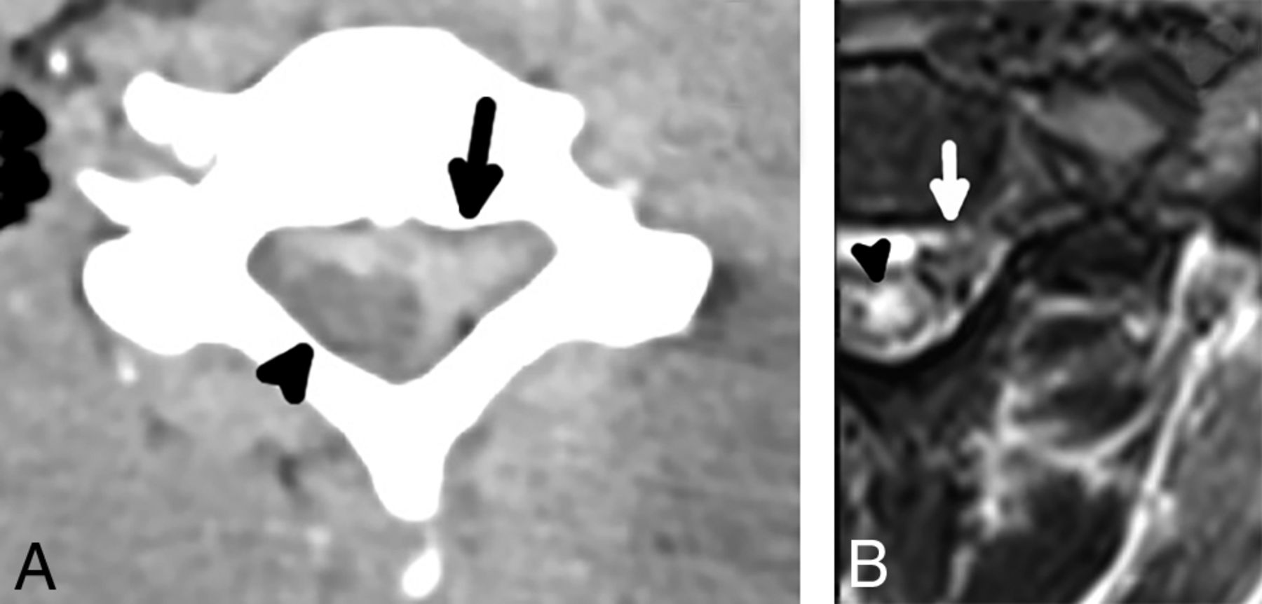
    A 42-year-old man with preganglionic BPI and extra-axial hemorrhage. An axial CT image (A) shows a large extra-axial hematoma centered at the left of the spinal canal (arrow), which appears to compress the eccentric spinal cord (arrowhead). An axial T2 STIR MR imaging (B) in the same patient performed a day later shows an extra-axial hematoma (arrow); the hematoma and motion artifacts on the examination limit the diagnostic quality, but multilevel nerve rootlet avulsions are evident on sequential images. There is also severely abnormal signal hyperintensity (arrowhead) and a probable focus of hemorrhage (focal T2 hypointenisty) in the spinal cord.

  • FIG 6.
    • Download figure
    • Open in new tab
    • Download powerpoint
    FIG 6.

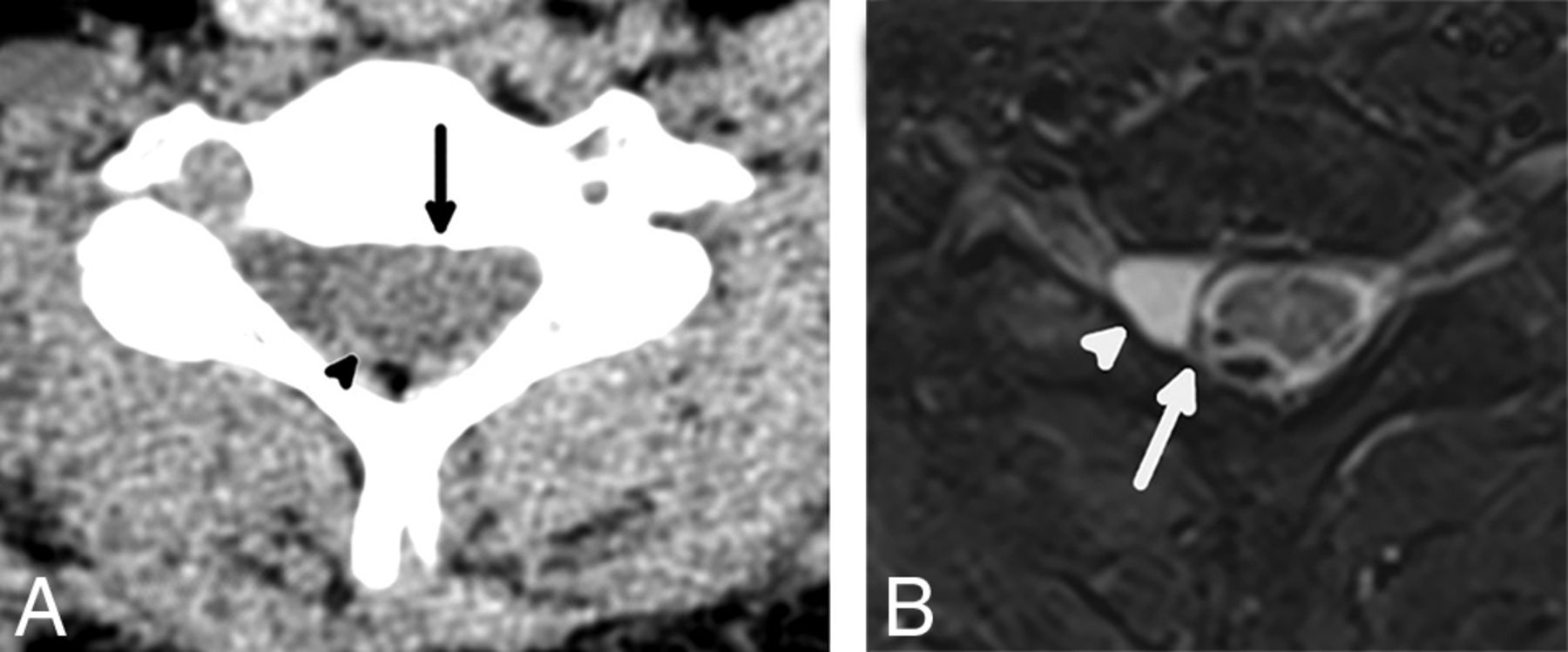
    A 26-year-old man with preganglionic BPI and eccentricity of the spinal cord. An axial CT image (A) shows subtle eccentricity of the spinal cord and thecal sac (arrow) with an asymmetric amount of CSF in the right aspect of the spinal canal, which is indicative of a pseudomeningocele. There is also thin extra-axial hemorrhage (arrowhead) along the posterior aspect of the spinal cord. An axial T2 STIR MR imaging performed a day later in the same patient (B) shows complete avulsion of the ventral and dorsal nerve rootlets with eccentricity of the spinal cord (arrow), which is lateralized to the left of the spinal canal, compatible with preganglionic BPI. A small pseudomeningocele is confirmed on MR imaging (arrowhead) and is a finding highly associated with nerve rootlet avulsion.

Tables

  • Figures
    • View popup
    Table 1:

    Patient demographics and mechanisms of injury by case group

    CharacteristicCases (n = 36)Controls (n = 50)
    Age (range) (mean) (yr)18–75 (38.7)18–73 (41.5)
    Sex
     Male2936
     Female714
    Mechanism of injury
     Motor vehicle collision2625
     Motorcycle collision51
     Fall from heighta38
     All-terrain vehicle accident20
     Motor vehicle vs pedestrian07
     Assault04
     Fall from standing03
     Fall from horse01
     Bicycle accident01
    • ↵a Fall from height = (>10 ft, 3.048 m).

    • View popup
    Table 2:

    Prevalence, sensitivities, specificities, and ORs of the CT findings

    FindingCases (n = 36)Controls (n = 50)Sensitivity (95% CI)Specificity (95% CI)OR, P Value (95% CI)
    Scalene muscle edema/enlargement34694.44 (81.86–98.46)88.00 (76.19–94.38)130.33; P < .001 (24.77–685.87)
    Interscalene fat pad effacement34594.44 (81.86–98.46)90.00 (78.64–95.65)153.00; P < .001 (27.97–836.89)
    First-rib fracture14738.89 (24.78–55.14)86.00 (73.81–93.05)3.91; P = .01 (1.38–11.09)
    Cervical lateral mass or transverse process fracture20455.56 (39.58–70.46)92.00 (81.16–96.85)14.38; P < .001 (4.27–48.45)
    Extra-axial spinal canal hemorrhage10027.78 (15.85–43.99)100.00 (92.86–100)40.02; P = .01 (2.26–709.90)
    Spinal cord eccentricity5013.89 (6.08–28.66)100.00 (92.86–100)17.63; P = .05 (0.9425–329.97)
PreviousNext
Back to top

In this issue

American Journal of Neuroradiology: 44 (8)
American Journal of Neuroradiology
Vol. 44, Issue 8
1 Aug 2023
  • Table of Contents
  • Index by author
  • Complete Issue (PDF)
Advertisement
Print
Download PDF
Email Article

Thank you for your interest in spreading the word on American Journal of Neuroradiology.

NOTE: We only request your email address so that the person you are recommending the page to knows that you wanted them to see it, and that it is not junk mail. We do not capture any email address.

Enter multiple addresses on separate lines or separate them with commas.
Reliable Initial Trauma CT Findings of Supraclavicular Brachial Plexus Injury in Patients Sustaining Blunt Injuries
(Your Name) has sent you a message from American Journal of Neuroradiology
(Your Name) thought you would like to see the American Journal of Neuroradiology web site.
CAPTCHA
This question is for testing whether or not you are a human visitor and to prevent automated spam submissions.
Cite this article
M.R. Povlow, J.R. Davis, A.M. Betts, S.M. Clayton, F.J. Cloran, J.K. Aden, J.L. Ritter
Reliable Initial Trauma CT Findings of Supraclavicular Brachial Plexus Injury in Patients Sustaining Blunt Injuries
American Journal of Neuroradiology Aug 2023, 44 (8) 951-958; DOI: 10.3174/ajnr.A7919

Citation Manager Formats

  • BibTeX
  • Bookends
  • EasyBib
  • EndNote (tagged)
  • EndNote 8 (xml)
  • Medlars
  • Mendeley
  • Papers
  • RefWorks Tagged
  • Ref Manager
  • RIS
  • Zotero
0 Responses
Respond to this article
Share
Bookmark this article
CT Findings for Supraclavicular Plexus Injury
M.R. Povlow, J.R. Davis, A.M. Betts, S.M. Clayton, F.J. Cloran, J.K. Aden, J.L. Ritter
American Journal of Neuroradiology Aug 2023, 44 (8) 951-958; DOI: 10.3174/ajnr.A7919
del.icio.us logo Twitter logo Facebook logo Mendeley logo
  • Tweet Widget
  • Facebook Like
  • Google Plus One
Purchase

Jump to section

  • Article
    • Abstract
    • ABBREVIATION:
    • MATERIALS AND METHODS
    • RESULTS
    • DISCUSSION
    • CONCLUSIONS
    • Acknowledgment
    • Footnotes
    • References
  • Figures & Data
  • Supplemental
  • Info & Metrics
  • Responses
  • References
  • PDF

Related Articles

  • PubMed
  • Google Scholar

Cited By...

  • No citing articles found.
  • Crossref
  • Google Scholar

This article has not yet been cited by articles in journals that are participating in Crossref Cited-by Linking.

More in this TOC Section

  • Diagnostic Performance of US in Neck Node NIRADS 2
  • Hydrops Herniation into the Semicircular Canals
  • ASL Sensitivity for Head and Neck Paraganglioma
Show more HEAD AND NECK IMAGING

Similar Articles

Advertisement

Indexed Content

  • Current Issue
  • Accepted Manuscripts
  • Article Preview
  • Past Issues
  • Editorials
  • Editor's Choice
  • Fellows' Journal Club
  • Letters to the Editor
  • Video Articles

Cases

  • Case Collection
  • Archive - Case of the Week
  • Archive - Case of the Month
  • Archive - Classic Case

More from AJNR

  • Trainee Corner
  • Imaging Protocols
  • MRI Safety Corner
  • Book Reviews

Multimedia

  • AJNR Podcasts
  • AJNR Scantastics

Resources

  • Turnaround Time
  • Submit a Manuscript
  • Submit a Video Article
  • Submit an eLetter to the Editor/Response
  • Manuscript Submission Guidelines
  • Statistical Tips
  • Fast Publishing of Accepted Manuscripts
  • Graphical Abstract Preparation
  • Imaging Protocol Submission
  • Evidence-Based Medicine Level Guide
  • Publishing Checklists
  • Author Policies
  • Become a Reviewer/Academy of Reviewers
  • News and Updates

About Us

  • About AJNR
  • Editorial Board
  • Editorial Board Alumni
  • Alerts
  • Permissions
  • Not an AJNR Subscriber? Join Now
  • Advertise with Us
  • Librarian Resources
  • Feedback
  • Terms and Conditions
  • AJNR Editorial Board Alumni

American Society of Neuroradiology

  • Not an ASNR Member? Join Now

© 2025 by the American Society of Neuroradiology All rights, including for text and data mining, AI training, and similar technologies, are reserved.
Print ISSN: 0195-6108 Online ISSN: 1936-959X

Powered by HighWire