Advertisement

Main menu

  • Home
  • Content
    • Current Issue
    • Accepted Manuscripts
    • Article Preview
    • Past Issue Archive
    • Video Articles
    • AJNR Case Collection
    • Case of the Week Archive
    • Case of the Month Archive
    • Classic Case Archive
  • Special Collections
    • AJNR Awards
    • Low-Field MRI
    • Alzheimer Disease
    • ASNR Foundation Special Collection
    • Photon-Counting CT
    • View All
  • Multimedia
    • AJNR Podcasts
    • AJNR SCANtastic
    • Trainee Corner
    • MRI Safety Corner
    • Imaging Protocols
  • For Authors
    • Submit a Manuscript
    • Submit a Video Article
    • Submit an eLetter to the Editor/Response
    • Manuscript Submission Guidelines
    • Statistical Tips
    • Fast Publishing of Accepted Manuscripts
    • Graphical Abstract Preparation
    • Imaging Protocol Submission
    • Author Policies
  • About Us
    • About AJNR
    • Editorial Board
    • Editorial Board Alumni
  • More
    • Become a Reviewer/Academy of Reviewers
    • Subscribers
    • Permissions
    • Alerts
    • Feedback
    • Advertisers
    • ASNR Home

User menu

  • Alerts
  • Log in

Search

  • Advanced search
American Journal of Neuroradiology

American Journal of Neuroradiology

ASHNR American Society of Functional Neuroradiology ASHNR American Society of Pediatric Neuroradiology ASSR
  • Alerts
  • Log in

Advanced Search

  • Home
  • Content
    • Current Issue
    • Accepted Manuscripts
    • Article Preview
    • Past Issue Archive
    • Video Articles
    • AJNR Case Collection
    • Case of the Week Archive
    • Case of the Month Archive
    • Classic Case Archive
  • Special Collections
    • AJNR Awards
    • Low-Field MRI
    • Alzheimer Disease
    • ASNR Foundation Special Collection
    • Photon-Counting CT
    • View All
  • Multimedia
    • AJNR Podcasts
    • AJNR SCANtastic
    • Trainee Corner
    • MRI Safety Corner
    • Imaging Protocols
  • For Authors
    • Submit a Manuscript
    • Submit a Video Article
    • Submit an eLetter to the Editor/Response
    • Manuscript Submission Guidelines
    • Statistical Tips
    • Fast Publishing of Accepted Manuscripts
    • Graphical Abstract Preparation
    • Imaging Protocol Submission
    • Author Policies
  • About Us
    • About AJNR
    • Editorial Board
    • Editorial Board Alumni
  • More
    • Become a Reviewer/Academy of Reviewers
    • Subscribers
    • Permissions
    • Alerts
    • Feedback
    • Advertisers
    • ASNR Home
  • Follow AJNR on Twitter
  • Visit AJNR on Facebook
  • Follow AJNR on Instagram
  • Join AJNR on LinkedIn
  • RSS Feeds

AJNR Awards, New Junior Editors, and more. Read the latest AJNR updates

Research ArticleExtracranial Vascular

Mapping of Anatomic Variants of the Proximal Vertebral Artery in Relation to Embryology

H.F. Bueno and E.A. Nimchinsky
American Journal of Neuroradiology August 2023, 44 (8) 943-950; DOI: https://doi.org/10.3174/ajnr.A7942
H.F. Bueno
aFrom the Department of Radiology, Rutgers-New Jersey Medical School, Newark, New Jersey
  • Find this author on Google Scholar
  • Find this author on PubMed
  • Search for this author on this site
  • ORCID record for H.F. Bueno
E.A. Nimchinsky
aFrom the Department of Radiology, Rutgers-New Jersey Medical School, Newark, New Jersey
  • Find this author on Google Scholar
  • Find this author on PubMed
  • Search for this author on this site
  • ORCID record for E.A. Nimchinsky
  • Article
  • Figures & Data
  • Info & Metrics
  • Responses
  • References
  • PDF
Loading

Article Figures & Data

Figures

  • Tables
  • FIG 1.
    • Download figure
    • Open in new tab
    • Download powerpoint
    FIG 1.

    Normal anatomy of the cervical VAs. Sagittal MIPs of the right (A) and left (B) VAs and both in the coronal plane (C), as well as a coronal 3D rendering (D) show the origin of both VAs from the SCAs (white arrows) and their entry into the FT at C6 (red arrows).
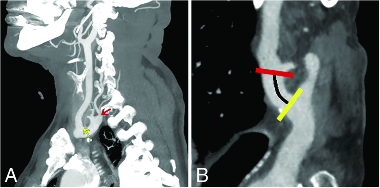
  • FIG 2.
    • Download figure
    • Open in new tab
    • Download powerpoint
    FIG 2.

    Quantification of the distance from the right SCA origin to the origin of the RVA. A, Sagittal MIPs show the origins of the RSCA (yellow arrow), defined as the distal wall of the origin of the right common carotid artery and the RVA (red arrow). B, Curved planar reformat shows a straightened SCA with estimation of the distance to the RVA origin along the SCA (black line indicates distance; yellow line, RSCA origin; red line, RVA origin).

  • FIG 3.
    • Download figure
    • Open in new tab
    • Download powerpoint
    FIG 3.

    RVA variants. RVA origin within 2 cm of the RSCA origin (white arrows) entering the FT (red arrows) at C3 (A), C4 (B), or C5 (C). D, The RVA originates from the distal aortic arch (white arrow), crosses along the upper thoracic vertebral bodies, and enters the FT at C7 (red arrow). Note that in this case, the left VA also enters the FT at C7 (green arrow), having originated as the fourth branch from the aortic arch (not shown).

  • FIG 4.
    • Download figure
    • Open in new tab
    • Download powerpoint
    FIG 4.

    Box-and-whisker plot showing the distance from the origin of the RSCA for right VAs entering the FT from C3 through C7. Although there is some overlap, high-entry VAs originate more proximally than arteries entering at C6. There is more heterogeneity in low-entry VAs, which may originate from the aortic arch (negative numbers on this figure) or along the RSCA. For this figure, all low-entry VAs are considered together, without regard for whether they share a common origin with the CCT.

  • FIG 5.
    • Download figure
    • Open in new tab
    • Download powerpoint
    FIG 5.

    LVA variants. LVA origin directly from the aortic arch, between the LCCA and LSCA origins (white arrows) entering the FT (red arrows) at C3 (A), C4 (B), or C5 (C). D, The LVA originates as the fourth branch from the arch (white arrow) and enters the FT at C7 (red arrow).

  • FIG 6.
    • Download figure
    • Open in new tab
    • Download powerpoint
    FIG 6.

    Multiple origins of the VAs. A, Two moieties originate proximally and distally along the LSCA. The moiety originating more distally (white arrow) at the expected VA origin enters the FT at C6 (red arrow), and the more proximal moiety (white arrowhead), originating within 2 cm of the RSCA, joins the first moiety at C4/C5 (red arrowhead), forming a fused artery that enters the FT at C4. This is the most common multiple-origin pattern. B, A similar pattern is noted on the left with the normal VA origin (white arrow), dominant in this case, entering the FT at C6 (red arrow) and the variant origin, originating from the arch between LCCA and LSCA (white arrowhead), joining at C4/C5 with a fused artery in the FT at C4 (red arrowhead). C, A rare subvariant, with 1 RVA moiety originating 8 mm from the right common carotid artery (not shown) and entering the FT at C3 (red arrowhead) and the other originating normally from the RSCA (white arrow) and entering at C6 (red arrow). D, An extremely rare variant, with the RVA having 3 origins, originating at 7, 11, and 14 mm along the RSCA and entering at C4 (red arrow), C5 (red arrowhead), and C6 (green arrow), respectively.
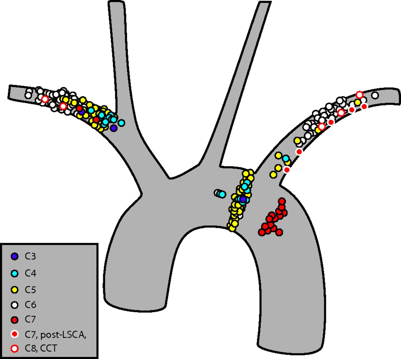
  • FIG 7.
    • Download figure
    • Open in new tab
    • Download powerpoint
    FIG 7.

    The origins of the VAs plotted against an idealized aortic arch. For this illustration, each VA origin was plotted in a 1D manner as a function of distance from the RSCA origin on the right or from the LSCA on the left. The position in any dimension other than that along the axis of the vessel is not based on anatomic data, and the distribution of vessel origins in the medial-lateral or craniocaudal dimension is used only to permit better visualization of the data points. Typical origins giving rise to VAs entering the FT at C6 are shown in white. Origins of vessels entering the FT at C3, C4, C5, and C7 are shown in purple, cyan, yellow, and red, respectively. Vessels originating along the posterior wall of the LSCA and entering at C7 are shown in red with a white outline. In addition, vessel origins entering at C7 whose vessel origin is shared with the CCT are shown in white with a red outline. Most of the high-entry vessels, which enter at C3–C5, originate in the proximal SCA on the right and directly from the arch or in the proximal subclavian artery on the left. Most of the low-entry vessels originate distal to or along the posterior wall of the LSCA in the region of the aortic isthmus.
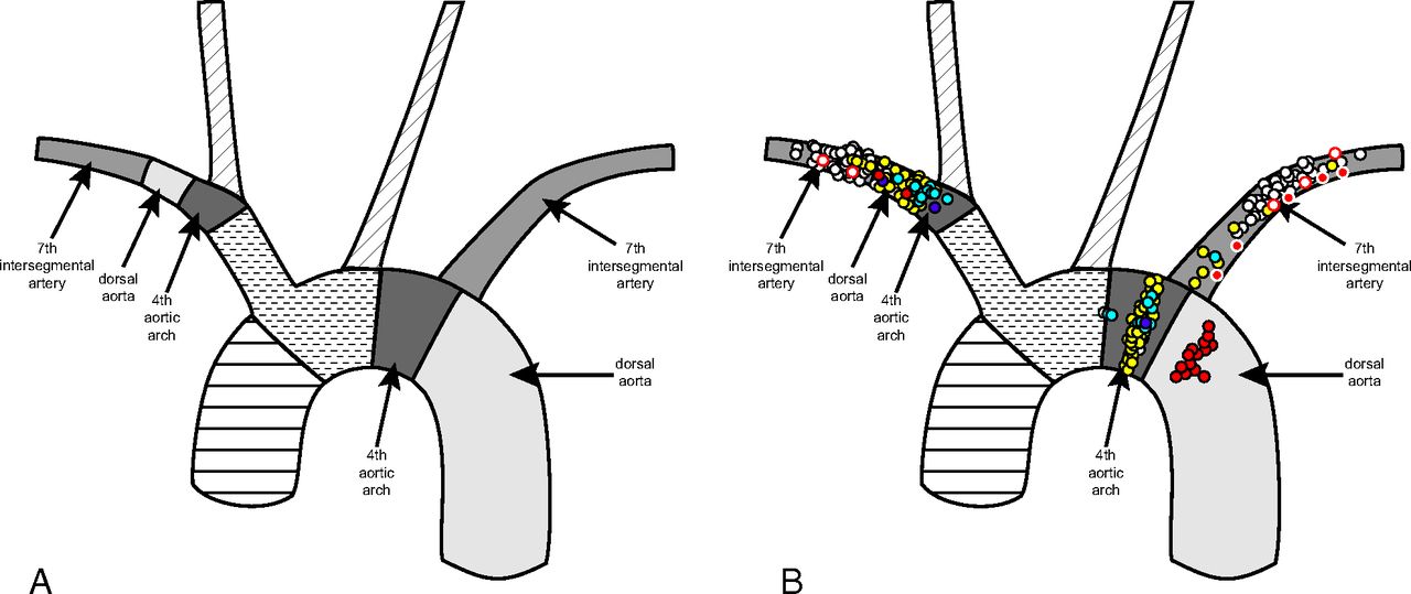
  • FIG 8.
    • Download figure
    • Open in new tab
    • Download powerpoint
    FIG 8.

    A, The embryologic origins of the aortic arch and proximal great vessels, adapted from Schoenwolf et al.31 B, VA origin map (from Fig 7) superimposed on the embryologic map. Note that while most VAs take origin from the seventh ISA segment of the SCA on both sides, the high-entry vessels take origin predominantly from the segments derived from the dorsal aorta and fourth aortic arch, and the low-entry vessels take origin predominantly from the distal aortic arch.

Tables

  • Figures
    • View popup
    Table 1:

    Distribution of variant VA origins and entries to the FT in a consecutive series of 493 casesa

    C4C5C6C7
    Right
     Proximal SCA62203
     SCA02482
     Arch0000
    Left
     SCA16272
     Archb43030
     SCA posterior wall0005
     4th branch arch0007
    • ↵a The 3 cases of duplicated origins in this sample are not included in this table. Note that no right VAs originating from the aortic arch, and no VAs on either side entering at C3 were seen in this sample.

    • ↵b Indicates the portion of the arch between the left common carotid and subclavian arteries.

    • View popup
    Table 2:

    Distribution of variant VA entries to the FT in an enriched sample of all 125 cases with variant VAsa

    RVA (%)LVA (%)
    Single origins
     C32 (3.6%)1 (1.2%)
     C414 (25.4%)11 (13.6%)
     C531 (56.4%)47 (55.6%)
     C77 (12.7%)24 (28.9%)
     Thoracic1 (1.8%)0 (0%)
    Multiple origins
     C3, C61 (8.3%)0 (0%)
     C4, C67 (58.3%)2 (16.7%)
     C5, C61 (8.3%)1 (8.3%)
     C4, C5, C61 (8.3%)0 (0%)
    • ↵a This sample includes the consecutive sample described earlier as well as an additional set of selected variants that were encountered outside of the sampling period.

PreviousNext
Back to top

In this issue

American Journal of Neuroradiology: 44 (8)
American Journal of Neuroradiology
Vol. 44, Issue 8
1 Aug 2023
  • Table of Contents
  • Index by author
  • Complete Issue (PDF)
Advertisement
Print
Download PDF
Email Article

Thank you for your interest in spreading the word on American Journal of Neuroradiology.

NOTE: We only request your email address so that the person you are recommending the page to knows that you wanted them to see it, and that it is not junk mail. We do not capture any email address.

Enter multiple addresses on separate lines or separate them with commas.
Mapping of Anatomic Variants of the Proximal Vertebral Artery in Relation to Embryology
(Your Name) has sent you a message from American Journal of Neuroradiology
(Your Name) thought you would like to see the American Journal of Neuroradiology web site.
CAPTCHA
This question is for testing whether or not you are a human visitor and to prevent automated spam submissions.
Cite this article
H.F. Bueno, E.A. Nimchinsky
Mapping of Anatomic Variants of the Proximal Vertebral Artery in Relation to Embryology
American Journal of Neuroradiology Aug 2023, 44 (8) 943-950; DOI: 10.3174/ajnr.A7942

Citation Manager Formats

  • BibTeX
  • Bookends
  • EasyBib
  • EndNote (tagged)
  • EndNote 8 (xml)
  • Medlars
  • Mendeley
  • Papers
  • RefWorks Tagged
  • Ref Manager
  • RIS
  • Zotero
0 Responses
Respond to this article
Share
Bookmark this article
Proximal Vertebral Artery Variants and Embryology
H.F. Bueno, E.A. Nimchinsky
American Journal of Neuroradiology Aug 2023, 44 (8) 943-950; DOI: 10.3174/ajnr.A7942
del.icio.us logo Twitter logo Facebook logo Mendeley logo
  • Tweet Widget
  • Facebook Like
  • Google Plus One
Purchase

Jump to section

  • Article
    • Abstract
    • ABBREVIATIONS:
    • MATERIALS AND METHODS
    • RESULTS
    • DISCUSSION
    • CONCLUSIONS
    • ACKNOWLEDGMENT
    • Footnotes
    • References
  • Figures & Data
  • Info & Metrics
  • Responses
  • References
  • PDF

Related Articles

  • No related articles found.
  • PubMed
  • Google Scholar

Cited By...

  • No citing articles found.
  • Crossref (2)
  • Google Scholar

This article has been cited by the following articles in journals that are participating in Crossref Cited-by Linking.

  • A type II proatlantal artery arising from the external carotid artery: A review of neurovascular anatomy
    Archit Bharathwaj Baskaran, Aditya Jhaveri, Olivia A. Kozel, Chibueze Ikechukwu Agwu, Sachin Kothari, Therese Dunne, Michael Charles Hurley, James E. Siegler
    Surgical Neurology International 2025 16
  • Common origin of the right vertebral artery and the right costocervical trunk diagnosed by magnetic resonance angiography
    Akira Uchino, Kenzo Minamimura
    Surgical and Radiologic Anatomy 2025 47 1

More in this TOC Section

  • High-Risk Plaque Features in Carotid MRI
  • Nonstenotic Carotid Plaques and Stroke Review
Show more EXTRACRANIAL VASCULAR

Similar Articles

Advertisement

Indexed Content

  • Current Issue
  • Accepted Manuscripts
  • Article Preview
  • Past Issues
  • Editorials
  • Editor's Choice
  • Fellows' Journal Club
  • Letters to the Editor
  • Video Articles

Cases

  • Case Collection
  • Archive - Case of the Week
  • Archive - Case of the Month
  • Archive - Classic Case

More from AJNR

  • Trainee Corner
  • Imaging Protocols
  • MRI Safety Corner
  • Book Reviews

Multimedia

  • AJNR Podcasts
  • AJNR Scantastics

Resources

  • Turnaround Time
  • Submit a Manuscript
  • Submit a Video Article
  • Submit an eLetter to the Editor/Response
  • Manuscript Submission Guidelines
  • Statistical Tips
  • Fast Publishing of Accepted Manuscripts
  • Graphical Abstract Preparation
  • Imaging Protocol Submission
  • Evidence-Based Medicine Level Guide
  • Publishing Checklists
  • Author Policies
  • Become a Reviewer/Academy of Reviewers
  • News and Updates

About Us

  • About AJNR
  • Editorial Board
  • Editorial Board Alumni
  • Alerts
  • Permissions
  • Not an AJNR Subscriber? Join Now
  • Advertise with Us
  • Librarian Resources
  • Feedback
  • Terms and Conditions
  • AJNR Editorial Board Alumni

American Society of Neuroradiology

  • Not an ASNR Member? Join Now

© 2025 by the American Society of Neuroradiology All rights, including for text and data mining, AI training, and similar technologies, are reserved.
Print ISSN: 0195-6108 Online ISSN: 1936-959X

Powered by HighWire