Advertisement

Main menu

  • Home
  • Content
    • Current Issue
    • Accepted Manuscripts
    • Article Preview
    • Past Issue Archive
    • Video Articles
    • AJNR Case Collection
    • Case of the Week Archive
    • Case of the Month Archive
    • Classic Case Archive
  • Special Collections
    • Advancing NeuroMRI with High-Relaxivity Contrast Agents
    • AJNR Awards
    • Low-Field MRI
    • Alzheimer Disease
    • ASNR Foundation Special Collection
    • Photon-Counting CT
    • View All
  • Multimedia
    • AJNR Podcasts
    • AJNR SCANtastic
    • Trainee Corner
    • MRI Safety Corner
    • Imaging Protocols
  • For Authors
    • Submit a Manuscript
    • Submit a Video Article
    • Submit an eLetter to the Editor/Response
    • Manuscript Submission Guidelines
    • Statistical Tips
    • Fast Publishing of Accepted Manuscripts
    • Graphical Abstract Preparation
    • Imaging Protocol Submission
    • Author Policies
  • About Us
    • About AJNR
    • Editorial Board
    • Editorial Board Alumni
  • More
    • Become a Reviewer/Academy of Reviewers
    • Subscribers
    • Permissions
    • Alerts
    • Feedback
    • Advertisers
    • ASNR Home

User menu

  • Alerts
  • Log in

Search

  • Advanced search
American Journal of Neuroradiology

American Journal of Neuroradiology

ASHNR American Society of Functional Neuroradiology ASHNR American Society of Pediatric Neuroradiology ASSR
  • Alerts
  • Log in

Advanced Search

  • Home
  • Content
    • Current Issue
    • Accepted Manuscripts
    • Article Preview
    • Past Issue Archive
    • Video Articles
    • AJNR Case Collection
    • Case of the Week Archive
    • Case of the Month Archive
    • Classic Case Archive
  • Special Collections
    • Advancing NeuroMRI with High-Relaxivity Contrast Agents
    • AJNR Awards
    • Low-Field MRI
    • Alzheimer Disease
    • ASNR Foundation Special Collection
    • Photon-Counting CT
    • View All
  • Multimedia
    • AJNR Podcasts
    • AJNR SCANtastic
    • Trainee Corner
    • MRI Safety Corner
    • Imaging Protocols
  • For Authors
    • Submit a Manuscript
    • Submit a Video Article
    • Submit an eLetter to the Editor/Response
    • Manuscript Submission Guidelines
    • Statistical Tips
    • Fast Publishing of Accepted Manuscripts
    • Graphical Abstract Preparation
    • Imaging Protocol Submission
    • Author Policies
  • About Us
    • About AJNR
    • Editorial Board
    • Editorial Board Alumni
  • More
    • Become a Reviewer/Academy of Reviewers
    • Subscribers
    • Permissions
    • Alerts
    • Feedback
    • Advertisers
    • ASNR Home
  • Follow AJNR on Twitter
  • Visit AJNR on Facebook
  • Follow AJNR on Instagram
  • Join AJNR on LinkedIn
  • RSS Feeds

AJNR Awards, New Junior Editors, and more. Read the latest AJNR updates


Improved Turnaround Times | Median time to first decision: 12 days

Research ArticleFunctional

Identification of the Language Network from Resting-State fMRI in Patients with Brain Tumors: How Accurate Are Experts?

S.K. Gujar, K. Manzoor, J. Wongsripuemtet, G. Wang, D. Ryan, S. Agarwal, M. Lindquist, B. Caffo, J.J. Pillai and H.I. Sair
American Journal of Neuroradiology March 2023, 44 (3) 274-282; DOI: https://doi.org/10.3174/ajnr.A7806
S.K. Gujar
aFrom the Division of Neuroradiology (S.K.G., K.M., J.W., D.R., S.A., J.J.P., H.I.S.), The Russell H. Morgan Department of Radiology and Radiological Science, Johns Hopkins University School of Medicine, Baltimore, Maryland
  • Find this author on Google Scholar
  • Find this author on PubMed
  • Search for this author on this site
  • ORCID record for S.K. Gujar
K. Manzoor
aFrom the Division of Neuroradiology (S.K.G., K.M., J.W., D.R., S.A., J.J.P., H.I.S.), The Russell H. Morgan Department of Radiology and Radiological Science, Johns Hopkins University School of Medicine, Baltimore, Maryland
  • Find this author on Google Scholar
  • Find this author on PubMed
  • Search for this author on this site
  • ORCID record for K. Manzoor
J. Wongsripuemtet
aFrom the Division of Neuroradiology (S.K.G., K.M., J.W., D.R., S.A., J.J.P., H.I.S.), The Russell H. Morgan Department of Radiology and Radiological Science, Johns Hopkins University School of Medicine, Baltimore, Maryland
  • Find this author on Google Scholar
  • Find this author on PubMed
  • Search for this author on this site
  • ORCID record for J. Wongsripuemtet
G. Wang
bDepartment of Biostatistics (G.W., M.L., B.C.)
  • Find this author on Google Scholar
  • Find this author on PubMed
  • Search for this author on this site
  • ORCID record for G. Wang
D. Ryan
aFrom the Division of Neuroradiology (S.K.G., K.M., J.W., D.R., S.A., J.J.P., H.I.S.), The Russell H. Morgan Department of Radiology and Radiological Science, Johns Hopkins University School of Medicine, Baltimore, Maryland
  • Find this author on Google Scholar
  • Find this author on PubMed
  • Search for this author on this site
  • ORCID record for D. Ryan
S. Agarwal
aFrom the Division of Neuroradiology (S.K.G., K.M., J.W., D.R., S.A., J.J.P., H.I.S.), The Russell H. Morgan Department of Radiology and Radiological Science, Johns Hopkins University School of Medicine, Baltimore, Maryland
  • Find this author on Google Scholar
  • Find this author on PubMed
  • Search for this author on this site
  • ORCID record for S. Agarwal
M. Lindquist
bDepartment of Biostatistics (G.W., M.L., B.C.)
  • Find this author on Google Scholar
  • Find this author on PubMed
  • Search for this author on this site
  • ORCID record for M. Lindquist
B. Caffo
bDepartment of Biostatistics (G.W., M.L., B.C.)
  • Find this author on Google Scholar
  • Find this author on PubMed
  • Search for this author on this site
  • ORCID record for B. Caffo
J.J. Pillai
aFrom the Division of Neuroradiology (S.K.G., K.M., J.W., D.R., S.A., J.J.P., H.I.S.), The Russell H. Morgan Department of Radiology and Radiological Science, Johns Hopkins University School of Medicine, Baltimore, Maryland
cDepartment of Neurosurgery (J.J.P.)
  • Find this author on Google Scholar
  • Find this author on PubMed
  • Search for this author on this site
  • ORCID record for J.J. Pillai
H.I. Sair
aFrom the Division of Neuroradiology (S.K.G., K.M., J.W., D.R., S.A., J.J.P., H.I.S.), The Russell H. Morgan Department of Radiology and Radiological Science, Johns Hopkins University School of Medicine, Baltimore, Maryland
dThe Malone Center for Engineering in Healthcare (H.I.S.), The Whiting School of Engineering, Johns Hopkins University, Baltimore, Maryland
  • Find this author on Google Scholar
  • Find this author on PubMed
  • Search for this author on this site
  • ORCID record for H.I. Sair
  • Article
  • Figures & Data
  • Info & Metrics
  • Responses
  • References
  • PDF
Loading

Article Figures & Data

Figures

  • Tables
  • FIG 1.
    • Download figure
    • Open in new tab
    • Download powerpoint
    FIG 1.

    Flow diagram of inclusion and exclusion criteria. SWG indicates Silent Word Generation; SC, Sentence Completion.

  • FIG 2.
    • Download figure
    • Open in new tab
    • Download powerpoint
    FIG 2.

    Correctness percentage by reviewer and ICA target components with their corresponding confidence intervals.
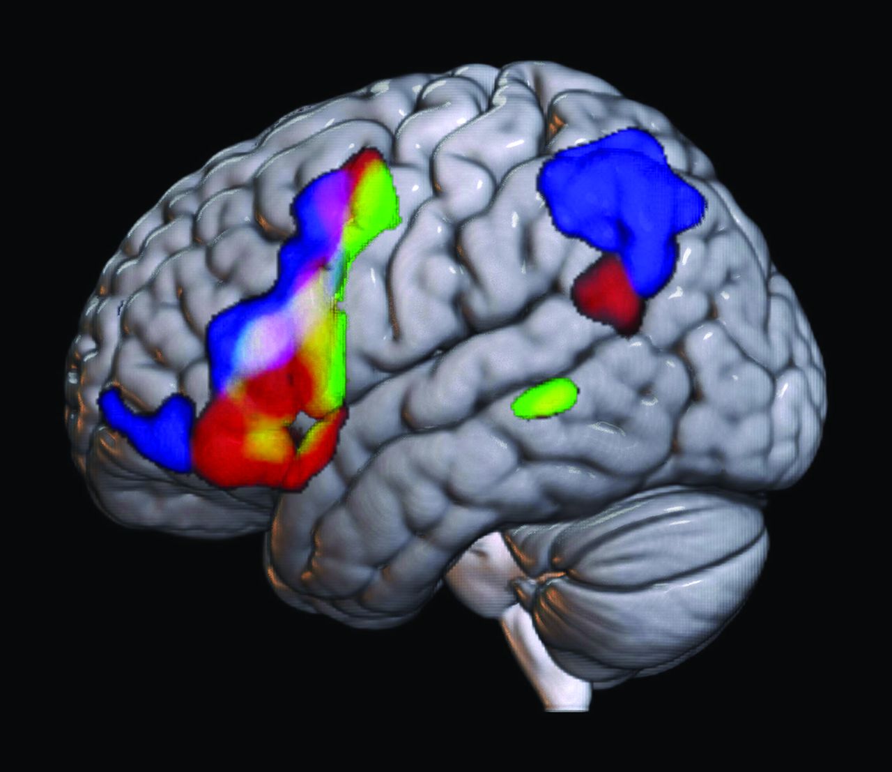
  • FIG 3.
    • Download figure
    • Open in new tab
    • Download powerpoint
    FIG 3.

    Comparison of task- versus rs-fMRI averaged across subjects. Green denotes average task-fMRI activation of the language system across all subjects. Red denotes the average spatial map of the rs-fMRI component correctly selected across subjects by all reviewers. Blue represents the average spatial map of the incorrectly selected rs-fMRI component across subjects by all reviewers. Overlays are additive: Yellow denotes overlapping voxels between green (task-fMRI) and red (correct rs-fMRI component). The white area in the inferior frontal gyrus along the inferior frontal sulcus denotes overlap across task-fMRI, incorrect rs-fMRI, and correct rs-fMRI. While some overlap is noted here, which may be a function of spatial smoothing, there is a clear distinction between the correct rs-fMRI versus incorrect rs-fMRI distribution.

Tables

  • Figures
    • View popup
    Table 1:

    Correctness percentage by reviewer and ICA target component

    ReviewerICA ComponentTop ChoicesNo. (% Correctness)95% CIs
    1Overall72.09%66.12%–77.39%
    120Top 128 (65.12%)49.01%–78.55%
    120Top 232 (74.42%)58.53%–85.96%
    120Top 333 (76.74%)61.00%–87.72%
    150Top 125 (58.14%)42.21%–72.63%
    150Top 233 (76.74%)61.00%–87.72%
    150Top 335 (81.40%)66.08%–91.08%
    2Overall50.39%44.13%–56.63%
    220Top 116 (37.21%)23.39%–53.28%
    220Top 223 (53.49%)37.83%–68.53%
    220Top 324 (55.81%)40.01%–70.59%
    250Top 115 (34.88%)21.45%–50.99%
    250Top 224 (55.81%)40.01%–70.59%
    250Top 328 (65.12%)49.01%–78.55%
    3Overall55.04%48.74%–61.18%
    320Top 122 (51.16%)35.68%–66.44%
    320Top 226 (60.47%)44.45%–74.63%
    320Top 326 (60.47%)44.45%–74.63%
    350Top 118 (41.86%)27.37%–57.79%
    350Top 224 (55.81%)40.01%–70.59%
    350Top 326 (60.47%)44.45%–74.63%
    • View popup
    Table 2:

    Difference in correctness percentages comparing the top choice with the top 2 and 3 choices

    ReviewerICAPair ComparisonNumber (Percentage Differences)P Valuea
    120Top 1 vs top 24 (8.30%).063
    20Top 1 vs top 35 (11.63%).031a
    20Top 2 vs top 31 (2.33%).500
    150Top 1 vs top 28 (18.60%).004a
    50Top 1 vs top 310 (23.26%)<.001a
    50Top 2 vs top 32 (4.65%).250
    220Top 1 vs top 27 (16.28%).008a
    20Top 1 vs top 38 (18.60%).004a
    20Top 2 vs top 31 (2.32%).500
    250Top 1 vs top 29 (20.93%).002a
    50Top 1 vs top 313 (30.23%)<.001a
    50Top 2 vs top 34 (9.30%).063
    320Top 1 vs top 24 (9.30%).063
    20Top 1 vs top 34 (9.30%).063
    20Top 2 vs top 30(0.00%)1.0
    350Top 1 vs top 26 (13.95%).016a
    50Top 1 vs top 38 (18.60%).004a
    50Top 2 vs top 32 (4.65%).250
    • ↵a P value from the 1-sided sign test.

    • View popup
    Table 3:

    Interrater (between reviewers) κ statistic by top choice groups

    Reviewer123
    Overall
     11.00.350.40
     20.351.00.16
     30.400.161.0
    Top choice
     11.00.470.21
     20.471.00.028
     30.210.0281.0
    Top 2 choices
     11.00.370.37
     20.371.00.27
     30.370.271.0
    Top 3 choices
     11.00.310.42
     20.311.00.12
     30.420.121.0
PreviousNext
Back to top

In this issue

American Journal of Neuroradiology: 44 (3)
American Journal of Neuroradiology
Vol. 44, Issue 3
1 Mar 2023
  • Table of Contents
  • Index by author
  • Complete Issue (PDF)
Advertisement
Print
Download PDF
Email Article

Thank you for your interest in spreading the word on American Journal of Neuroradiology.

NOTE: We only request your email address so that the person you are recommending the page to knows that you wanted them to see it, and that it is not junk mail. We do not capture any email address.

Enter multiple addresses on separate lines or separate them with commas.
Identification of the Language Network from Resting-State fMRI in Patients with Brain Tumors: How Accurate Are Experts?
(Your Name) has sent you a message from American Journal of Neuroradiology
(Your Name) thought you would like to see the American Journal of Neuroradiology web site.
CAPTCHA
This question is for testing whether or not you are a human visitor and to prevent automated spam submissions.
Cite this article
S.K. Gujar, K. Manzoor, J. Wongsripuemtet, G. Wang, D. Ryan, S. Agarwal, M. Lindquist, B. Caffo, J.J. Pillai, H.I. Sair
Identification of the Language Network from Resting-State fMRI in Patients with Brain Tumors: How Accurate Are Experts?
American Journal of Neuroradiology Mar 2023, 44 (3) 274-282; DOI: 10.3174/ajnr.A7806

Citation Manager Formats

  • BibTeX
  • Bookends
  • EasyBib
  • EndNote (tagged)
  • EndNote 8 (xml)
  • Medlars
  • Mendeley
  • Papers
  • RefWorks Tagged
  • Ref Manager
  • RIS
  • Zotero
0 Responses
Respond to this article
Share
Bookmark this article
Language Network Accuracy from Resting-State fMRI
S.K. Gujar, K. Manzoor, J. Wongsripuemtet, G. Wang, D. Ryan, S. Agarwal, M. Lindquist, B. Caffo, J.J. Pillai, H.I. Sair
American Journal of Neuroradiology Mar 2023, 44 (3) 274-282; DOI: 10.3174/ajnr.A7806
del.icio.us logo Twitter logo Facebook logo Mendeley logo
  • Tweet Widget
  • Facebook Like
  • Google Plus One
Purchase

Jump to section

  • Article
    • Abstract
    • ABBREVIATIONS:
    • MATERIALS AND METHODS
    • RESULTS
    • DISCUSSION
    • CONCLUSIONS
    • Footnotes
    • References
  • Figures & Data
  • Info & Metrics
  • Responses
  • References
  • PDF

Related Articles

  • No related articles found.
  • PubMed
  • Google Scholar

Cited By...

  • No citing articles found.
  • Crossref
  • Google Scholar

This article has not yet been cited by articles in journals that are participating in Crossref Cited-by Linking.

More in this TOC Section

  • Kurtosis and Epileptogenic Tubers: A Pilot Study
  • Glutaric Aciduria Type 1: DK vs. Conventional MRI
  • Perifocal Edema in Meningiomas&Brain Connectivity
Show more Functional

Similar Articles

Advertisement

Indexed Content

  • Current Issue
  • Accepted Manuscripts
  • Article Preview
  • Past Issues
  • Editorials
  • Editor's Choice
  • Fellows' Journal Club
  • Letters to the Editor
  • Video Articles

Cases

  • Case Collection
  • Archive - Case of the Week
  • Archive - Case of the Month
  • Archive - Classic Case

More from AJNR

  • Trainee Corner
  • Imaging Protocols
  • MRI Safety Corner
  • Book Reviews

Multimedia

  • AJNR Podcasts
  • AJNR Scantastics

Resources

  • Turnaround Time
  • Submit a Manuscript
  • Submit a Video Article
  • Submit an eLetter to the Editor/Response
  • Manuscript Submission Guidelines
  • Statistical Tips
  • Fast Publishing of Accepted Manuscripts
  • Graphical Abstract Preparation
  • Imaging Protocol Submission
  • Evidence-Based Medicine Level Guide
  • Publishing Checklists
  • Author Policies
  • Become a Reviewer/Academy of Reviewers
  • News and Updates

About Us

  • About AJNR
  • Editorial Board
  • Editorial Board Alumni
  • Alerts
  • Permissions
  • Not an AJNR Subscriber? Join Now
  • Advertise with Us
  • Librarian Resources
  • Feedback
  • Terms and Conditions
  • AJNR Editorial Board Alumni

American Society of Neuroradiology

  • Not an ASNR Member? Join Now

© 2025 by the American Society of Neuroradiology All rights, including for text and data mining, AI training, and similar technologies, are reserved.
Print ISSN: 0195-6108 Online ISSN: 1936-959X

Powered by HighWire