Advertisement

Main menu

  • Home
  • Content
    • Current Issue
    • Accepted Manuscripts
    • Article Preview
    • Past Issue Archive
    • Video Articles
    • AJNR Case Collection
    • Case of the Week Archive
    • Case of the Month Archive
    • Classic Case Archive
  • Special Collections
    • AJNR Awards
    • Low-Field MRI
    • Alzheimer Disease
    • ASNR Foundation Special Collection
    • Photon-Counting CT
    • View All
  • Multimedia
    • AJNR Podcasts
    • AJNR SCANtastic
    • Trainee Corner
    • MRI Safety Corner
    • Imaging Protocols
  • For Authors
    • Submit a Manuscript
    • Submit a Video Article
    • Submit an eLetter to the Editor/Response
    • Manuscript Submission Guidelines
    • Statistical Tips
    • Fast Publishing of Accepted Manuscripts
    • Graphical Abstract Preparation
    • Imaging Protocol Submission
    • Author Policies
  • About Us
    • About AJNR
    • Editorial Board
    • Editorial Board Alumni
  • More
    • Become a Reviewer/Academy of Reviewers
    • Subscribers
    • Permissions
    • Alerts
    • Feedback
    • Advertisers
    • ASNR Home

User menu

  • Alerts
  • Log in

Search

  • Advanced search
American Journal of Neuroradiology

American Journal of Neuroradiology

ASHNR American Society of Functional Neuroradiology ASHNR American Society of Pediatric Neuroradiology ASSR
  • Alerts
  • Log in

Advanced Search

  • Home
  • Content
    • Current Issue
    • Accepted Manuscripts
    • Article Preview
    • Past Issue Archive
    • Video Articles
    • AJNR Case Collection
    • Case of the Week Archive
    • Case of the Month Archive
    • Classic Case Archive
  • Special Collections
    • AJNR Awards
    • Low-Field MRI
    • Alzheimer Disease
    • ASNR Foundation Special Collection
    • Photon-Counting CT
    • View All
  • Multimedia
    • AJNR Podcasts
    • AJNR SCANtastic
    • Trainee Corner
    • MRI Safety Corner
    • Imaging Protocols
  • For Authors
    • Submit a Manuscript
    • Submit a Video Article
    • Submit an eLetter to the Editor/Response
    • Manuscript Submission Guidelines
    • Statistical Tips
    • Fast Publishing of Accepted Manuscripts
    • Graphical Abstract Preparation
    • Imaging Protocol Submission
    • Author Policies
  • About Us
    • About AJNR
    • Editorial Board
    • Editorial Board Alumni
  • More
    • Become a Reviewer/Academy of Reviewers
    • Subscribers
    • Permissions
    • Alerts
    • Feedback
    • Advertisers
    • ASNR Home
  • Follow AJNR on Twitter
  • Visit AJNR on Facebook
  • Follow AJNR on Instagram
  • Join AJNR on LinkedIn
  • RSS Feeds

AJNR Awards, New Junior Editors, and more. Read the latest AJNR updates

Research ArticleHead & Neck
Open Access

The Impact of Interactive MRI-Based Radiologist Review on Radiotherapy Target Volume Delineation in Head and Neck Cancer

D. Adjogatse, I. Petkar, M. Reis Ferreira, A. Kong, M. Lei, C. Thomas, S.F. Barrington, C. Dudau, P. Touska, T. Guerrero Urbano and S.E.J. Connor
American Journal of Neuroradiology February 2023, 44 (2) 192-198; DOI: https://doi.org/10.3174/ajnr.A7773
D. Adjogatse
aFrom the Departments of Oncology (D.A., I.P., M.R.F., A.K., M.L., T.G.U.)
dSchool of Biomedical Engineering and Imaging Sciences (D.A., C.T., S.E.J.C.)
  • Find this author on Google Scholar
  • Find this author on PubMed
  • Search for this author on this site
  • ORCID record for D. Adjogatse
I. Petkar
aFrom the Departments of Oncology (D.A., I.P., M.R.F., A.K., M.L., T.G.U.)
  • Find this author on Google Scholar
  • Find this author on PubMed
  • Search for this author on this site
M. Reis Ferreira
aFrom the Departments of Oncology (D.A., I.P., M.R.F., A.K., M.L., T.G.U.)
  • Find this author on Google Scholar
  • Find this author on PubMed
  • Search for this author on this site
  • ORCID record for M. Reis Ferreira
A. Kong
aFrom the Departments of Oncology (D.A., I.P., M.R.F., A.K., M.L., T.G.U.)
  • Find this author on Google Scholar
  • Find this author on PubMed
  • Search for this author on this site
  • ORCID record for A. Kong
M. Lei
aFrom the Departments of Oncology (D.A., I.P., M.R.F., A.K., M.L., T.G.U.)
  • Find this author on Google Scholar
  • Find this author on PubMed
  • Search for this author on this site
  • ORCID record for M. Lei
C. Thomas
bMedical Physics (C.T.)
dSchool of Biomedical Engineering and Imaging Sciences (D.A., C.T., S.E.J.C.)
  • Find this author on Google Scholar
  • Find this author on PubMed
  • Search for this author on this site
  • ORCID record for C. Thomas
S.F. Barrington
eKing’s College London and Guy’s and St Thomas’ PET Centre (S.F.B.), School of Biomedical Engineering and Imaging Sciences, King’s College London, King’s Health Partners, London, UK
  • Find this author on Google Scholar
  • Find this author on PubMed
  • Search for this author on this site
  • ORCID record for S.F. Barrington
C. Dudau
cRadiology (C.D., P.T., S.E.J.C.), Guy’s and St Thomas’ National Health Service Foundation Trust, London, UK
fDepartment of Neurororadiology (C.D., S.E.J.C.), King’s College Hospital, London, UK
  • Find this author on Google Scholar
  • Find this author on PubMed
  • Search for this author on this site
  • ORCID record for C. Dudau
P. Touska
cRadiology (C.D., P.T., S.E.J.C.), Guy’s and St Thomas’ National Health Service Foundation Trust, London, UK
  • Find this author on Google Scholar
  • Find this author on PubMed
  • Search for this author on this site
  • ORCID record for P. Touska
T. Guerrero Urbano
aFrom the Departments of Oncology (D.A., I.P., M.R.F., A.K., M.L., T.G.U.)
gFaculty of Dentistry, Oral and Craniofacial Sciences (T.G.U.), King’s College London, London, UK
  • Find this author on Google Scholar
  • Find this author on PubMed
  • Search for this author on this site
S.E.J. Connor
cRadiology (C.D., P.T., S.E.J.C.), Guy’s and St Thomas’ National Health Service Foundation Trust, London, UK
dSchool of Biomedical Engineering and Imaging Sciences (D.A., C.T., S.E.J.C.)
fDepartment of Neurororadiology (C.D., S.E.J.C.), King’s College Hospital, London, UK
  • Find this author on Google Scholar
  • Find this author on PubMed
  • Search for this author on this site
  • ORCID record for S.E.J. Connor
  • Article
  • Figures & Data
  • Supplemental
  • Info & Metrics
  • Responses
  • References
  • PDF
Loading

Article Figures & Data

Figures

  • FIG 1.
    • Download figure
    • Open in new tab
    • Download powerpoint
    FIG 1.

    Examples of volume amendments made at peer review. Selected images show the difference in GTVonc and GTVrad delineations. A and B, T2-weighted axial images show an intermediate-signal left piriform fossa tumor (arrows). Following review of the diffusion-weighted imaging (b = 1000) (single arrow) (C), the contouring on T1-weighted gadolinium-enhanced axial image is expanded from GTVonc (red) to GTVrad (green) (D). T2-weighted axial image demonstrates multiple bilateral lymph nodes. The left submandibular gland (arrow) was initially included in the GTVonc (red) because it was isointense to other pathologic lymph nodes. F, Diffusion-weighted imaging (b = 1000) aided in the identification of the lower signal submandibular gland; hence, it was excluded from GTVrad (green).

  • FIG 2.
    • Download figure
    • Open in new tab
    • Download powerpoint
    FIG 2.

    Factors contributing to volume amendments: GTVT and GTVN. 1Changes made following joint discussion between the oncologist and radiologist, eg, inclusion of lymph nodes with borderline pathologic changes. 2Diffusion-weighted MR imaging sequences. 3Tumor volumes described as complex by the oncologist and radiologist at the time of peer review. Includes skull base disease with perineural spread, septate tumor, and postexcisional biopsy changes.
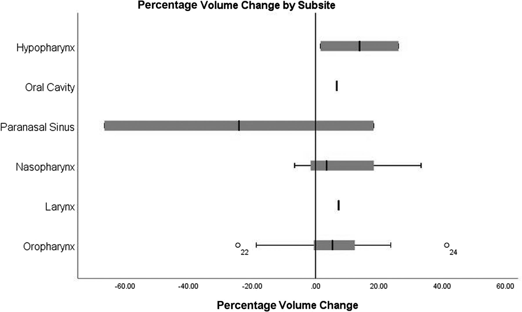
  • FIG 3.
    • Download figure
    • Open in new tab
    • Download powerpoint
    FIG 3.

    Boxplots of percentage volume change for GTVT by subsite. Case number 22 and 24 (highlighted as superscript numbers on the box plot of oropharyngeal cancer cases) are outliers in terms of percentage volume change. Case 22: the GTVT was reduced by 24.5% on radiologist review due to the removal of normal oral tongue and normal parapharyngeal fat. Case 24; GTVT increased by 41.4% on radiologist review due to imaging misinterpretation of abnormal deep mucosal disease extent.

PreviousNext
Back to top

In this issue

American Journal of Neuroradiology: 44 (2)
American Journal of Neuroradiology
Vol. 44, Issue 2
1 Feb 2023
  • Table of Contents
  • Index by author
  • Complete Issue (PDF)
Advertisement
Print
Download PDF
Email Article

Thank you for your interest in spreading the word on American Journal of Neuroradiology.

NOTE: We only request your email address so that the person you are recommending the page to knows that you wanted them to see it, and that it is not junk mail. We do not capture any email address.

Enter multiple addresses on separate lines or separate them with commas.
The Impact of Interactive MRI-Based Radiologist Review on Radiotherapy Target Volume Delineation in Head and Neck Cancer
(Your Name) has sent you a message from American Journal of Neuroradiology
(Your Name) thought you would like to see the American Journal of Neuroradiology web site.
CAPTCHA
This question is for testing whether or not you are a human visitor and to prevent automated spam submissions.
Cite this article
D. Adjogatse, I. Petkar, M. Reis Ferreira, A. Kong, M. Lei, C. Thomas, S.F. Barrington, C. Dudau, P. Touska, T. Guerrero Urbano, S.E.J. Connor
The Impact of Interactive MRI-Based Radiologist Review on Radiotherapy Target Volume Delineation in Head and Neck Cancer
American Journal of Neuroradiology Feb 2023, 44 (2) 192-198; DOI: 10.3174/ajnr.A7773

Citation Manager Formats

  • BibTeX
  • Bookends
  • EasyBib
  • EndNote (tagged)
  • EndNote 8 (xml)
  • Medlars
  • Mendeley
  • Papers
  • RefWorks Tagged
  • Ref Manager
  • RIS
  • Zotero
0 Responses
Respond to this article
Share
Bookmark this article
Interactive MRI Review Impact Radiotherapy Targets
D. Adjogatse, I. Petkar, M. Reis Ferreira, A. Kong, M. Lei, C. Thomas, S.F. Barrington, C. Dudau, P. Touska, T. Guerrero Urbano, S.E.J. Connor
American Journal of Neuroradiology Feb 2023, 44 (2) 192-198; DOI: 10.3174/ajnr.A7773
del.icio.us logo Twitter logo Facebook logo Mendeley logo
  • Tweet Widget
  • Facebook Like
  • Google Plus One
Purchase

Jump to section

  • Article
    • Abstract
    • ABBREVIATIONS:
    • MATERIALS AND METHODS
    • RESULTS
    • DISCUSSION
    • CONCLUSIONS
    • Footnotes
    • References
  • Figures & Data
  • Supplemental
  • Info & Metrics
  • Responses
  • References
  • PDF

Related Articles

  • PubMed
  • Google Scholar

Cited By...

  • No citing articles found.
  • Crossref (5)
  • Google Scholar

This article has been cited by the following articles in journals that are participating in Crossref Cited-by Linking.

  • PET/MR Imaging in Head and Neck Cancer
    Minerva Becker, Claudio de Vito, Nicolas Dulguerov, Habib Zaidi
    Magnetic Resonance Imaging Clinics of North America 2023 31 4
  • Delta radiomics to track radiation response in lung tumors receiving stereotactic magnetic resonance-guided radiotherapy
    Yining Zha, Zezhong Ye, Anna Zapaishchykova, John He, Shu-Hui Hsu, Jonathan E. Leeman, Kelly J. Fitzgerald, David E. Kozono, Raymond H. Mak, Hugo J.W.L. Aerts, Benjamin H. Kann
    Physics and Imaging in Radiation Oncology 2024 31
  • A systematic review and meta-analysis on the impact of institutional peer review in radiation oncology
    Jane Jomy, Rachel Lu, Radha Sharma, Ke Xin Lin, David C. Chen, Jeff Winter, Srinivas Raman
    Radiotherapy and Oncology 2025 202
  • PET/MR Imaging in Head and Neck Cancer
    Minerva Becker, Claudio de Vito, Nicolas Dulguerov, Habib Zaidi
    PET Clinics 2025 20 3
  • Radiologist Involvement in Radiation Oncology Peer Review
    Ryan T. Hughes, Niema B. Razavian, Sydney Smith, Ralph B. D’Agostino, Paul M. Bunch, Janardhana Ponnatapura, Trevor J. Royce, James D. Ververs, Chandylen L. Nightingale, Kathryn E. Weaver, Michael K. Farris
    JAMA Network Open 2024 7 12

More in this TOC Section

  • Chondrosarcoma vs Synovial Chondromatosis: Imaging
  • WHO Classification Update: Nasal&Skull Base Tumors
  • Peritumoral Signal in Vestibular Schwannomas
Show more Head & Neck

Similar Articles

Advertisement

Indexed Content

  • Current Issue
  • Accepted Manuscripts
  • Article Preview
  • Past Issues
  • Editorials
  • Editor's Choice
  • Fellows' Journal Club
  • Letters to the Editor
  • Video Articles

Cases

  • Case Collection
  • Archive - Case of the Week
  • Archive - Case of the Month
  • Archive - Classic Case

More from AJNR

  • Trainee Corner
  • Imaging Protocols
  • MRI Safety Corner
  • Book Reviews

Multimedia

  • AJNR Podcasts
  • AJNR Scantastics

Resources

  • Turnaround Time
  • Submit a Manuscript
  • Submit a Video Article
  • Submit an eLetter to the Editor/Response
  • Manuscript Submission Guidelines
  • Statistical Tips
  • Fast Publishing of Accepted Manuscripts
  • Graphical Abstract Preparation
  • Imaging Protocol Submission
  • Evidence-Based Medicine Level Guide
  • Publishing Checklists
  • Author Policies
  • Become a Reviewer/Academy of Reviewers
  • News and Updates

About Us

  • About AJNR
  • Editorial Board
  • Editorial Board Alumni
  • Alerts
  • Permissions
  • Not an AJNR Subscriber? Join Now
  • Advertise with Us
  • Librarian Resources
  • Feedback
  • Terms and Conditions
  • AJNR Editorial Board Alumni

American Society of Neuroradiology

  • Not an ASNR Member? Join Now

© 2025 by the American Society of Neuroradiology All rights, including for text and data mining, AI training, and similar technologies, are reserved.
Print ISSN: 0195-6108 Online ISSN: 1936-959X

Powered by HighWire