
Article Figures & Data
Figures
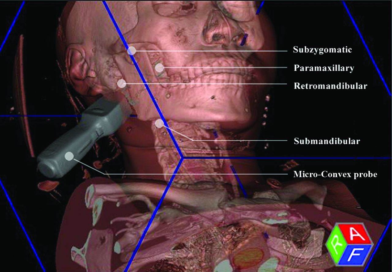
- FIG 1.
Schematic of US fusion–guided biopsy, with the use of a Micro-Convex probe and 4 available needle approaches.
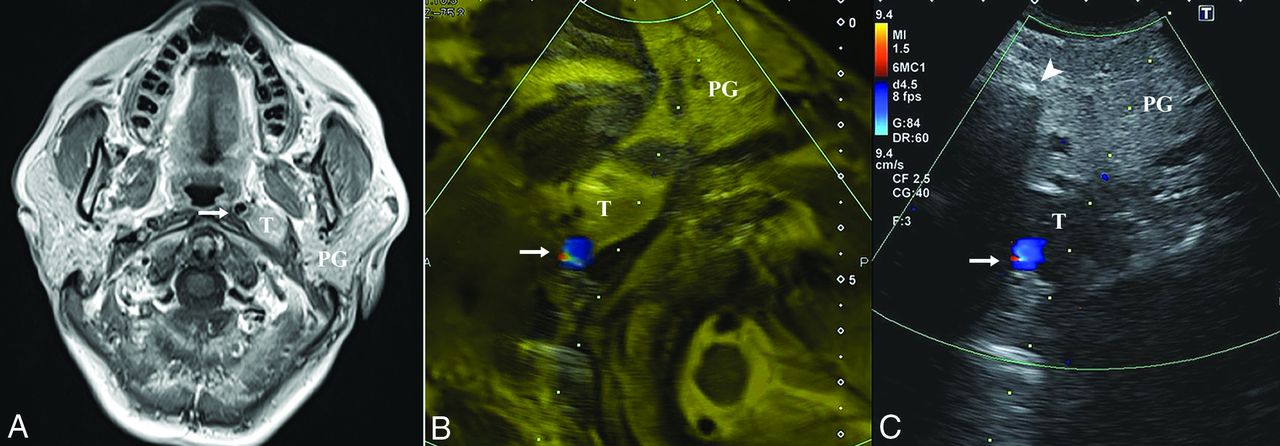
- FIG 2.
A 58-year-old woman without clinical presentation (patient No. 2). A, Axial T1-weighted postcontrast MR image shows a left parapharyngeal lesion with heterogeneous hyperintensity and a demarcated border. The target lesion is located deep in the parotid gland, and the ICA (arrow) is displaced medially. After a successful fusion of MR imaging (B) and US (C), MR imaging facilitates the accurate localization of the target lesion, which is ill-defined on US, with the ICA (arrow) confirmed on the color Doppler mode and the overlay mode (yellow mask). The dotted line indicates the expected needle path via the retromandibular approach. The histopathologic yield was schwannoma. T indicates target lesion; PG, parotid gland.
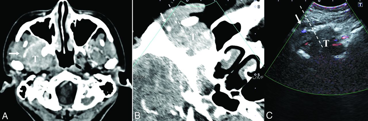
- FIG 3.
A 30-year-old woman with maxillofacial swelling, pain and dizziness for 2 years (patient No. 3). A, The axial contrast-enhanced CT image shows a homogeneously enhancing lesion involving the infratemporal fossa and pterygopalatine fossa. A branch of the maxillary artery runs superficially around the lesion (arrow). After a successful fusion of CT (B) and US (C), we can locate the target lesion that is occult on US due to the acoustic shadow of the mandible. Color Doppler US helps to avoid the maxillary artery (arrow). The dashed line indicates the expected needle path via the subzygomatic approach. Histopathology demonstrated meningioma (meningothelial subtype, World Health Organization grade I). T indicates target lesion.
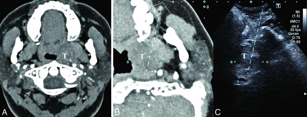
- FIG 4.
A 32-year-old woman with aural fullness, tinnitus, and hearing loss for 1 year (patient No. 10). A, The axial contrast-enhanced CT image shows a parapharyngeal lesion at the level of the alveolar ridge with mild and heterogeneous enhancement, constricting the auditory tube. After successful fusion with CT (B), a heterogeneous low-echoic lesion can be identified and located on US. C, The paramaxillary approach is chosen (dashed line). Histopathology demonstrated lymphatic malformation. T indicates target lesion.
- FIG 5.
A 39-year-old man with headache for 1 month (patient No. 14). A multiplanar image of US–PET/CT fusion with the patient’s head tilted to the contralateral side. A, The axial plain CT shows an ill-defined lesion centered in the infratemporal fossa with bony destruction. B, The axial fused [18F] FDG-PET/CT reveals a high focal uptake. C, The target lesion is occult behind the acoustic shadow of the coronoid process on US, but a selective biopsy can be guided under fusion images. The subzygomatic approach is chosen (dashed line). Histopathology revealed nasopharyngeal carcinoma. T indicates target lesion.


Jump to section
Related Articles
Cited By...
- No citing articles found.