
Article Figures & Data
Figures
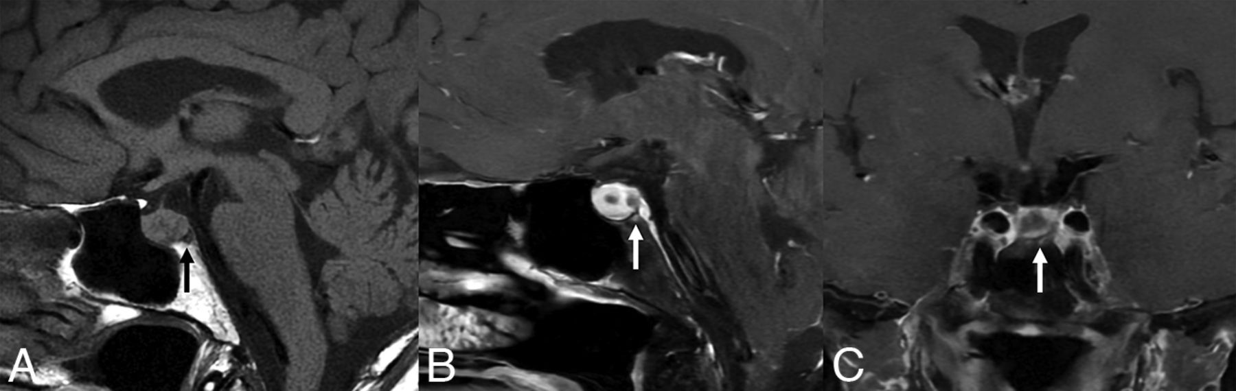
- FIG 1.
A 72-year-old woman who presented with fatigue and was found to have a bilobed pituitary lesion, preoperatively favored to be a pituitary adenoma. A, Precontrast T1-weighted imaging shows focal hypointense signal in the posterior basisphenoid bone marrow (arrows). Postcontrast imaging shows corresponding enhancement (B and C). Pathology confirmed an rRCC.
- FIG 2.
A 38-year-old woman presented with headaches for 6 months. MR imaging shows a sellar/suprasellar mass with a septated cystic lesion in the posterior aspect of the sella, preoperatively favored to represent a pituitary adenoma. Postcontrast MR imaging shows basisphenoid bone marrow (arrows) enhancement (B and C) with corresponding edema (D). There was no intrinsic basisphenoid T1-hyperintense signal on the precontrast imaging (A). Pathology confirmed the cystic lesion to be an rRCC. The enlarged suprasellar lesion was biopsied and found to be mixed inflammatory infiltrate and fibrosis, thought to represent inflammatory hypophysitis secondary to the rRCC.
- FIG 3.
A 39-year-old woman with a history of gamma knife therapy to the sella for an undiagnosed lesion at another institution presented with an enlarging cystic pituitary lesion. T2-weighted imaging (A) shows a hypointense nodule and edema of the basisphenoid bone marrow (arrows). This area has corresponding T1-hypointense signal on precontrast imaging (B) and enhancement (C). Pathology confirmed an rRCC.
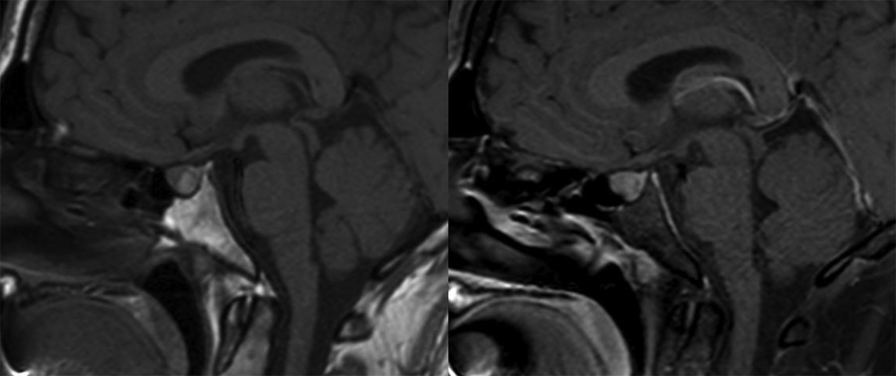
- FIG 4.
Example of a pathology-proved nonruptured RCC. The precontrast T1-weighted (left) images show normal signal of the basisphenoid bone marrow edema below the T1-hyperintense RCC. The fat-saturated postcontrasted image (right) shows normal basisphenoid bone marrow without abnormal enhancement.


Tables
Patient Age (yr) Sex Prolactin (μg/L) Presenting Symptom Preop Diagnosis Follow-Up 1 51 Female 33.2 HA Adenoma No recurrence at 6 mo 2 47 Female n/a HA, panhypopituitary RCC Recurrence at 3 yr 3 38 Female 2.7 HA Adenoma No recurrence at 4 yr 4 39 Female 22.4 Enlarging lesion RCC No recurrence at 1 mo 5 30 Female 36.5 Diabetes insipidus, vision changes Adenoma No recurrence at 2 mo 6 72 Female 42.4 Fatigue Adenoma No recurrence at 2 mo 7 33 Female 113.2 Hyperprolactinemia Adenoma No recurrence at 2 mo Note:—HA indicates headache; Preop, preoperative; n/a, not available.
Patient Max Dimension (mm) Bone Marrow Enhancement T2 Signal T2 Dark Nodule T1 Hyperintense Signal Location 1 11.5 Focal Hyperintense No No Midline 2 14.0 Focal Hypointense No Yes Midline 3 10.8 Diffuse Heterogeneous No No Midline 4 10.8 Diffuse Hyperintense Yes Yes Off-midline 5 11.1 Diffuse Hyperintense Yes No Midline 6 8.2 Focal Heterogeneous No No Replaces entire pituitary 7 12.2 Diffuse Hyperintense Yes No Replaces entire pituitary Note:—Max indicates maximum.


