Advertisement

Main menu

  • Home
  • Content
    • Current Issue
    • Accepted Manuscripts
    • Article Preview
    • Past Issue Archive
    • Video Articles
    • AJNR Case Collection
    • Case of the Week Archive
    • Case of the Month Archive
    • Classic Case Archive
  • Special Collections
    • AJNR Awards
    • Low-Field MRI
    • Alzheimer Disease
    • ASNR Foundation Special Collection
    • Photon-Counting CT
    • View All
  • Multimedia
    • AJNR Podcasts
    • AJNR SCANtastic
    • Trainee Corner
    • MRI Safety Corner
    • Imaging Protocols
  • For Authors
    • Submit a Manuscript
    • Submit a Video Article
    • Submit an eLetter to the Editor/Response
    • Manuscript Submission Guidelines
    • Statistical Tips
    • Fast Publishing of Accepted Manuscripts
    • Graphical Abstract Preparation
    • Imaging Protocol Submission
    • Author Policies
  • About Us
    • About AJNR
    • Editorial Board
    • Editorial Board Alumni
  • More
    • Become a Reviewer/Academy of Reviewers
    • Subscribers
    • Permissions
    • Alerts
    • Feedback
    • Advertisers
    • ASNR Home

User menu

  • Alerts
  • Log in

Search

  • Advanced search
American Journal of Neuroradiology

American Journal of Neuroradiology

ASHNR American Society of Functional Neuroradiology ASHNR American Society of Pediatric Neuroradiology ASSR
  • Alerts
  • Log in

Advanced Search

  • Home
  • Content
    • Current Issue
    • Accepted Manuscripts
    • Article Preview
    • Past Issue Archive
    • Video Articles
    • AJNR Case Collection
    • Case of the Week Archive
    • Case of the Month Archive
    • Classic Case Archive
  • Special Collections
    • AJNR Awards
    • Low-Field MRI
    • Alzheimer Disease
    • ASNR Foundation Special Collection
    • Photon-Counting CT
    • View All
  • Multimedia
    • AJNR Podcasts
    • AJNR SCANtastic
    • Trainee Corner
    • MRI Safety Corner
    • Imaging Protocols
  • For Authors
    • Submit a Manuscript
    • Submit a Video Article
    • Submit an eLetter to the Editor/Response
    • Manuscript Submission Guidelines
    • Statistical Tips
    • Fast Publishing of Accepted Manuscripts
    • Graphical Abstract Preparation
    • Imaging Protocol Submission
    • Author Policies
  • About Us
    • About AJNR
    • Editorial Board
    • Editorial Board Alumni
  • More
    • Become a Reviewer/Academy of Reviewers
    • Subscribers
    • Permissions
    • Alerts
    • Feedback
    • Advertisers
    • ASNR Home
  • Follow AJNR on Twitter
  • Visit AJNR on Facebook
  • Follow AJNR on Instagram
  • Join AJNR on LinkedIn
  • RSS Feeds

AJNR Awards, New Junior Editors, and more. Read the latest AJNR updates

Research ArticleHead and Neck Imaging

Differentiation between Chondrosarcoma and Synovial Chondromatosis of the Temporomandibular Joint Using CT and MR Imaging

B.G. Jang, K.H. Huh, H.G. Yeom, J.H. Kang, J.E. Kim, H.J. Yoon, W.J. Yi, M.S. Heo and S.S. Lee
American Journal of Neuroradiology October 2023, 44 (10) 1176-1183; DOI: https://doi.org/10.3174/ajnr.A7980
B.G. Jang
aFrom the Department of Oral and Maxillofacial Radiology and Dental Research Institute (B.G.J., K.H.H., J.E.K., H.J.Y., W.J.Y., M.S.H., S.S.L.), School of Dentistry, Seoul National University, Seoul, Korea
  • Find this author on Google Scholar
  • Find this author on PubMed
  • Search for this author on this site
  • ORCID record for B.G. Jang
K.H. Huh
aFrom the Department of Oral and Maxillofacial Radiology and Dental Research Institute (B.G.J., K.H.H., J.E.K., H.J.Y., W.J.Y., M.S.H., S.S.L.), School of Dentistry, Seoul National University, Seoul, Korea
  • Find this author on Google Scholar
  • Find this author on PubMed
  • Search for this author on this site
  • ORCID record for K.H. Huh
H.G. Yeom
bDepartment of Oral and Maxillofacial Radiology and Wonkwang Dental Research Institute (H.G.Y.), School of Dentistry, Wonkwang University, Iksan, Korea
  • Find this author on Google Scholar
  • Find this author on PubMed
  • Search for this author on this site
  • ORCID record for H.G. Yeom
J.H. Kang
cDepartment of Oral and Maxillofacial Radiology (J.H.K.), Seoul National University Dental Hospital, Seoul, Korea
  • Find this author on Google Scholar
  • Find this author on PubMed
  • Search for this author on this site
  • ORCID record for J.H. Kang
J.E. Kim
aFrom the Department of Oral and Maxillofacial Radiology and Dental Research Institute (B.G.J., K.H.H., J.E.K., H.J.Y., W.J.Y., M.S.H., S.S.L.), School of Dentistry, Seoul National University, Seoul, Korea
  • Find this author on Google Scholar
  • Find this author on PubMed
  • Search for this author on this site
  • ORCID record for J.E. Kim
H.J. Yoon
aFrom the Department of Oral and Maxillofacial Radiology and Dental Research Institute (B.G.J., K.H.H., J.E.K., H.J.Y., W.J.Y., M.S.H., S.S.L.), School of Dentistry, Seoul National University, Seoul, Korea
  • Find this author on Google Scholar
  • Find this author on PubMed
  • Search for this author on this site
  • ORCID record for H.J. Yoon
W.J. Yi
aFrom the Department of Oral and Maxillofacial Radiology and Dental Research Institute (B.G.J., K.H.H., J.E.K., H.J.Y., W.J.Y., M.S.H., S.S.L.), School of Dentistry, Seoul National University, Seoul, Korea
  • Find this author on Google Scholar
  • Find this author on PubMed
  • Search for this author on this site
  • ORCID record for W.J. Yi
M.S. Heo
aFrom the Department of Oral and Maxillofacial Radiology and Dental Research Institute (B.G.J., K.H.H., J.E.K., H.J.Y., W.J.Y., M.S.H., S.S.L.), School of Dentistry, Seoul National University, Seoul, Korea
  • Find this author on Google Scholar
  • Find this author on PubMed
  • Search for this author on this site
  • ORCID record for M.S. Heo
S.S. Lee
aFrom the Department of Oral and Maxillofacial Radiology and Dental Research Institute (B.G.J., K.H.H., J.E.K., H.J.Y., W.J.Y., M.S.H., S.S.L.), School of Dentistry, Seoul National University, Seoul, Korea
  • Find this author on Google Scholar
  • Find this author on PubMed
  • Search for this author on this site
  • ORCID record for S.S. Lee
  • Article
  • Figures & Data
  • Supplemental
  • Info & Metrics
  • Responses
  • References
  • PDF
Loading

Article Figures & Data

Figures

  • Tables
  • FIG 1.
    • Download figure
    • Open in new tab
    • Download powerpoint
    FIG 1.

    Schematic drawings of the degree of infiltration into the tendon of the LPM by a mass in the pterygoid fovea. Complete and partial infiltration is shown in A and B, respectively. For such cases, a score of 1 is assigned. No infiltration with an intact LPM attachment is shown in C, in which case a score of zero is assigned. MC indicates mandibular condyle.

  • FIG 2.
    • Download figure
    • Open in new tab
    • Download powerpoint
    FIG 2.

    CS of the right TMJ in 54-year-old woman. Axial MDCT with bone and soft-tissue windows (A and B). T2-weighted MR (C) and contrast-enhanced T1-weighted MR (D) images demonstrate a large mass infiltrating the tendon of the lateral pterygoid muscle (asterisk) in the pterygoid fovea and show septalike internal enhancement. Stippled calcification and severe destruction of the mandibular condyle with a spiculate periosteal reaction are revealed (A and B). Note the lesion center on the mandibular condyle. Sagittal MDCT (E) and T2-weighted MR images (F) show the eroded mandibular condyle and the intact articular eminence. Note the disc (arrow) located between them. This mass showed all 9 high-risk imaging features for CS and received a composite score of 9.

  • FIG 3.
    • Download figure
    • Open in new tab
    • Download powerpoint
    FIG 3.

    CS of the left TMJ in 44-year-old woman. Axial MDCT (A), fat-suppressed T2-weighted MR (B), and contrast-enhanced T1-weighted MR (C) images reveal a mass surrounding the mandibular condyle. Sagittal MDCT (D) and contrast-enhanced T1-weighted MR images (E) demonstrate the mass center in the joint space. Lesion centered on the superior joint space (arrows), destruction and sclerosis of the articular eminence/glenoid fossa, absence of periosteal reaction, and the relatively small lesion size favor SC. On the other hand, destruction of the mandibular condyle, infiltration into the tendon (arrowhead) of the lateral pterygoid muscle (asterisk), stippled calcification, and internal enhancement favor CS. A histopathologic examination after mass resection resulted in a diagnosis of CS. This TMJ mass revealed 4 high-risk imaging features and a composite score of 4. It was not easy to differentiate CS from SC by imaging features.
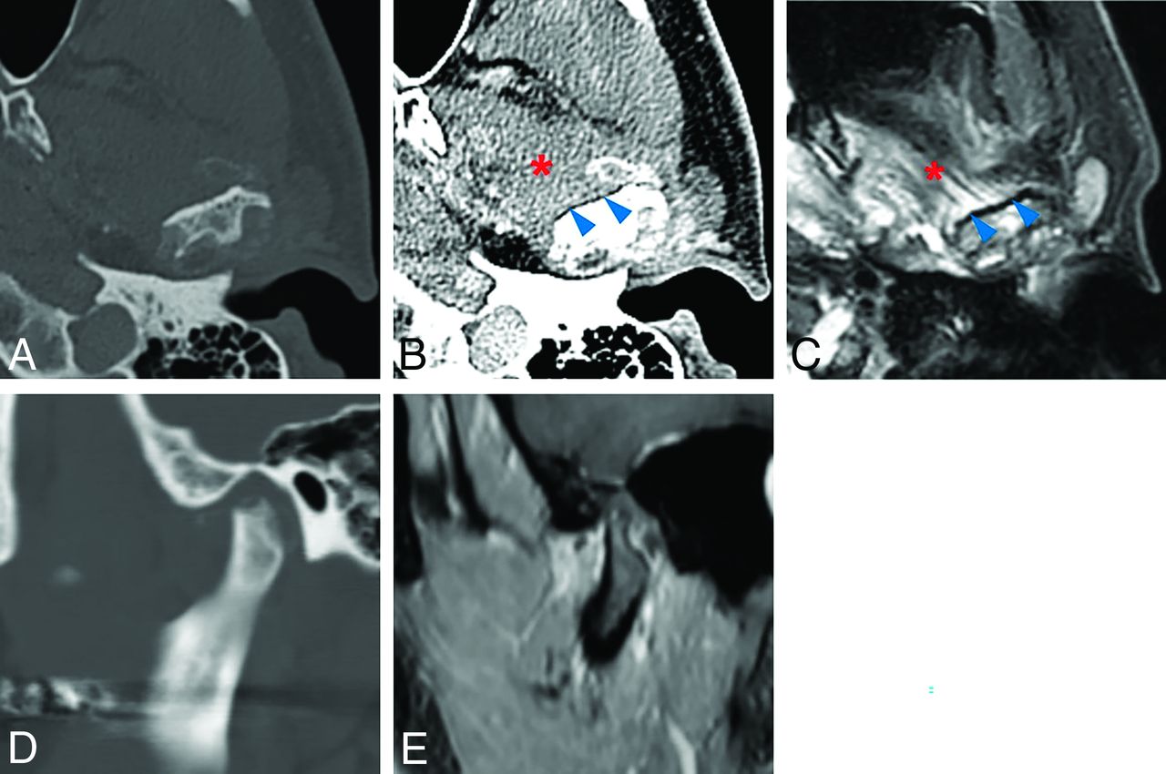
  • FIG 4.
    • Download figure
    • Open in new tab
    • Download powerpoint
    FIG 4.

    SC of the left TMJ in 52-year-old man. Axial MDCT (A and B) and fat-suppressed T2-weighted MR images (C) show a mass surrounding and eroding the mandibular condyle. Sagittal MDCT (D) and contrast-enhanced T1-weighted MR images (E) reveal the mass to be centered on the joint space. Ring-and-arc calcification, intact attachment of tendon (blue arrowheads) of the lateral pterygoid muscle (red asterisk), lesion center on the joint space, and a relatively small lesion size favor an imaging diagnosis of SC. The result of a preoperative incisional biopsy was CS, but the final histopathologic diagnosis was changed to SC after mass resection. This TMJ mass showed 3 high-risk imaging features and received a composite score of 3.

  • FIG 5.
    • Download figure
    • Open in new tab
    • Download powerpoint
    FIG 5.

    ROC curves for qualitative and quantitative variables show that the best discriminating values for differentiating CS from SC are the following: Mandibular condyle for lesion center, Presence for destruction of the mandibular condyle, Absence for destruction of the articular eminence/glenoid fossa, Absence for sclerosis of the articular eminence/glenoid fossa, Presence for infiltration into the tendon of lateral pterygoid muscle, Stippled pattern of calcification, Presence for periosteal reaction, Presence for internal enhancement, and 30.5 mm for lesion size. Of the 9 variables, except for pattern of calcification and mean lesion size, the remaining 7 variables were dichotomous qualitative variables with discriminating values determined at 1 of the 2 characteristics. On the other hand, mean lesion size was a quantitative variable, and the discriminating value was determined at the point of 30.5 mm, which revealed the maximum Youden index among multiple points. Last, the pattern of calcification, the 5 patterns, namely absence, stippled, flocculent, ring-and-arc, and popcorn, were considered as a dimensional continuum in ascending order and converted into an ordinal quantitative variable. The ROC curve was then constructed, and the discriminating value was derived at the point of stippled calcification, which indicates that CS showed a higher prevalence in absence or stippled calcification, and SC, in the other calcification patterns.

  • FIG 6.
    • Download figure
    • Open in new tab
    • Download powerpoint
    FIG 6.

    The ROC curve of the composite score for distinguishing CS from SC. The score was assessed by assigning 1 point for each of the following high-risk imaging features for chondrosarcoma: lesion center on the mandibular condyle, destruction of the mandibular condyle, no destruction of the articular eminence/glenoid fossa, no sclerosis of the articular eminence/glenoid fossa, infiltration into the tendon of the lateral pterygoid muscle, absence or stippled calcification, periosteal reaction, internal enhancement, and ≥30.5 mm lesion size. The ROC analysis demonstrated that the best cutoff value for differentiating CS from SC was +4 points (Youden index = 0.917, AUC = 0.986; 95% CI, 0.950–1.000).

Tables

  • Figures
    • View popup
    Table 1:

    Clinical features of CS and SCa

    Independent VariablesTotal (n = 47)CS (n = 12)SC (n = 35)P Value
    Ageb (mean) (yr)49.3 (SD, 13.5)50.3 (SD, 15.6)48.9 (SD, 13.2).756
    Sexc.659
     Male7 (14.9)1 (8.3)6 (17.1)
     Female40 (85.1)11 (91.7)29 (82.9)
    Sided.679
     Right25 (53.2)7 (58.3)18 (51.4)
     Left22 (46.8)5 (41.7)17 (48.6)
    Chief complaint
     Swellingd11 (23.4)5 (41.7)6 (17.1).118
     Painc43 (91.5)11 (91.7)32 (91.4)1.000
     Trismusc20 (42.6)8 (66.7)12 (34.3).089
    • ↵a Values are the number of patients with percentage in parentheses or the mean ± SD.

    • ↵b Student t test.

    • ↵c Fisher exact test.

    • ↵d χ2test.

    • View popup
    Table 2:

    Imaging features for CS and SCa

    Independent VariablesTotal (n = 47)CS (n = 12)SC (n = 35)P ValueInterobserver Agreement
    Lesion center<.001b,c.832f
     Joint space40 (85.1)5 (41.7)35 (100)
     Mandibular condyle7 (14.9)7 (58.3)0 (0)
    Destruction of the mandibular condyle31 (66.0)11 (91.7)20 (57.1).037b,c.905f
    Sclerosis of the mandibular condyle23 (48.9)8 (66.7)15 (42.9).193c.872f
    Destruction of the articular eminence/glenoid fossa31 (66.0)4 (33.3)27 (77.1).012b,c.856f
    Sclerosis of the articular eminence/glenoid fossa29 (61.7)2 (16.7)27 (77.1)<.001b,c.861f
    Infiltration into the tendon of the LPM17 (36.2)12 (100.0)5 (14.3)<.001b,c.908f
    Calcification37 (78.7)8 (66.7)29 (82.9).251c.828f
    Pattern of calcification.008b,d.835f
     Absence10 (21.3)4 (33.3)6 (17.1)
     Stippled9 (19.1)6 (50.0)3 (8.6)
     Flocculent4 (8.5)0 (0.0)4 (11.4)
     Ring-and-arc9 (19.1)0 (0.0)9 (25.7)
     Popcorn15 (31.9)2 (16.7)13 (37.1)
    Periosteal reaction12 (25.5)8 (66.7)4 (11.4).001b,c.827f
    Osteophyte10 (21.3)2 (16.7)8 (22.9)1.000c.873f
    Peripheral enhancement19 (79.2)11 (91.7)8 (66.7).317c.864f
    Internal enhancement15 (62.5)12 (100.0)3 (25.0)<.001b,c.830f
    Lesion size28.5 (SD, 7.4)37.1 (SD, 8.2)25.6 (SD, 4.2)<.001b,e.932g
    X/Z ratio1.6 (SD, 0.3)1.5 (SD, 0.2)1.7 (SD, 0.3).067e.928g
    • a Values are the number of patients with percentages in parentheses or the mean ± SD.

    • ↵b Statistically significant.

    • ↵c Fisher exact test.

    • d Linear-by-linear association test.

    • ↵e Student t test.

    • ↵f Cohen κ coefficient.

    • ↵g Intraclass correlation coefficient.

  • Table 3: Diagnostic performance of each qualitative and quantitative parameter that showed a statistically significant difference for differentiating CS from SCa

    Independent VariablesPrevalenceSensitivity (%)Specificity (%)Accuracy (%)PPV (%)NPV (%)AUC (95% CI)
    Lesion center (mandibular condyle)CS 7/12 SC 0/3558.3100.089.4100.087.50.792 (0.611–0.972)
    Destruction of the mandibular condyle (presence)CS 11/12 SC 20/3591.742.955.335.593.80.673 (0.510–0.835)
    Destruction of articular eminence/glenoid fossa (absence)CS 8/12 SC 8/3566.777.174.550.087.10.719 (0.542–0.896)
    Sclerosis of the articular eminence/glenoid fossa (absence)CS 10/12 SC 8/3583.377.178.755.693.10.802 (0.654–0.950)
    Infiltration into the tendon of the LPM (presence)CS 12/12 SC 5/35100.085.789.470.6100.00.929 (0.855–1.000)
    Calcification (absence or stippled)CS 10/12 SC 9/3583.374.376.652.692.90.729 (0.556–0.901)
    Periosteal reaction (presence)CS 8/12 SC 4/3566.788.683.066.788.60.776 (0.604–0.948)
    Internal enhancement (presence)CS 12/12 SC 3/12100.075.087.580.0100.00.875 (0.719–1.000)
    Mean lesion size (≥ 30.5 mm)CS 10/12 SC 5/3583.385.785.166.793.80.889 (0.773–1.000)
    • ↵a The items in parentheses correspond to the characteristics of CS, and 1 point was assigned if relevant imaging features in the parentheses were present.

PreviousNext
Back to top

In this issue

American Journal of Neuroradiology: 44 (10)
American Journal of Neuroradiology
Vol. 44, Issue 10
1 Oct 2023
  • Table of Contents
  • Index by author
  • Complete Issue (PDF)
Advertisement
Print
Download PDF
Email Article

Thank you for your interest in spreading the word on American Journal of Neuroradiology.

NOTE: We only request your email address so that the person you are recommending the page to knows that you wanted them to see it, and that it is not junk mail. We do not capture any email address.

Enter multiple addresses on separate lines or separate them with commas.
Differentiation between Chondrosarcoma and Synovial Chondromatosis of the Temporomandibular Joint Using CT and MR Imaging
(Your Name) has sent you a message from American Journal of Neuroradiology
(Your Name) thought you would like to see the American Journal of Neuroradiology web site.
CAPTCHA
This question is for testing whether or not you are a human visitor and to prevent automated spam submissions.
Cite this article
B.G. Jang, K.H. Huh, H.G. Yeom, J.H. Kang, J.E. Kim, H.J. Yoon, W.J. Yi, M.S. Heo, S.S. Lee
Differentiation between Chondrosarcoma and Synovial Chondromatosis of the Temporomandibular Joint Using CT and MR Imaging
American Journal of Neuroradiology Oct 2023, 44 (10) 1176-1183; DOI: 10.3174/ajnr.A7980

Citation Manager Formats

  • BibTeX
  • Bookends
  • EasyBib
  • EndNote (tagged)
  • EndNote 8 (xml)
  • Medlars
  • Mendeley
  • Papers
  • RefWorks Tagged
  • Ref Manager
  • RIS
  • Zotero
0 Responses
Respond to this article
Share
Bookmark this article
Chondrosarcoma vs Synovial Chondromatosis: Imaging
B.G. Jang, K.H. Huh, H.G. Yeom, J.H. Kang, J.E. Kim, H.J. Yoon, W.J. Yi, M.S. Heo, S.S. Lee
American Journal of Neuroradiology Oct 2023, 44 (10) 1176-1183; DOI: 10.3174/ajnr.A7980
del.icio.us logo Twitter logo Facebook logo Mendeley logo
  • Tweet Widget
  • Facebook Like
  • Google Plus One
Purchase

Jump to section

  • Article
    • Abstract
    • ABBREVIATIONS:
    • MATERIALS AND METHODS
    • RESULTS
    • DISCUSSION
    • CONCLUSIONS
    • Acknowledgments
    • Footnotes
    • References
  • Figures & Data
  • Supplemental
  • Info & Metrics
  • Responses
  • References
  • PDF

Related Articles

  • PubMed
  • Google Scholar

Cited By...

  • No citing articles found.
  • Crossref
  • Google Scholar

This article has not yet been cited by articles in journals that are participating in Crossref Cited-by Linking.

More in this TOC Section

  • Hydrops Herniation into the Semicircular Canals
  • ASL Sensitivity for Head and Neck Paraganglioma
  • Post SRS Peritumoral Hyperintense Signal of VSs
Show more Head and Neck Imaging

Similar Articles

Advertisement

Indexed Content

  • Current Issue
  • Accepted Manuscripts
  • Article Preview
  • Past Issues
  • Editorials
  • Editor's Choice
  • Fellows' Journal Club
  • Letters to the Editor
  • Video Articles

Cases

  • Case Collection
  • Archive - Case of the Week
  • Archive - Case of the Month
  • Archive - Classic Case

More from AJNR

  • Trainee Corner
  • Imaging Protocols
  • MRI Safety Corner
  • Book Reviews

Multimedia

  • AJNR Podcasts
  • AJNR Scantastics

Resources

  • Turnaround Time
  • Submit a Manuscript
  • Submit a Video Article
  • Submit an eLetter to the Editor/Response
  • Manuscript Submission Guidelines
  • Statistical Tips
  • Fast Publishing of Accepted Manuscripts
  • Graphical Abstract Preparation
  • Imaging Protocol Submission
  • Evidence-Based Medicine Level Guide
  • Publishing Checklists
  • Author Policies
  • Become a Reviewer/Academy of Reviewers
  • News and Updates

About Us

  • About AJNR
  • Editorial Board
  • Editorial Board Alumni
  • Alerts
  • Permissions
  • Not an AJNR Subscriber? Join Now
  • Advertise with Us
  • Librarian Resources
  • Feedback
  • Terms and Conditions
  • AJNR Editorial Board Alumni

American Society of Neuroradiology

  • Not an ASNR Member? Join Now

© 2025 by the American Society of Neuroradiology All rights, including for text and data mining, AI training, and similar technologies, are reserved.
Print ISSN: 0195-6108 Online ISSN: 1936-959X

Powered by HighWire