
Article Figures & Data
Figures
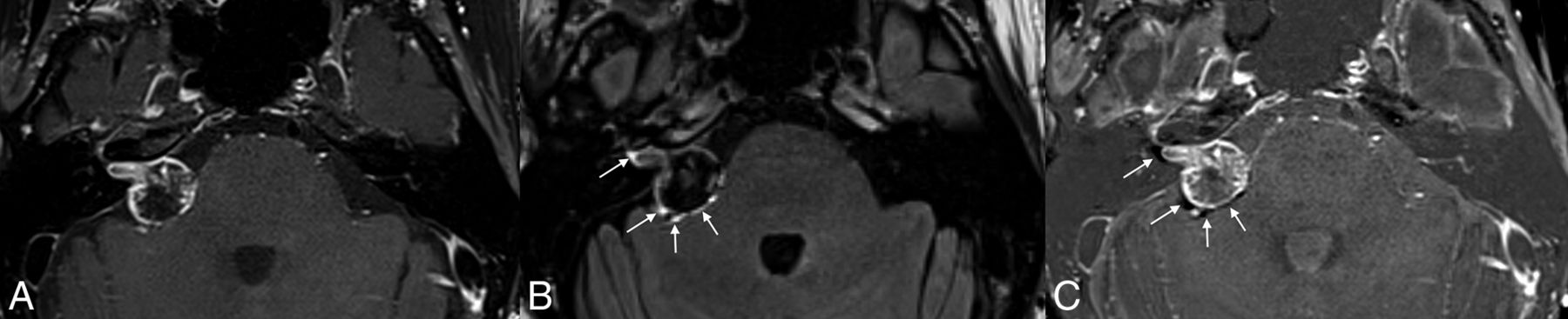
- FIG 1.
Example of a peritumoral halo on postcontrast FLAIR images. Postcontrast fat-saturated T1-weighted image (A) demonstrates a vestibular schwannoma extending from the right IAC into the CPA. Postcontrast FLAIR image (B) shows a discontinuous peritumoral hyperintense halo that extends beyond the tumor margins on postcontrast T1-weighted images (arrows). Corresponding subtraction FLAIR image (C) confirms the presence of a halo (arrows).
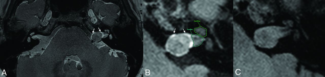
- FIG 2.
Multiple findings of possible gadolinium leakage from a vestibular schwannoma. Fused axial T2 SPACE and postcontrast T2 FLAIR image (A) shows a vestibular schwannoma centered in the left porus acusticus with a peritumoral halo (arrows) that extends past the solid tumor borders. Hyperintense signal was observed in the ipsilateral IAC fundus and asymmetrically elevated signal in the ipsilateral cochlea on the postcontrast T2 FLAIR image (B). Noncontrast FLAIR image on a prior study (C) does not show abnormal peritumoral signal, indicating that this finding should not represent peritumoral edema.


Tables
Sizes and incidence of multiple imaging findings
Findings Size Vestibular schwannoma (mean) (mm) 5.8 (SD, 3.1 ) Fundal cleft (mean) (mm) 2.3 (SD, 1.8) Peritumoral halo (mean) (mm) 1.0 (SD, 0.2) Incidence Peritumoral halo (No.) (%) 18/20 (90%) Hyperintense signal in fundal cleft (No.) (%) 13/16 (82.3%) Elevated intracochlear signal on postcontrast FLAIR (No.) (%) 10/20 (50.0%)


Jump to section
Related Articles
Cited By...
- Peritumoral Hyperintense Signal on Postcontrast FLAIR Images Surrounding Vestibular Schwannomas Following Stereotactic Radiosurgery
- Vestibular Schwannoma-Related Increased Labyrinthine Postgadolinium 3D-FLAIR Signal Intensity and Association with Hearing Impairment
- The "Outline Sign": Thin Hyperenhancing Perimeter as an MR Imaging Feature of Meningioma. A Useful Tool in the Temporal Bone Region for Differentiating Meningiomas from Schwannomas and Paragangliomas
- Imaging Findings Post-Stereotactic Radiosurgery for Vestibular Schwannoma: A Primer for the Radiologist