Advertisement

Main menu

  • Home
  • Content
    • Current Issue
    • Accepted Manuscripts
    • Article Preview
    • Past Issue Archive
    • Video Articles
    • AJNR Case Collection
    • Case of the Week Archive
    • Case of the Month Archive
    • Classic Case Archive
  • Special Collections
    • AJNR Awards
    • Low-Field MRI
    • Alzheimer Disease
    • ASNR Foundation Special Collection
    • Photon-Counting CT
    • View All
  • Multimedia
    • AJNR Podcasts
    • AJNR SCANtastic
    • Trainee Corner
    • MRI Safety Corner
    • Imaging Protocols
  • For Authors
    • Submit a Manuscript
    • Submit a Video Article
    • Submit an eLetter to the Editor/Response
    • Manuscript Submission Guidelines
    • Statistical Tips
    • Fast Publishing of Accepted Manuscripts
    • Graphical Abstract Preparation
    • Imaging Protocol Submission
    • Author Policies
  • About Us
    • About AJNR
    • Editorial Board
    • Editorial Board Alumni
  • More
    • Become a Reviewer/Academy of Reviewers
    • Subscribers
    • Permissions
    • Alerts
    • Feedback
    • Advertisers
    • ASNR Home

User menu

  • Alerts
  • Log in

Search

  • Advanced search
American Journal of Neuroradiology

American Journal of Neuroradiology

ASHNR American Society of Functional Neuroradiology ASHNR American Society of Pediatric Neuroradiology ASSR
  • Alerts
  • Log in

Advanced Search

  • Home
  • Content
    • Current Issue
    • Accepted Manuscripts
    • Article Preview
    • Past Issue Archive
    • Video Articles
    • AJNR Case Collection
    • Case of the Week Archive
    • Case of the Month Archive
    • Classic Case Archive
  • Special Collections
    • AJNR Awards
    • Low-Field MRI
    • Alzheimer Disease
    • ASNR Foundation Special Collection
    • Photon-Counting CT
    • View All
  • Multimedia
    • AJNR Podcasts
    • AJNR SCANtastic
    • Trainee Corner
    • MRI Safety Corner
    • Imaging Protocols
  • For Authors
    • Submit a Manuscript
    • Submit a Video Article
    • Submit an eLetter to the Editor/Response
    • Manuscript Submission Guidelines
    • Statistical Tips
    • Fast Publishing of Accepted Manuscripts
    • Graphical Abstract Preparation
    • Imaging Protocol Submission
    • Author Policies
  • About Us
    • About AJNR
    • Editorial Board
    • Editorial Board Alumni
  • More
    • Become a Reviewer/Academy of Reviewers
    • Subscribers
    • Permissions
    • Alerts
    • Feedback
    • Advertisers
    • ASNR Home
  • Follow AJNR on Twitter
  • Visit AJNR on Facebook
  • Follow AJNR on Instagram
  • Join AJNR on LinkedIn
  • RSS Feeds

AJNR Awards, New Junior Editors, and more. Read the latest AJNR updates

Research ArticleHead & Neck

Differentiation of Skull Base Chondrosarcomas, Chordomas, and Metastases: Utility of DWI and Dynamic Contrast-Enhanced Perfusion MR Imaging

Y. Ota, E. Liao, A.A. Capizzano, A. Baba, R. Kurokawa, M. Kurokawa and A. Srinivasan
American Journal of Neuroradiology September 2022, 43 (9) 1325-1332; DOI: https://doi.org/10.3174/ajnr.A7607
Y. Ota
aFrom the Division of Neuroradiology (Y.O., E.L., A.A.C., A.B., R.K., M.K., A.S.), Department of Radiology, University of Michigan, Ann Arbor, Michigan
  • Find this author on Google Scholar
  • Find this author on PubMed
  • Search for this author on this site
  • ORCID record for Y. Ota
E. Liao
aFrom the Division of Neuroradiology (Y.O., E.L., A.A.C., A.B., R.K., M.K., A.S.), Department of Radiology, University of Michigan, Ann Arbor, Michigan
  • Find this author on Google Scholar
  • Find this author on PubMed
  • Search for this author on this site
  • ORCID record for E. Liao
A.A. Capizzano
aFrom the Division of Neuroradiology (Y.O., E.L., A.A.C., A.B., R.K., M.K., A.S.), Department of Radiology, University of Michigan, Ann Arbor, Michigan
  • Find this author on Google Scholar
  • Find this author on PubMed
  • Search for this author on this site
  • ORCID record for A.A. Capizzano
A. Baba
aFrom the Division of Neuroradiology (Y.O., E.L., A.A.C., A.B., R.K., M.K., A.S.), Department of Radiology, University of Michigan, Ann Arbor, Michigan
bDepartment of Radiology (A.B.), Jikei University School of Medicine Ringgold standard institution, Tokyo, Japan
  • Find this author on Google Scholar
  • Find this author on PubMed
  • Search for this author on this site
  • ORCID record for A. Baba
R. Kurokawa
aFrom the Division of Neuroradiology (Y.O., E.L., A.A.C., A.B., R.K., M.K., A.S.), Department of Radiology, University of Michigan, Ann Arbor, Michigan
cDepartment of Radiology (R.K.), The University of Tokyo Hospital, Tokyo, Japan
  • Find this author on Google Scholar
  • Find this author on PubMed
  • Search for this author on this site
  • ORCID record for R. Kurokawa
M. Kurokawa
aFrom the Division of Neuroradiology (Y.O., E.L., A.A.C., A.B., R.K., M.K., A.S.), Department of Radiology, University of Michigan, Ann Arbor, Michigan
dDepartment of Radiology (M.K.), Tokyo Metropolitan Cancer and Infectious Diseases Center Komagome Hospital Ringgold standard institution, Bunkyo-ku, Japan
  • Find this author on Google Scholar
  • Find this author on PubMed
  • Search for this author on this site
  • ORCID record for M. Kurokawa
A. Srinivasan
aFrom the Division of Neuroradiology (Y.O., E.L., A.A.C., A.B., R.K., M.K., A.S.), Department of Radiology, University of Michigan, Ann Arbor, Michigan
  • Find this author on Google Scholar
  • Find this author on PubMed
  • Search for this author on this site
  • ORCID record for A. Srinivasan
  • Article
  • Figures & Data
  • Info & Metrics
  • Responses
  • References
  • PDF
Loading

Article Figures & Data

Figures

  • Tables
  • FIG 1.
    • Download figure
    • Open in new tab
    • Download powerpoint
    FIG 1.

    Box-and-whisker plots of DWI and DCE-MR imaging parameters with the Kruskal-Wallis H test and post hoc test with the Bonferroni correction are shown. The boundaries of boxes represent 25th and 75th percentiles, and the lines in boxes indicate medians.
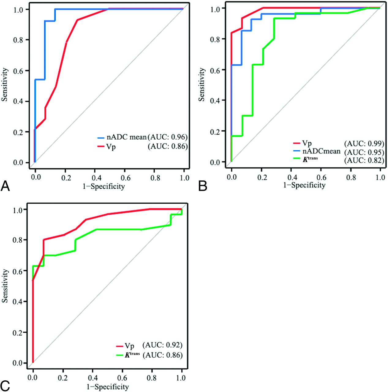
  • FIG 2.
    • Download figure
    • Open in new tab
    • Download powerpoint
    FIG 2.

    Receiver operating characteristic curves of chondrosarcoma versus chordoma (A), chondrosarcoma versus metastasis (B), and chordoma versus metastasis (C) are shown.
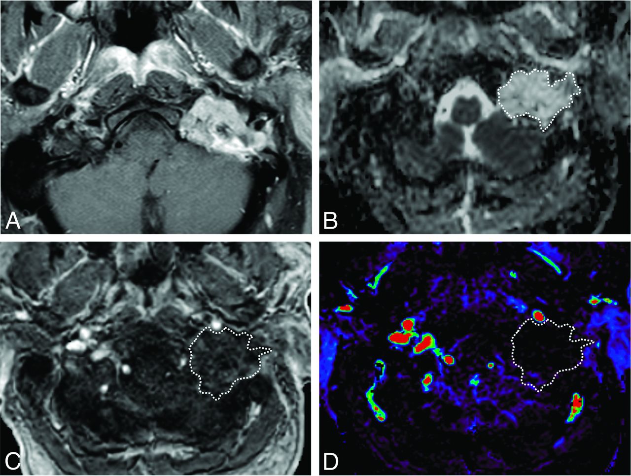
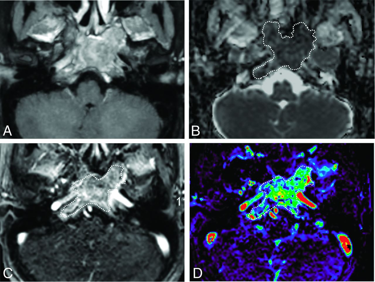
  • FIG 3.
    • Download figure
    • Open in new tab
    • Download powerpoint
    FIG 3.

    A 56-year-old man with a chondrosarcoma in the left petrous apex. A, Axial fat-saturated postcontrast T1-weighted image shows a heterogeneously enhancing mass in the left petrous apex. B, An ROI was placed on the ADC map, and the nADCmean was calculated. Mean ADC and ADC values in the medulla were 2.05, and 0.80 × 10−3 mm2/s, respectively. The nADCmean was 2.56. C, An ROI was placed on the permeability map, and Vp, Ktrans, and Ve were calculated. D, Vp is 0.01.
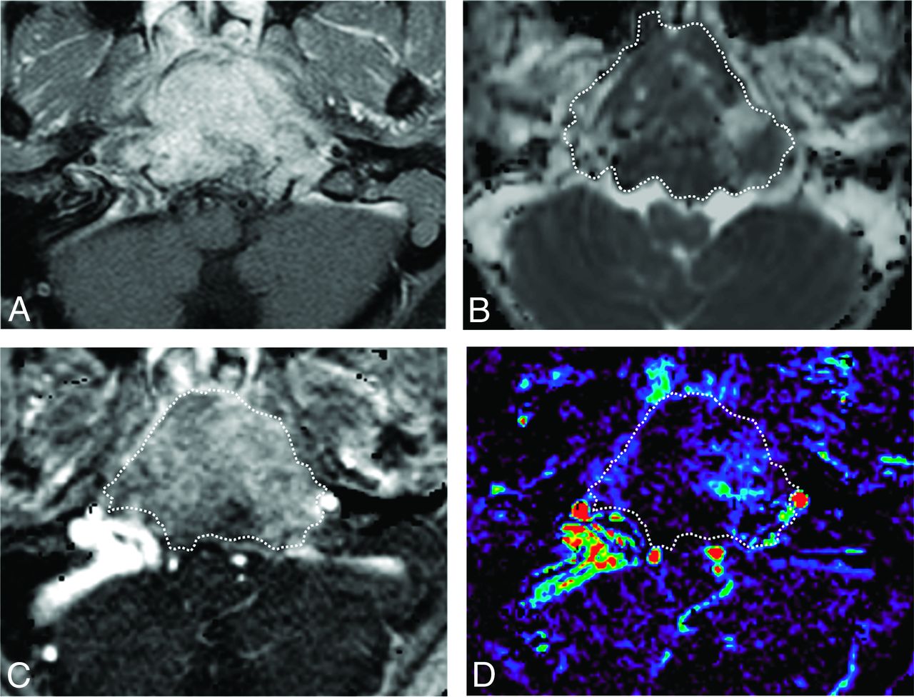
  • FIG 4.
    • Download figure
    • Open in new tab
    • Download powerpoint
    FIG 4.

    A 16-year-old boy with a chordoma in the clivus. A, Axial fat-saturated postcontrast T1-weighted image shows a heterogeneously enhancing mass in the clivus. B, An ROI was placed on the ADC map, and the nADCmean was 1.5. C, An ROI was placed on the permeability map, and Vp, Ktrans, and Ve were calculated. D, Vp is 0.08.

  • FIG 5.
    • Download figure
    • Open in new tab
    • Download powerpoint
    FIG 5.

    A 39-year-old woman with a metastatic breast cancer lesion in the clivus. A, Axial fat-saturated postcontrast T1-weighted image shows a heterogeneously enhancing mass in the clivus. B, An ROI was placed on the ADC map, and the nADCmean was 0.97. C, An ROI was placed on the permeability map, and Vp, Ktrans, and Ve were calculated. D, Vp showed 0.23.

Tables

  • Figures
    • View popup
    Table 1:

    Patient demographicsa

    ChondrosarcomaChordomaMetastasis
    No. of patients151430
    Sex (male/total)9/156/1411/30
    Age (yr)62 (46–70)47 (32–58)61 (47–70)
    Maximum axial diameter (mm)29.5 (24–36)33 (24–40)18.5 (15–23)
    Primary cancer typeNANA11 Breast, 6 lung, 6 malignant melanoma,4 head and neck, 2 sarcoma, 1 kidney
    • Note:—NA indicates not applicable.

    • ↵a Values are presented as the median (IQR).

    • View popup
    Table 2:

    Conventional imaging characteristics of chondrosarcomas, chordomas, and metastases

    ChondrosarcomaChordomaMetastasisP Value
    Main location (petrous bone:clivus)9:61:135:25Pa = .08Pb = .14Pc = .65
    Cystic/necrotic changes8/157/1410/30Pa = 1.0Pb = .22Pc = .33
    T2 hyperintensity or iso-/hypointensity relative to the brain parenchyma (T2 hyperintensity/total)10/159/149/30Pa = 1.0Pb = .027Pc = .049
    Enhancement patterns (heterogeneous/total)15/1514/1426/30Pa = 1.0Pb = .29Pc = .29
    Other metastatic lesions in the head and neckNANA5/30NA
    • Note:—Pa is obtained from a comparison between chondrosarcoma and chordoma, Pb is from a comparison between chondrosarcoma and metastasis, and Pc is from a comparison between chordoma versus metastasis.

    • View popup
    Table 3:

    DWI and DCE-MR imaging parameters of chondrosarcoma, chordoma, and metastasis using the Kruskal-Wallis H test and the post hoc test with Bonferroni correctiona

    ChondrosarcomaChordomaMetastasisP ValueaP Valueb
    Chondrosarcoma vs ChordomaChondrosarcoma vs MetastasisChordoma vs Metastasis
    nADCmean2.44 (2.27–2.71)1.30 (1.07–1.73)1.39 (1.21–1.70)<.001<.001<.0011.0
    Vp0.015 (0.01–0.028)0.045 (0.04–0.068)0.12 (0.09–0.14)<.001.003<.001<.001
    Ve0.25 (0.15–0.31)0.35 (0.30–0.40)0.48 (0.34–0.63).007.14.022.17
    Ktrans (minute−1)0.08 (0.06–0.17)0.16 (0.13–0.20)0.39 (0.20–0.63)<.001.16.002.002
    • Note:—Pa is from Kruskal–Wallis H test. Pb is adjusted for pair-wise comparison by Bonferroni correction.

    • ↵a Numbers in parentheses indicate interquartile range.

    • View popup
    Table 4:

    Diagnostic performance of DCE-MR imaging parameters for each tumor comparisona

    ParametersChondrosarcoma vs ChordomaChondrosarcoma vs MetastasisChordoma vs Metastasis
    nADCmeanVpnADCmeanVpKtrans (minute−1)VpKtrans (minute−1)
    Cutoff1.990.032.090.060.120.090.30
    Sensitivity1.0 (0.68–1)0.93 (0.66–0.99)0.93 (0.76–0.99)0.93 (0.78–0.99)0.93 (0.78–0.99)0.80 (0.61–0.92)0.60 (0.41–0.77)
    Specificity0.86 (0.57–0.98)0.71 (0.42–0.92)0.87 (0.60–0.98)0.93 (0.66–0.9)0.71 (0.42–0.92)0.93 (0.66–1)1.0 (0.68–1)
    PPV0.88 (0.62–0.98)0.77 (0.50–0.93)0.93 (0.76–0.99)0.97 (0.82–1)0.88 (0.71–0.97)0.96 (0.80–1)1.0 (0.74–1)
    NPV1.0 (0.64–1)0.91 (0.59–0.99)0.87 (0.60–0.98)0.87 (0.60–0.98)0.83 (0.52–0.98)0.68 (0.43–0.87)0.54 (0.33–0.73)
    Accuracy0.93 (0.78–0.99)0.82 (0.63–0.94)0.91 (0.77–0.97)0.93 (0.81–0.99)0.86 (0.73–0.95)0.84 (0.70–0.93)0.73 (0.57–0.85)
    AUC0.96 (0.90–1)0.86 (0.72–1)0.95 (0.88–1)0.99 (0.96–1)0.82 (0.67–0.98)0.92 (0.84– 1)0.82 (0.70–0.95)
    • Note:—PPV indicates positive predictive value; NPV, negative predictive value.

    • ↵a Numbers in parentheses represent 95% CIs.

PreviousNext
Back to top

In this issue

American Journal of Neuroradiology: 43 (9)
American Journal of Neuroradiology
Vol. 43, Issue 9
1 Sep 2022
  • Table of Contents
  • Index by author
  • Complete Issue (PDF)
Advertisement
Print
Download PDF
Email Article

Thank you for your interest in spreading the word on American Journal of Neuroradiology.

NOTE: We only request your email address so that the person you are recommending the page to knows that you wanted them to see it, and that it is not junk mail. We do not capture any email address.

Enter multiple addresses on separate lines or separate them with commas.
Differentiation of Skull Base Chondrosarcomas, Chordomas, and Metastases: Utility of DWI and Dynamic Contrast-Enhanced Perfusion MR Imaging
(Your Name) has sent you a message from American Journal of Neuroradiology
(Your Name) thought you would like to see the American Journal of Neuroradiology web site.
CAPTCHA
This question is for testing whether or not you are a human visitor and to prevent automated spam submissions.
Cite this article
Y. Ota, E. Liao, A.A. Capizzano, A. Baba, R. Kurokawa, M. Kurokawa, A. Srinivasan
Differentiation of Skull Base Chondrosarcomas, Chordomas, and Metastases: Utility of DWI and Dynamic Contrast-Enhanced Perfusion MR Imaging
American Journal of Neuroradiology Sep 2022, 43 (9) 1325-1332; DOI: 10.3174/ajnr.A7607

Citation Manager Formats

  • BibTeX
  • Bookends
  • EasyBib
  • EndNote (tagged)
  • EndNote 8 (xml)
  • Medlars
  • Mendeley
  • Papers
  • RefWorks Tagged
  • Ref Manager
  • RIS
  • Zotero
0 Responses
Respond to this article
Share
Bookmark this article
Skull Base Chordomas, Chondrosarcomas, Metastases
Y. Ota, E. Liao, A.A. Capizzano, A. Baba, R. Kurokawa, M. Kurokawa, A. Srinivasan
American Journal of Neuroradiology Sep 2022, 43 (9) 1325-1332; DOI: 10.3174/ajnr.A7607
del.icio.us logo Twitter logo Facebook logo Mendeley logo
  • Tweet Widget
  • Facebook Like
  • Google Plus One
Purchase

Jump to section

  • Article
    • Abstract
    • ABBREVIATIONS:
    • MATERIALS AND METHODS
    • RESULTS
    • DISCUSSION
    • CONCLUSIONS
    • Footnotes
    • References
  • Figures & Data
  • Info & Metrics
  • Responses
  • References
  • PDF

Related Articles

  • No related articles found.
  • PubMed
  • Google Scholar

Cited By...

  • No citing articles found.
  • Crossref (8)
  • Google Scholar

This article has been cited by the following articles in journals that are participating in Crossref Cited-by Linking.

  • DWI and Dynamic Contrast-enhanced Perfusion MRI for Differentiation of Common Skull Base Tumors
    Nahyun Jo
    Radiology: Imaging Cancer 2023 5 2
  • Evaluation of cranial nerve involvement in chordomas and chondrosarcomas: a retrospective imaging study
    Tatsushi Oura, Taro Shimono, Daisuke Horiuchi, Takeo Goto, Hirotaka Takita, Taro Tsukamoto, Hiroyuki Tatekawa, Daiju Ueda, Shu Matsushita, Yasuhito Mitsuyama, Natsuko Atsukawa, Yukio Miki
    Neuroradiology 2024 66 6
  • Freiburg Neuropathology Case Conference
    E. Wogram, F. Schlunk, M. J. Shah, M. Prinz, H. Urbach, D. Erny, C. A. Taschner
    Clinical Neuroradiology 2023 33 2
  • Imaging of the skull base and orbital tumors
    Masafumi Sakai, Takashi Hiyama, Hirofumi Kuno, Tatsushi Kobayashi, Takahito Nakajima
    Japanese Journal of Radiology 2025 43 2
  • Low-dose GBCA administration for brain tumour dynamic contrast enhanced MRI: a feasibility study
    Daniel Lewis, Ka-Loh Li, Mueez Waqar, David J. Coope, Omar N. Pathmanaban, Andrew T. King, Ibrahim Djoukhadar, Sha Zhao, Timothy F. Cootes, Alan Jackson, Xiaoping Zhu
    Scientific Reports 2024 14 1
  • Neuroradiology of Brain Tumors
    Fabio Maria Triulzi
    2023
  • Precise differentiation between jugular foramen paragangliomas and metastases: utility of diffusion-weighted and dynamic contrast-enhanced magnetic resonance imaging
    Yoshiaki Ota, Guillaume Curaudeau, Eric Liao, Jayapalli Bapuraj, Akira Baba, Gaurang Shah, Ashok Srinivasan
    Neuroradiology 2023 65 4
  • Skull base “intrinsic” bony mass lesions: conventional, diffusion and perfusion imaging with a proposed imaging approach
    Lamya Eissa, Rim Aly Bastawi
    Egyptian Journal of Radiology and Nuclear Medicine 2024 55 1

More in this TOC Section

Head & Neck

  • NI-RADS for HEAD&NECK Cancer Recurrence
  • Chondrosarcoma vs Synovial Chondromatosis: Imaging
  • WHO Classification Update: Nasal&Skull Base Tumors
Show more Head & Neck

Functional

  • Kurtosis and Epileptogenic Tubers: A Pilot Study
  • Glutaric Aciduria Type 1: DK vs. Conventional MRI
  • Multiparametric MRI in PEDS Pontine Glioma
Show more Functional

Similar Articles

Advertisement

Indexed Content

  • Current Issue
  • Accepted Manuscripts
  • Article Preview
  • Past Issues
  • Editorials
  • Editor's Choice
  • Fellows' Journal Club
  • Letters to the Editor
  • Video Articles

Cases

  • Case Collection
  • Archive - Case of the Week
  • Archive - Case of the Month
  • Archive - Classic Case

More from AJNR

  • Trainee Corner
  • Imaging Protocols
  • MRI Safety Corner

Multimedia

  • AJNR Podcasts
  • AJNR Scantastics

Resources

  • Turnaround Time
  • Submit a Manuscript
  • Submit a Video Article
  • Submit an eLetter to the Editor/Response
  • Manuscript Submission Guidelines
  • Statistical Tips
  • Fast Publishing of Accepted Manuscripts
  • Graphical Abstract Preparation
  • Imaging Protocol Submission
  • Evidence-Based Medicine Level Guide
  • Publishing Checklists
  • Author Policies
  • Become a Reviewer/Academy of Reviewers
  • News and Updates

About Us

  • About AJNR
  • Editorial Board
  • Editorial Board Alumni
  • Alerts
  • Permissions
  • Not an AJNR Subscriber? Join Now
  • Advertise with Us
  • Librarian Resources
  • Feedback
  • Terms and Conditions
  • AJNR Editorial Board Alumni

American Society of Neuroradiology

  • Not an ASNR Member? Join Now

© 2025 by the American Society of Neuroradiology All rights, including for text and data mining, AI training, and similar technologies, are reserved.
Print ISSN: 0195-6108 Online ISSN: 1936-959X

Powered by HighWire