Advertisement

Main menu

  • Home
  • Content
    • Current Issue
    • Accepted Manuscripts
    • Article Preview
    • Past Issue Archive
    • Video Articles
    • AJNR Case Collection
    • Case of the Week Archive
    • Case of the Month Archive
    • Classic Case Archive
  • Special Collections
    • Advancing NeuroMRI with High-Relaxivity Contrast Agents
    • AJNR Awards
    • Low-Field MRI
    • Alzheimer Disease
    • ASNR Foundation Special Collection
    • Photon-Counting CT
    • View All
  • Multimedia
    • AJNR Podcasts
    • AJNR SCANtastic
    • Trainee Corner
    • MRI Safety Corner
    • Imaging Protocols
  • For Authors
    • Submit a Manuscript
    • Submit a Video Article
    • Submit an eLetter to the Editor/Response
    • Manuscript Submission Guidelines
    • Statistical Tips
    • Fast Publishing of Accepted Manuscripts
    • Graphical Abstract Preparation
    • Imaging Protocol Submission
    • Author Policies
  • About Us
    • About AJNR
    • Editorial Board
    • Editorial Board Alumni
  • More
    • Become a Reviewer/Academy of Reviewers
    • Subscribers
    • Permissions
    • Alerts
    • Feedback
    • Advertisers
    • ASNR Home

User menu

  • Alerts
  • Log in

Search

  • Advanced search
American Journal of Neuroradiology

American Journal of Neuroradiology

ASHNR American Society of Functional Neuroradiology ASHNR American Society of Pediatric Neuroradiology ASSR
  • Alerts
  • Log in

Advanced Search

  • Home
  • Content
    • Current Issue
    • Accepted Manuscripts
    • Article Preview
    • Past Issue Archive
    • Video Articles
    • AJNR Case Collection
    • Case of the Week Archive
    • Case of the Month Archive
    • Classic Case Archive
  • Special Collections
    • Advancing NeuroMRI with High-Relaxivity Contrast Agents
    • AJNR Awards
    • Low-Field MRI
    • Alzheimer Disease
    • ASNR Foundation Special Collection
    • Photon-Counting CT
    • View All
  • Multimedia
    • AJNR Podcasts
    • AJNR SCANtastic
    • Trainee Corner
    • MRI Safety Corner
    • Imaging Protocols
  • For Authors
    • Submit a Manuscript
    • Submit a Video Article
    • Submit an eLetter to the Editor/Response
    • Manuscript Submission Guidelines
    • Statistical Tips
    • Fast Publishing of Accepted Manuscripts
    • Graphical Abstract Preparation
    • Imaging Protocol Submission
    • Author Policies
  • About Us
    • About AJNR
    • Editorial Board
    • Editorial Board Alumni
  • More
    • Become a Reviewer/Academy of Reviewers
    • Subscribers
    • Permissions
    • Alerts
    • Feedback
    • Advertisers
    • ASNR Home
  • Follow AJNR on Twitter
  • Visit AJNR on Facebook
  • Follow AJNR on Instagram
  • Join AJNR on LinkedIn
  • RSS Feeds

AJNR Awards, New Junior Editors, and more. Read the latest AJNR updates


Improved Turnaround Times | Median time to first decision: 12 days

Research ArticleHead and Neck Imaging

Normalized Parameters of Dynamic Contrast-Enhanced Perfusion MRI and DWI-ADC for Differentiation between Posttreatment Changes and Recurrence in Head and Neck Cancer

A. Baba, R. Kurokawa, E. Rawie, M. Kurokawa, Y. Ota and A. Srinivasan
American Journal of Neuroradiology August 2022, 43 (8) 1184-1189; DOI: https://doi.org/10.3174/ajnr.A7567
A. Baba
aFrom the Division of Neuroradiology (A.B., R.K., M.K., Y.O., A.S.), Department of Radiology, University of Michigan, Ann Arbor, Michigan
  • Find this author on Google Scholar
  • Find this author on PubMed
  • Search for this author on this site
  • ORCID record for A. Baba
R. Kurokawa
aFrom the Division of Neuroradiology (A.B., R.K., M.K., Y.O., A.S.), Department of Radiology, University of Michigan, Ann Arbor, Michigan
  • Find this author on Google Scholar
  • Find this author on PubMed
  • Search for this author on this site
  • ORCID record for R. Kurokawa
E. Rawie
bDepartment of Radiology (E.R.), Brooke Army Medical Center, San Antonio, Texas
  • Find this author on Google Scholar
  • Find this author on PubMed
  • Search for this author on this site
  • ORCID record for E. Rawie
M. Kurokawa
aFrom the Division of Neuroradiology (A.B., R.K., M.K., Y.O., A.S.), Department of Radiology, University of Michigan, Ann Arbor, Michigan
  • Find this author on Google Scholar
  • Find this author on PubMed
  • Search for this author on this site
  • ORCID record for M. Kurokawa
Y. Ota
aFrom the Division of Neuroradiology (A.B., R.K., M.K., Y.O., A.S.), Department of Radiology, University of Michigan, Ann Arbor, Michigan
  • Find this author on Google Scholar
  • Find this author on PubMed
  • Search for this author on this site
  • ORCID record for Y. Ota
A. Srinivasan
aFrom the Division of Neuroradiology (A.B., R.K., M.K., Y.O., A.S.), Department of Radiology, University of Michigan, Ann Arbor, Michigan
  • Find this author on Google Scholar
  • Find this author on PubMed
  • Search for this author on this site
  • ORCID record for A. Srinivasan
  • Article
  • Figures & Data
  • Supplemental
  • Info & Metrics
  • Responses
  • References
  • PDF
Loading

Article Figures & Data

Figures

  • Tables
  • FIG 1.
    • Download figure
    • Open in new tab
    • Download powerpoint
    FIG 1.

    A case of head and neck cancer recurrence. A 72-year-old man after an operation and radiation therapy for squamous cell carcinoma of the floor of the mouth. The left side of the floor of the mouth shows some high signal intensity (arrows) on T2WI (A) and mild enhancement (dotted arrows) on postcontrast fat-suppressed T1WI (B). The ADC map (C) shows low signal (arrowheads), with an nADCmean of 1.38. DCE-MR imaging (D, Vp; E, Ve; F, Ktrans) shows increased parameters (thick arrows), with an nVp of 7.33, nVe of 3.93, and nKtrans of 5.44.

  • FIG 2.
    • Download figure
    • Open in new tab
    • Download powerpoint
    FIG 2.

    Benign posttreatment change of head and neck cancer in a 64-year-old woman after surgery and radiation therapy for mucoepidermoid carcinoma of the left parotid gland. The left parotid surgical bed shows low signal intensity (arrow) on T2WI (A) and mild enhancement (dotted arrow) on postcontrast fat-suppressed T1WI (B). The ADC map (C) shows no prominent low signal (arrowhead), with an nADCmean of 1.75. DCE-MR imaging (D, Vp; E, Ve; F, Ktrans) shows no increased parameters (thick arrows) with an nVp of 0.33, nVe of 0.33, and nKtrans of 0.03.
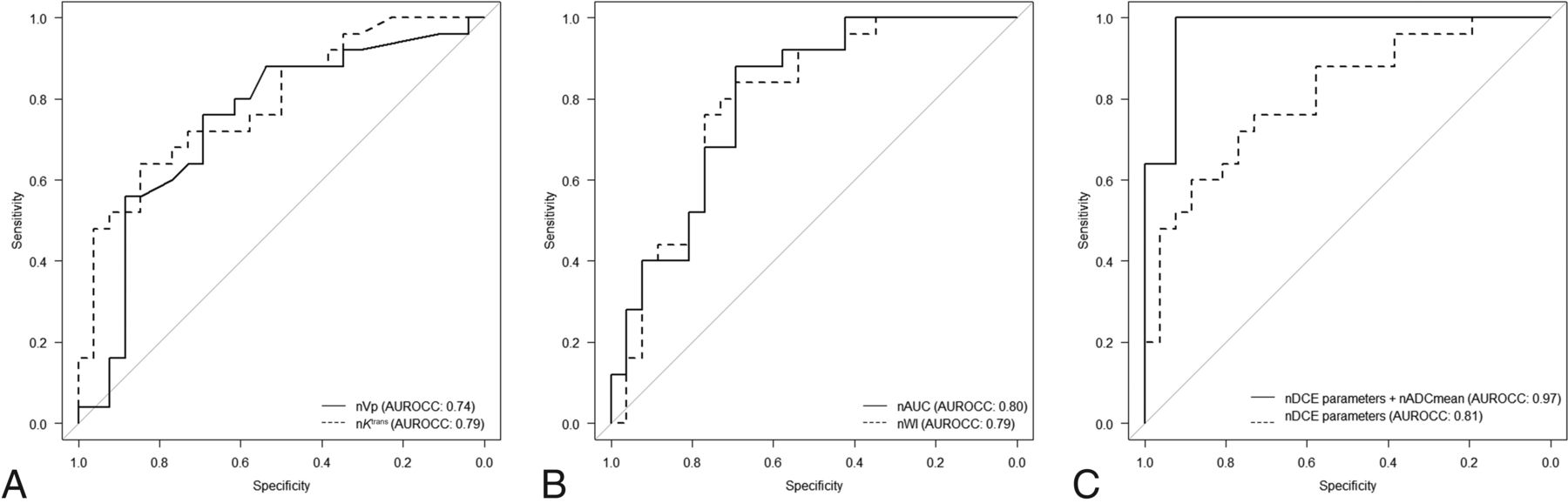
  • FIG 3.
    • Download figure
    • Open in new tab
    • Download powerpoint
    FIG 3.

    Receiver operating characteristic curves. AUROCCs for nVp (solid line) and nKtrans (dashed line) are 0.74 and 0.79, respectively (A). AUROCCs for nAUC (solid line) and nWI (dashed line) are 0.80 and 0.79, respectively (B). AUROCCs for the combination of normalized DCE-MR imaging parameters (nVp, nKtrans, nAUC, and nWI) (dashed line) and the combination of normalized DCE-MR imaging parameters and nADCmean (solid line) are 0.81 and 0.97, respectively (C).

Tables

  • Figures
  • DCE-MR imaging and ADC parameters

    Recurrence (n = 25)
    (Median [Range])
    Benign Posttreatment Change
    (n = 26) (Median [Range])
    P Value
    DCE parameters
     Vp0.09 (0.02–0.22)0.05 (0.01–0.14)
     Reference Vp0.01 (0.01–0.04)0.02 (0.01–0.05)
     nVp7.00 (0.50–17.33)2.02 (0.33–14.33).003a
     Ve0.39 (0.07–0.98)0.28 (0.01–1.08)
     Reference Ve0.14 (0.01–0.26)0.19 (0.07–0.67)
     nVe2.55 (0.32–97.66)1.56 (0.01–6.35).014
     Ktrans0.25 (0.06–1.02)0.13 (0.01–0.39)
     Reference Ktrans0.08 (0.01–0.15)0.11 (0.04–0.33)
     nKtrans3.87 (0.66–101.00)1.27 (0.03–8.00)<.001a
     Kep0.61 (0.39–2.03)0.50 (0.16–2.12)
     Reference Kep0.48 (0.02–1.36)0.55 (0.32–1.06)
     nKep1.45 (0.47–26.17)0.96 (0.28–2.69).009
     AUC28,265 (3523–427,571)11,887 (537–335,025)
     Reference AUC7119 (615–188,345)7890 (3530–185,938)
     nAUC3.34 (1.31–20.81)1.39 (0.01–9.96)<.001a
     WI1.03 (0.29–2102.33)0.70 (0.06–38.00)
     Reference WI0.38 (0.02–696.00)0.33 (0.07–21.24)
     nWI4.13 (0.99–20.17)1.70 (0.07–32.48)<.001a
     WO0.68 (0.01–472.33)0.57 (0.01–18.99)
     Reference WO0.08 (0.01–117.00)0.43 (0.02–27.28)
     nWO3.78 (0.31–292.88)1.35 (0–109.00).016
     ADC value1.01 (0.55–1.30)1.61 (1.02–2.53)
     Reference ADC0.79 (0.56–0.94)0.78 (0.67–0.99)
     nADCmean1.31 (0.77–1.65)2.07 (1.39–3.04)<.001a
    • ↵a Statistically significant.

PreviousNext
Back to top

In this issue

American Journal of Neuroradiology: 43 (8)
American Journal of Neuroradiology
Vol. 43, Issue 8
1 Aug 2022
  • Table of Contents
  • Index by author
  • Complete Issue (PDF)
Advertisement
Print
Download PDF
Email Article

Thank you for your interest in spreading the word on American Journal of Neuroradiology.

NOTE: We only request your email address so that the person you are recommending the page to knows that you wanted them to see it, and that it is not junk mail. We do not capture any email address.

Enter multiple addresses on separate lines or separate them with commas.
Normalized Parameters of Dynamic Contrast-Enhanced Perfusion MRI and DWI-ADC for Differentiation between Posttreatment Changes and Recurrence in Head and Neck Cancer
(Your Name) has sent you a message from American Journal of Neuroradiology
(Your Name) thought you would like to see the American Journal of Neuroradiology web site.
CAPTCHA
This question is for testing whether or not you are a human visitor and to prevent automated spam submissions.
Cite this article
A. Baba, R. Kurokawa, E. Rawie, M. Kurokawa, Y. Ota, A. Srinivasan
Normalized Parameters of Dynamic Contrast-Enhanced Perfusion MRI and DWI-ADC for Differentiation between Posttreatment Changes and Recurrence in Head and Neck Cancer
American Journal of Neuroradiology Aug 2022, 43 (8) 1184-1189; DOI: 10.3174/ajnr.A7567

Citation Manager Formats

  • BibTeX
  • Bookends
  • EasyBib
  • EndNote (tagged)
  • EndNote 8 (xml)
  • Medlars
  • Mendeley
  • Papers
  • RefWorks Tagged
  • Ref Manager
  • RIS
  • Zotero
0 Responses
Respond to this article
Share
Bookmark this article
MRI and DWI-ADC in HEAD&NECK Cancer Recurrence
A. Baba, R. Kurokawa, E. Rawie, M. Kurokawa, Y. Ota, A. Srinivasan
American Journal of Neuroradiology Aug 2022, 43 (8) 1184-1189; DOI: 10.3174/ajnr.A7567
del.icio.us logo Twitter logo Facebook logo Mendeley logo
  • Tweet Widget
  • Facebook Like
  • Google Plus One
Purchase

Jump to section

  • Article
    • Abstract
    • ABBREVIATIONS:
    • MATERIALS AND METHODS
    • RESULTS
    • DISCUSSION
    • CONCLUSIONS
    • Footnotes
    • References
  • Figures & Data
  • Supplemental
  • Info & Metrics
  • Responses
  • References
  • PDF

Related Articles

  • PubMed
  • Google Scholar

Cited By...

  • Time-Saving 3D MR Imaging Protocols with Millimeter and Submillimeter Isotropic Spatial Resolution for Face and Neck Imaging as Implemented at a Single-Site Major Referral Center
  • Crossref
  • Google Scholar

This article has not yet been cited by articles in journals that are participating in Crossref Cited-by Linking.

More in this TOC Section

Head and Neck Imaging

  • Diagnostic Performance of US in Neck Node NIRADS 2
  • Hydrops Herniation into the Semicircular Canals
  • ASL Sensitivity for Head and Neck Paraganglioma
Show more Head and Neck Imaging

Functional

  • Kurtosis and Epileptogenic Tubers: A Pilot Study
  • Glutaric Aciduria Type 1: DK vs. Conventional MRI
  • Multiparametric MRI in PEDS Pontine Glioma
Show more Functional

Similar Articles

Advertisement

Indexed Content

  • Current Issue
  • Accepted Manuscripts
  • Article Preview
  • Past Issues
  • Editorials
  • Editor's Choice
  • Fellows' Journal Club
  • Letters to the Editor
  • Video Articles

Cases

  • Case Collection
  • Archive - Case of the Week
  • Archive - Case of the Month
  • Archive - Classic Case

More from AJNR

  • Trainee Corner
  • Imaging Protocols
  • MRI Safety Corner
  • Book Reviews

Multimedia

  • AJNR Podcasts
  • AJNR Scantastics

Resources

  • Turnaround Time
  • Submit a Manuscript
  • Submit a Video Article
  • Submit an eLetter to the Editor/Response
  • Manuscript Submission Guidelines
  • Statistical Tips
  • Fast Publishing of Accepted Manuscripts
  • Graphical Abstract Preparation
  • Imaging Protocol Submission
  • Evidence-Based Medicine Level Guide
  • Publishing Checklists
  • Author Policies
  • Become a Reviewer/Academy of Reviewers
  • News and Updates

About Us

  • About AJNR
  • Editorial Board
  • Editorial Board Alumni
  • Alerts
  • Permissions
  • Not an AJNR Subscriber? Join Now
  • Advertise with Us
  • Librarian Resources
  • Feedback
  • Terms and Conditions
  • AJNR Editorial Board Alumni

American Society of Neuroradiology

  • Not an ASNR Member? Join Now

© 2025 by the American Society of Neuroradiology All rights, including for text and data mining, AI training, and similar technologies, are reserved.
Print ISSN: 0195-6108 Online ISSN: 1936-959X

Powered by HighWire