
Article Figures & Data
Figures
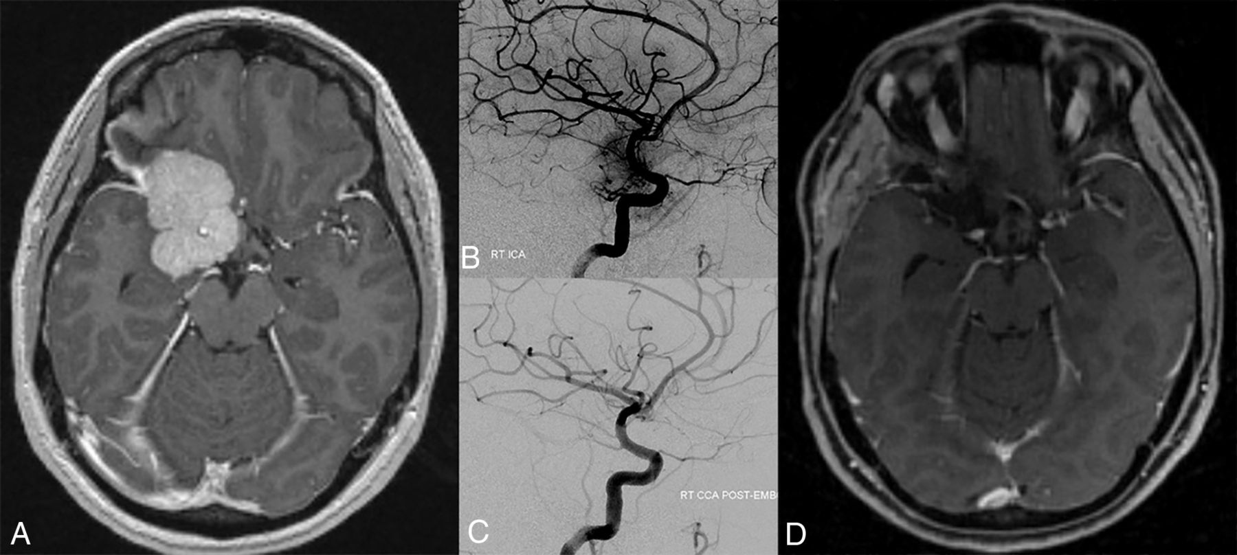
- FIG 1.
A patient with a large right sphenoid wing meningioma with encasement of the ICA visible on a T1-weighted, axial, postcontrast image (A). Right ICA DSA injection in a lateral view (B) demonstrates the supply to the tumor from an enlarged meningohypophyseal trunk. After selective embolization through both the meningohypophyseal trunk and indirectly from the middle meningeal artery, a final right common carotid artery injection in a lateral view (C) demonstrates interval 90% tumor embolization. Complete gross resection of the meningioma is visible on the T1-weighted, axial, postcontrast image (D). RT indicates right; CCA, common carotid artery; EMB, embolization.
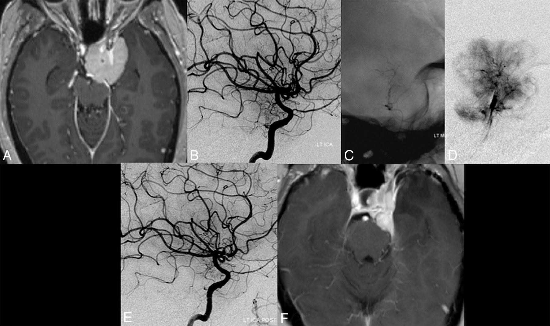
- FIG 2.
A patient with a Meckel cave meningioma also with involvement of the cavernous sinus visible on a T1-weighted, axial, postcontrast image (A). Right ICA DSA injection in lateral view (B) demonstrates the supply to the tumor from an enlarged inferolateral trunk from the left ICA. Inferolateral trunk catheterization is performed with a Headway Duo over a Synchro-14 microguidewire (C and D). MICRO DSA performed through a microcatheter on a lateral view (unsubtracted, E, and subtracted, F) better demonstrates the selective supply to the meningioma. Final lateral view ICA injection (G) demonstrates marked interval reduction of blush in the region of the tumor. Complete gross resection of the meningioma is visible on a T1-weighted, axial, postcontrast image (H). LT indicates left.
- FIG 3.
A patient with a right petroclival meningioma as shown on MR imaging T2 (A) and postcontrast T1 (B). A right ICA injection lateral view DSA (C) demonstrates a large blush supplied by the meningohypophyseal trunk. Selective catheterization and DSA (D) with a Marathon microcatheter (Medtronic), followed by embolization with 45- to 150- and 150- to 250-μm particles. Final right ICA injection, lateral view DSA (E) demonstrates resolution of the previously seen blush. Final CT (F) postsurgical resection demonstrates complete excision with a small amount of retraction injury in the right temporal lobe.
- FIG 4.
A patient with a left anterior clinoid meningioma as shown on postcontrast T1 MR imaging (A). A left ICA injection, lateral view DSA (B), demonstrates a large blush supplied by the meningohypophyseal trunk. Selective catheterization with unsubtracted (C) and subtracted (D) views with a Marathon microcatheter, followed by embolization with 45- to 150-μm particles. A final left ICA injection, lateral view DSA (E) demonstrates 60% embolization of the tumor. A final MR imaging T1 postcontrast (F) postsurgical resection demonstrates partial excision of the mass.


Jump to section
Related Articles
Cited By...
- Transophthalmic Artery Embolization of Anterior Skull Base Meningiomas: Technical Case Series
- Embolization of Posterior Fossa Meningiomas Supplied with Meningohypophyseal Trunk by Using n-BCA and Dual Balloon Protection
- Tumor Embolization via the Meningohypophyseal and Inferolateral Trunk in Patients with Skull Base Tumors Using the Distal Balloon Protection Technique
- Outcomes of Preoperative Transophthalmic Artery Embolization of Meningiomas: A Systematic Review with a Focus on Embolization Agent