Advertisement

Main menu

  • Home
  • Content
    • Current Issue
    • Accepted Manuscripts
    • Article Preview
    • Past Issue Archive
    • Video Articles
    • AJNR Case Collection
    • Case of the Week Archive
    • Case of the Month Archive
    • Classic Case Archive
  • Special Collections
    • AJNR Awards
    • Low-Field MRI
    • Alzheimer Disease
    • ASNR Foundation Special Collection
    • Photon-Counting CT
    • View All
  • Multimedia
    • AJNR Podcasts
    • AJNR SCANtastic
    • Trainee Corner
    • MRI Safety Corner
    • Imaging Protocols
  • For Authors
    • Submit a Manuscript
    • Submit a Video Article
    • Submit an eLetter to the Editor/Response
    • Manuscript Submission Guidelines
    • Statistical Tips
    • Fast Publishing of Accepted Manuscripts
    • Graphical Abstract Preparation
    • Imaging Protocol Submission
    • Author Policies
  • About Us
    • About AJNR
    • Editorial Board
    • Editorial Board Alumni
  • More
    • Become a Reviewer/Academy of Reviewers
    • Subscribers
    • Permissions
    • Alerts
    • Feedback
    • Advertisers
    • ASNR Home

User menu

  • Alerts
  • Log in

Search

  • Advanced search
American Journal of Neuroradiology

American Journal of Neuroradiology

ASHNR American Society of Functional Neuroradiology ASHNR American Society of Pediatric Neuroradiology ASSR
  • Alerts
  • Log in

Advanced Search

  • Home
  • Content
    • Current Issue
    • Accepted Manuscripts
    • Article Preview
    • Past Issue Archive
    • Video Articles
    • AJNR Case Collection
    • Case of the Week Archive
    • Case of the Month Archive
    • Classic Case Archive
  • Special Collections
    • AJNR Awards
    • Low-Field MRI
    • Alzheimer Disease
    • ASNR Foundation Special Collection
    • Photon-Counting CT
    • View All
  • Multimedia
    • AJNR Podcasts
    • AJNR SCANtastic
    • Trainee Corner
    • MRI Safety Corner
    • Imaging Protocols
  • For Authors
    • Submit a Manuscript
    • Submit a Video Article
    • Submit an eLetter to the Editor/Response
    • Manuscript Submission Guidelines
    • Statistical Tips
    • Fast Publishing of Accepted Manuscripts
    • Graphical Abstract Preparation
    • Imaging Protocol Submission
    • Author Policies
  • About Us
    • About AJNR
    • Editorial Board
    • Editorial Board Alumni
  • More
    • Become a Reviewer/Academy of Reviewers
    • Subscribers
    • Permissions
    • Alerts
    • Feedback
    • Advertisers
    • ASNR Home
  • Follow AJNR on Twitter
  • Visit AJNR on Facebook
  • Follow AJNR on Instagram
  • Join AJNR on LinkedIn
  • RSS Feeds

AJNR Awards, New Junior Editors, and more. Read the latest AJNR updates

Research ArticleSpine Imaging and Spine Image-Guided Interventions

Diffuse Calvarial Hyperostosis and Spontaneous Intracranial Hypotension: A Case-Control Study

J.C. Babcock, D.R. Johnson, J.C. Benson, D.K. Kim, P.H. Luetmer, D.P. Shlapak, C.P. Cross, M.P. Johnson, J.K. Cutsforth-Gregory and C.M. Carr
American Journal of Neuroradiology July 2022, 43 (7) 978-983; DOI: https://doi.org/10.3174/ajnr.A7557
J.C. Babcock
aFrom the Department of Radiology, Mayo Clinic, Rochester, Minnesota
  • Find this author on Google Scholar
  • Find this author on PubMed
  • Search for this author on this site
  • ORCID record for J.C. Babcock
D.R. Johnson
aFrom the Department of Radiology, Mayo Clinic, Rochester, Minnesota
  • Find this author on Google Scholar
  • Find this author on PubMed
  • Search for this author on this site
  • ORCID record for D.R. Johnson
J.C. Benson
aFrom the Department of Radiology, Mayo Clinic, Rochester, Minnesota
  • Find this author on Google Scholar
  • Find this author on PubMed
  • Search for this author on this site
  • ORCID record for J.C. Benson
D.K. Kim
aFrom the Department of Radiology, Mayo Clinic, Rochester, Minnesota
  • Find this author on Google Scholar
  • Find this author on PubMed
  • Search for this author on this site
  • ORCID record for D.K. Kim
P.H. Luetmer
aFrom the Department of Radiology, Mayo Clinic, Rochester, Minnesota
  • Find this author on Google Scholar
  • Find this author on PubMed
  • Search for this author on this site
  • ORCID record for P.H. Luetmer
D.P. Shlapak
aFrom the Department of Radiology, Mayo Clinic, Rochester, Minnesota
  • Find this author on Google Scholar
  • Find this author on PubMed
  • Search for this author on this site
  • ORCID record for D.P. Shlapak
C.P. Cross
aFrom the Department of Radiology, Mayo Clinic, Rochester, Minnesota
  • Find this author on Google Scholar
  • Find this author on PubMed
  • Search for this author on this site
  • ORCID record for C.P. Cross
M.P. Johnson
aFrom the Department of Radiology, Mayo Clinic, Rochester, Minnesota
  • Find this author on Google Scholar
  • Find this author on PubMed
  • Search for this author on this site
  • ORCID record for M.P. Johnson
J.K. Cutsforth-Gregory
aFrom the Department of Radiology, Mayo Clinic, Rochester, Minnesota
  • Find this author on Google Scholar
  • Find this author on PubMed
  • Search for this author on this site
  • ORCID record for J.K. Cutsforth-Gregory
C.M. Carr
aFrom the Department of Radiology, Mayo Clinic, Rochester, Minnesota
  • Find this author on Google Scholar
  • Find this author on PubMed
  • Search for this author on this site
  • ORCID record for C.M. Carr
  • Article
  • Figures & Data
  • Info & Metrics
  • Responses
  • References
  • PDF
Loading

Article Figures & Data

Figures

  • Tables
  • FIG 1.
    • Download figure
    • Open in new tab
    • Download powerpoint
    FIG 1.

    Flow chart defining the process of selecting the subject inclusion cohort. EMR indicates electronic medical record.
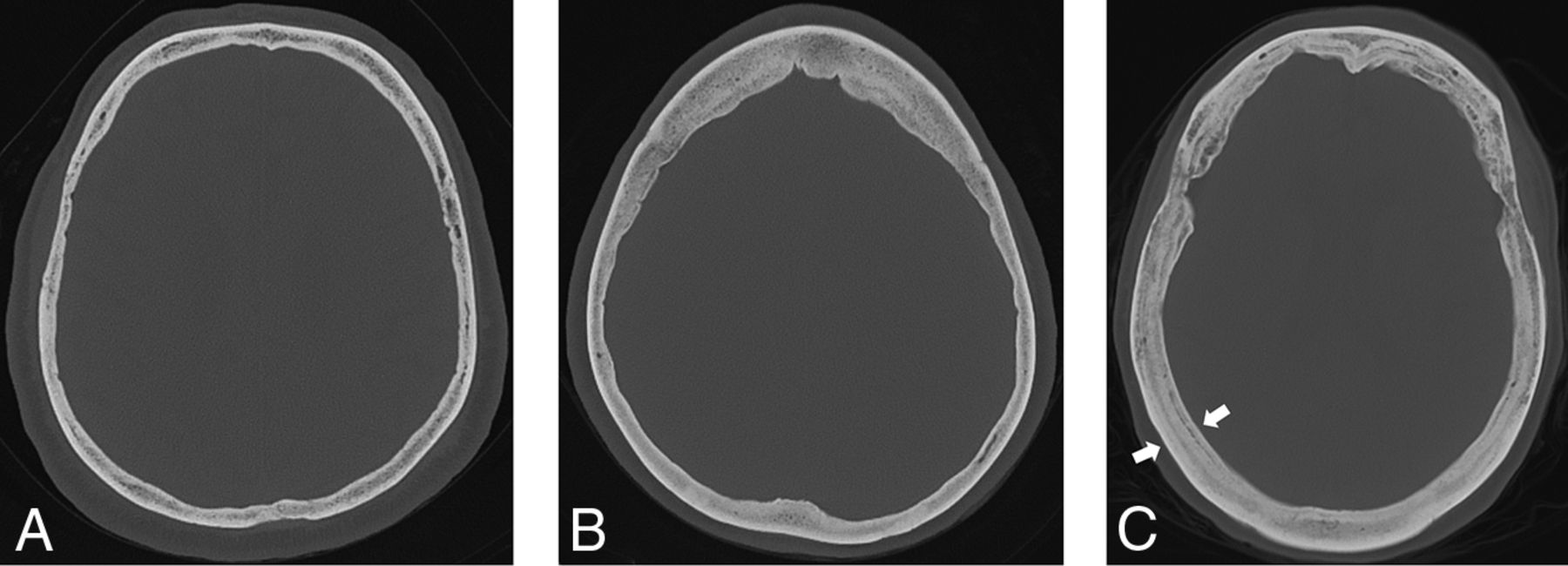
  • FIG 2.
    • Download figure
    • Open in new tab
    • Download powerpoint
    FIG 2.

    Hyperostosis examples. Normal calvarial thickness (A). Axial bone kernel and bone window CT image with a representative example of normal calvarial thickness. Diffuse calvarial hyperostosis (B). Axial bone kernel and bone window CT image demonstrates diffuse thickening of the calvaria. Layered calvarial hyperostosis (C). Axial bone kernel and bone window CT image demonstrates calvarial thickening with discrete enlargement of the inner and outer tables (white arrows), producing a layered appearance.

  • FIG 3.
    • Download figure
    • Open in new tab
    • Download powerpoint
    FIG 3.

    Example of calvarial thickness measurements obtained in the same patient in the axial and coronal planes. Full-field (A) and zoomed (B) axial bone kernel and bone window CT images demonstrate a sample axial thickness measurement obtained 25°–35° off midline. Full-field (C) and zoomed (D) coronal bone kernel and bone window CT images demonstrate a sample coronal thickness measurement obtained 35°–45° off midline.

  • FIG 4.
    • Download figure
    • Open in new tab
    • Download powerpoint
    FIG 4.

    Development of hyperostosis in a 67-year-old man with 2 decades of waxing and waning SIH symptoms with a history of remote CSF leak at C2–C4. A, Axial CT head image at 45 years of age shows qualitatively normal calvarial thickness. B, Sagittal T1-weighted MR image at 48 years of age demonstrates severe brain sag and a suggestion of developing layered hyperostosis. Diffuse pachymeningeal thickening and enhancement are also present (not shown). C, The most recent axial head CT at 67 years of age demonstrates new layered calvarial hyperostosis.

  • FIG 5.
    • Download figure
    • Open in new tab
    • Download powerpoint
    FIG 5.

    A 53-year-old man who developed refractory papilledema and rebound intracranial hypertension following repair of a CSF-venous fistula at T8–T9 at 51 years of age. Rebound intracranial hypertension symptoms began 3 weeks following treatment. Sagittal T1-weighted (A) and axial FLAIR (B) MR images at 29 years of age demonstrate brain sag with normal baseline qualitative calvarial thickness, respectively. Sagittal T1-weighted (C) and axial T2-weighted (D) MR images at 51 years of age demonstrate improvement in brain sag with new posterior globe flattening indicative of papilledema, respectively. E, Preoperative stereotactic CT image before ventriculoperitoneal shunt placement demonstrates new layered calvarial hyperostosis.

Tables

  • Figures
    • View popup
    Table 1:

    Subject and control group demographics

    Subject (n = 166)Control (n = 321)P Value
    Age (mean) (SD)54.5 (12.8)54.4 (12.9).902
    Male sex63 (38.0%)118 (36.8%).796
    CT studies46 (27.7%)87 (27.1%).886
    MR imaging studies120 (72.3%)234 (72.9%)
    • View popup
    Table 2:

    Symptom duration of the SIH cohorta

    PresentAbsentP Value
    Overall hyperostosisbn = 64
    36 months (12–78)
    n = 102
    22 months (8–49)
    .171
    Diffuse hyperostosisn = 11
    36 months (7–80)
    n = 155
    25 months (9–64)
    .798
    Layered hyperostosisn = 53
    36 months(12–77)
    n = 113
    22 months(8–51)
    .197
    • a Numbers in parentheses are interquartile range (25th and 75th percentiles) of symptom duration in months.

    • b Diffuse and layered.

PreviousNext
Back to top

In this issue

American Journal of Neuroradiology: 43 (7)
American Journal of Neuroradiology
Vol. 43, Issue 7
1 Jul 2022
  • Table of Contents
  • Index by author
  • Complete Issue (PDF)
Advertisement
Print
Download PDF
Email Article

Thank you for your interest in spreading the word on American Journal of Neuroradiology.

NOTE: We only request your email address so that the person you are recommending the page to knows that you wanted them to see it, and that it is not junk mail. We do not capture any email address.

Enter multiple addresses on separate lines or separate them with commas.
Diffuse Calvarial Hyperostosis and Spontaneous Intracranial Hypotension: A Case-Control Study
(Your Name) has sent you a message from American Journal of Neuroradiology
(Your Name) thought you would like to see the American Journal of Neuroradiology web site.
CAPTCHA
This question is for testing whether or not you are a human visitor and to prevent automated spam submissions.
Cite this article
J.C. Babcock, D.R. Johnson, J.C. Benson, D.K. Kim, P.H. Luetmer, D.P. Shlapak, C.P. Cross, M.P. Johnson, J.K. Cutsforth-Gregory, C.M. Carr
Diffuse Calvarial Hyperostosis and Spontaneous Intracranial Hypotension: A Case-Control Study
American Journal of Neuroradiology Jul 2022, 43 (7) 978-983; DOI: 10.3174/ajnr.A7557

Citation Manager Formats

  • BibTeX
  • Bookends
  • EasyBib
  • EndNote (tagged)
  • EndNote 8 (xml)
  • Medlars
  • Mendeley
  • Papers
  • RefWorks Tagged
  • Ref Manager
  • RIS
  • Zotero
0 Responses
Respond to this article
Share
Bookmark this article
Calvarial Hyperostosis and CSF Hypotension Study
J.C. Babcock, D.R. Johnson, J.C. Benson, D.K. Kim, P.H. Luetmer, D.P. Shlapak, C.P. Cross, M.P. Johnson, J.K. Cutsforth-Gregory, C.M. Carr
American Journal of Neuroradiology Jul 2022, 43 (7) 978-983; DOI: 10.3174/ajnr.A7557
del.icio.us logo Twitter logo Facebook logo Mendeley logo
  • Tweet Widget
  • Facebook Like
  • Google Plus One
Purchase

Jump to section

  • Article
    • Abstract
    • ABBREVIATION:
    • MATERIALS AND METHODS
    • RESULTS
    • DISCUSSION
    • CONCLUSIONS
    • Footnotes
    • References
  • Figures & Data
  • Info & Metrics
  • Responses
  • References
  • PDF

Related Articles

  • No related articles found.
  • PubMed
  • Google Scholar

Cited By...

  • Spinal CSF Leaks: The Neuroradiologist Transforming Care
  • Likelihood of Discovering a CSF Leak Based on Intracranial MRI Findings in Patients without a Spinal Longitudinal Extradural Collection: A New Probabilistic Scoring System
  • The Monro-Kellie Doctrine: A Review and Call for Revision
  • Crossref (12)
  • Google Scholar

This article has been cited by the following articles in journals that are participating in Crossref Cited-by Linking.

  • The Monro-Kellie Doctrine: A Review and Call for Revision
    J.C. Benson, A.A. Madhavan, J.K. Cutsforth-Gregory, D.R. Johnson, C.M. Carr
    American Journal of Neuroradiology 2023 44 1
  • Likelihood of Discovering a CSF Leak Based on Intracranial MRI Findings in Patients without a Spinal Longitudinal Extradural Collection: A New Probabilistic Scoring System
    John C. Benson, Ajay A. Madhavan, Ian T. Mark, Jeremy K. Cutsforth-Gregory, Waleed Brinjikji, Jared T. Verdoorn
    American Journal of Neuroradiology 2023 44 11
  • Spinal CSF Leaks: The Neuroradiologist Transforming Care
    Mark D. Mamlouk, Andrew L. Callen, Ajay A. Madhavan, Niklas Lützen, Lalani Carlton Jones, Ian T. Mark, Waleed Brinjikji, John C. Benson, Jared T. Verdoorn, D.K. Kim, Timothy J. Amrhein, Linda Gray, William P. Dillon, Marcel M. Maya, Thien J. Huynh, Vinil N. Shah, Tomas Dobrocky, Eike I. Piechowiak, Joseph Levi Chazen, Michael D. Malinzak, Jessica L. Houk, Peter G. Kranz
    American Journal of Neuroradiology 2024 45 11
  • Unusual neuroimaging findings in spontaneous intracranial hypotension
    Andrew L. Callen, William P. Dillon, Vinil N. Shah
    Neuroradiology 2023 65 5
  • Clinical Features and Diagnosis of Spontaneous Intracranial Hypotension
    Jill C. Rau, Jeremy K. Cutsforth-Gregory
    CONTINUUM: Lifelong Learning in Neurology 2025 31 3
  • Clinical Neuroradiology
    Lalani Carlton Jones, Joanna Bladowska, Andrew Martin, Mario Muto, Charles Romanowski, Daniel J. Warren
    2024
  • Démarche décisionnelle devant une anomalie de la voûte du crâne
    François Lafourcade, Magali Raveneau, Annick Sevely, Sofia Patsoura, Hubert Basselerie, Franck Lapègue, Jean Darcourt, Fabrice Bonneville, Nicolas Sans, Marie Faruch-Bilfeld
    Journal d'imagerie diagnostique et interventionnelle 2023 6 5
  • Radiopaedia.org
    Frank Gaillard, Liz Silverstone, Magdi Mahsoub
    2008
  • Radiopaedia.org
    Aqeel Alameer, Rohit Sharma, Frank Gaillard
    2022
  • Suspected Calvarial Hyperostosis Syndrome Causing Different Ophthalmological Signs in Two Young Labrador Retrievers—Case Report
    Andrea Steinmetz, Stefan Kohl
    Veterinary Ophthalmology 2025

More in this TOC Section

  • Management Outcomes For VO Spine Biopsy
  • Cone Beam CT Myelography
  • Post-Procedural Brachial Neuritis Features
Show more SPINE IMAGING AND SPINE IMAGE-GUIDED INTERVENTIONS

Similar Articles

Advertisement

Indexed Content

  • Current Issue
  • Accepted Manuscripts
  • Article Preview
  • Past Issues
  • Editorials
  • Editor's Choice
  • Fellows' Journal Club
  • Letters to the Editor
  • Video Articles

Cases

  • Case Collection
  • Archive - Case of the Week
  • Archive - Case of the Month
  • Archive - Classic Case

More from AJNR

  • Trainee Corner
  • Imaging Protocols
  • MRI Safety Corner
  • Book Reviews

Multimedia

  • AJNR Podcasts
  • AJNR Scantastics

Resources

  • Turnaround Time
  • Submit a Manuscript
  • Submit a Video Article
  • Submit an eLetter to the Editor/Response
  • Manuscript Submission Guidelines
  • Statistical Tips
  • Fast Publishing of Accepted Manuscripts
  • Graphical Abstract Preparation
  • Imaging Protocol Submission
  • Evidence-Based Medicine Level Guide
  • Publishing Checklists
  • Author Policies
  • Become a Reviewer/Academy of Reviewers
  • News and Updates

About Us

  • About AJNR
  • Editorial Board
  • Editorial Board Alumni
  • Alerts
  • Permissions
  • Not an AJNR Subscriber? Join Now
  • Advertise with Us
  • Librarian Resources
  • Feedback
  • Terms and Conditions
  • AJNR Editorial Board Alumni

American Society of Neuroradiology

  • Not an ASNR Member? Join Now

© 2025 by the American Society of Neuroradiology All rights, including for text and data mining, AI training, and similar technologies, are reserved.
Print ISSN: 0195-6108 Online ISSN: 1936-959X

Powered by HighWire