Advertisement

Main menu

  • Home
  • Content
    • Current Issue
    • Accepted Manuscripts
    • Article Preview
    • Past Issue Archive
    • Video Articles
    • AJNR Case Collection
    • Case of the Week Archive
    • Case of the Month Archive
    • Classic Case Archive
  • Special Collections
    • AJNR Awards
    • Low-Field MRI
    • Alzheimer Disease
    • ASNR Foundation Special Collection
    • Photon-Counting CT
    • View All
  • Multimedia
    • AJNR Podcasts
    • AJNR SCANtastic
    • Trainee Corner
    • MRI Safety Corner
    • Imaging Protocols
  • For Authors
    • Submit a Manuscript
    • Submit a Video Article
    • Submit an eLetter to the Editor/Response
    • Manuscript Submission Guidelines
    • Statistical Tips
    • Fast Publishing of Accepted Manuscripts
    • Graphical Abstract Preparation
    • Imaging Protocol Submission
    • Author Policies
  • About Us
    • About AJNR
    • Editorial Board
    • Editorial Board Alumni
  • More
    • Become a Reviewer/Academy of Reviewers
    • Subscribers
    • Permissions
    • Alerts
    • Feedback
    • Advertisers
    • ASNR Home

User menu

  • Alerts
  • Log in

Search

  • Advanced search
American Journal of Neuroradiology

American Journal of Neuroradiology

ASHNR American Society of Functional Neuroradiology ASHNR American Society of Pediatric Neuroradiology ASSR
  • Alerts
  • Log in

Advanced Search

  • Home
  • Content
    • Current Issue
    • Accepted Manuscripts
    • Article Preview
    • Past Issue Archive
    • Video Articles
    • AJNR Case Collection
    • Case of the Week Archive
    • Case of the Month Archive
    • Classic Case Archive
  • Special Collections
    • AJNR Awards
    • Low-Field MRI
    • Alzheimer Disease
    • ASNR Foundation Special Collection
    • Photon-Counting CT
    • View All
  • Multimedia
    • AJNR Podcasts
    • AJNR SCANtastic
    • Trainee Corner
    • MRI Safety Corner
    • Imaging Protocols
  • For Authors
    • Submit a Manuscript
    • Submit a Video Article
    • Submit an eLetter to the Editor/Response
    • Manuscript Submission Guidelines
    • Statistical Tips
    • Fast Publishing of Accepted Manuscripts
    • Graphical Abstract Preparation
    • Imaging Protocol Submission
    • Author Policies
  • About Us
    • About AJNR
    • Editorial Board
    • Editorial Board Alumni
  • More
    • Become a Reviewer/Academy of Reviewers
    • Subscribers
    • Permissions
    • Alerts
    • Feedback
    • Advertisers
    • ASNR Home
  • Follow AJNR on Twitter
  • Visit AJNR on Facebook
  • Follow AJNR on Instagram
  • Join AJNR on LinkedIn
  • RSS Feeds

AJNR Awards, New Junior Editors, and more. Read the latest AJNR updates

Research ArticleNeurovascular/Stroke Imaging

RAPID CT Perfusion–Based Relative CBF Identifies Good Collateral Status Better Than Hypoperfusion Intensity Ratio, CBV-Index, and Time-to-Maximum in Anterior Circulation Stroke

A. Potreck, E. Scheidecker, C.S. Weyland, U. Neuberger, C. Herweh, M.A. Möhlenbruch, M. Chen, S. Nagel, M. Bendszus and F. Seker
American Journal of Neuroradiology July 2022, 43 (7) 960-965; DOI: https://doi.org/10.3174/ajnr.A7542
A. Potreck
aFrom the Departments of Neuroradiology (A.P., E.S., C.S.W., U.N., C.H., M.A.M., M.B., F.S.)
  • Find this author on Google Scholar
  • Find this author on PubMed
  • Search for this author on this site
  • ORCID record for A. Potreck
E. Scheidecker
aFrom the Departments of Neuroradiology (A.P., E.S., C.S.W., U.N., C.H., M.A.M., M.B., F.S.)
  • Find this author on Google Scholar
  • Find this author on PubMed
  • Search for this author on this site
C.S. Weyland
aFrom the Departments of Neuroradiology (A.P., E.S., C.S.W., U.N., C.H., M.A.M., M.B., F.S.)
  • Find this author on Google Scholar
  • Find this author on PubMed
  • Search for this author on this site
  • ORCID record for C.S. Weyland
U. Neuberger
aFrom the Departments of Neuroradiology (A.P., E.S., C.S.W., U.N., C.H., M.A.M., M.B., F.S.)
  • Find this author on Google Scholar
  • Find this author on PubMed
  • Search for this author on this site
  • ORCID record for U. Neuberger
C. Herweh
aFrom the Departments of Neuroradiology (A.P., E.S., C.S.W., U.N., C.H., M.A.M., M.B., F.S.)
  • Find this author on Google Scholar
  • Find this author on PubMed
  • Search for this author on this site
  • ORCID record for C. Herweh
M.A. Möhlenbruch
aFrom the Departments of Neuroradiology (A.P., E.S., C.S.W., U.N., C.H., M.A.M., M.B., F.S.)
  • Find this author on Google Scholar
  • Find this author on PubMed
  • Search for this author on this site
  • ORCID record for M.A. Möhlenbruch
M. Chen
bDepartment of Neurology (M.C., S.N.), Heidelberg University Hospital, Heidelberg, Germany
  • Find this author on Google Scholar
  • Find this author on PubMed
  • Search for this author on this site
  • ORCID record for M. Chen
S. Nagel
bDepartment of Neurology (M.C., S.N.), Heidelberg University Hospital, Heidelberg, Germany
  • Find this author on Google Scholar
  • Find this author on PubMed
  • Search for this author on this site
  • ORCID record for S. Nagel
M. Bendszus
aFrom the Departments of Neuroradiology (A.P., E.S., C.S.W., U.N., C.H., M.A.M., M.B., F.S.)
  • Find this author on Google Scholar
  • Find this author on PubMed
  • Search for this author on this site
  • ORCID record for M. Bendszus
F. Seker
aFrom the Departments of Neuroradiology (A.P., E.S., C.S.W., U.N., C.H., M.A.M., M.B., F.S.)
  • Find this author on Google Scholar
  • Find this author on PubMed
  • Search for this author on this site
  • ORCID record for F. Seker
  • Article
  • Figures & Data
  • Supplemental
  • Info & Metrics
  • Responses
  • References
  • PDF
Loading

Article Figures & Data

Figures

  • Tables
  • FIG 1.
    • Download figure
    • Open in new tab
    • Download powerpoint
    FIG 1.

    Noncontrast CT, conventional CTA, and dynamic CTA for 2 exemplary patients with acute ischemic stroke. Patient A (left column) underwent imaging within 293 minutes from symptom onset, and patient B (right column), within 284 minutes from symptom onset. Both patients had an ASPECTS of 10 on noncontrast CT (subfigures A1 and B1). Conventional CTA reveals an acute occlusion of the M1 segment of the left MCA (indicated by the arrows on subfigures A2 and B2) for both patients. On dynamic CTA, patient A had good collateral supply, and arterial contrast-enhancement was almost synchronous, compared with the unaffected right hemisphere (early arterial phase [A3]; parenchymal phase [A4]; late venous phase [A5]). In contrast, patient B exhibited poor collateral supply on dynamic CTA with delayed and reduced arterial enhancement (reduced and delayed contrast-enhancement by 2 phases compared with the contralateral hemisphere [B3–B5]).

  • FIG 2.
    • Download figure
    • Open in new tab
    • Download powerpoint
    FIG 2.

    Tmax maps (A1 and B1) and CBF maps (A2 and B2) for 2 patients with either good (patient A, left column) or poor collateral supply (patient B, right column). Tmax lesion sizes, HIR, and CBF lesion sizes are all considerably smaller for patient A with good collateral supply compared with patient B with poor collateral supply. See Fig 1 for the corresponding noncontrast CT, CTA, and dynamic CTA for the same patients.
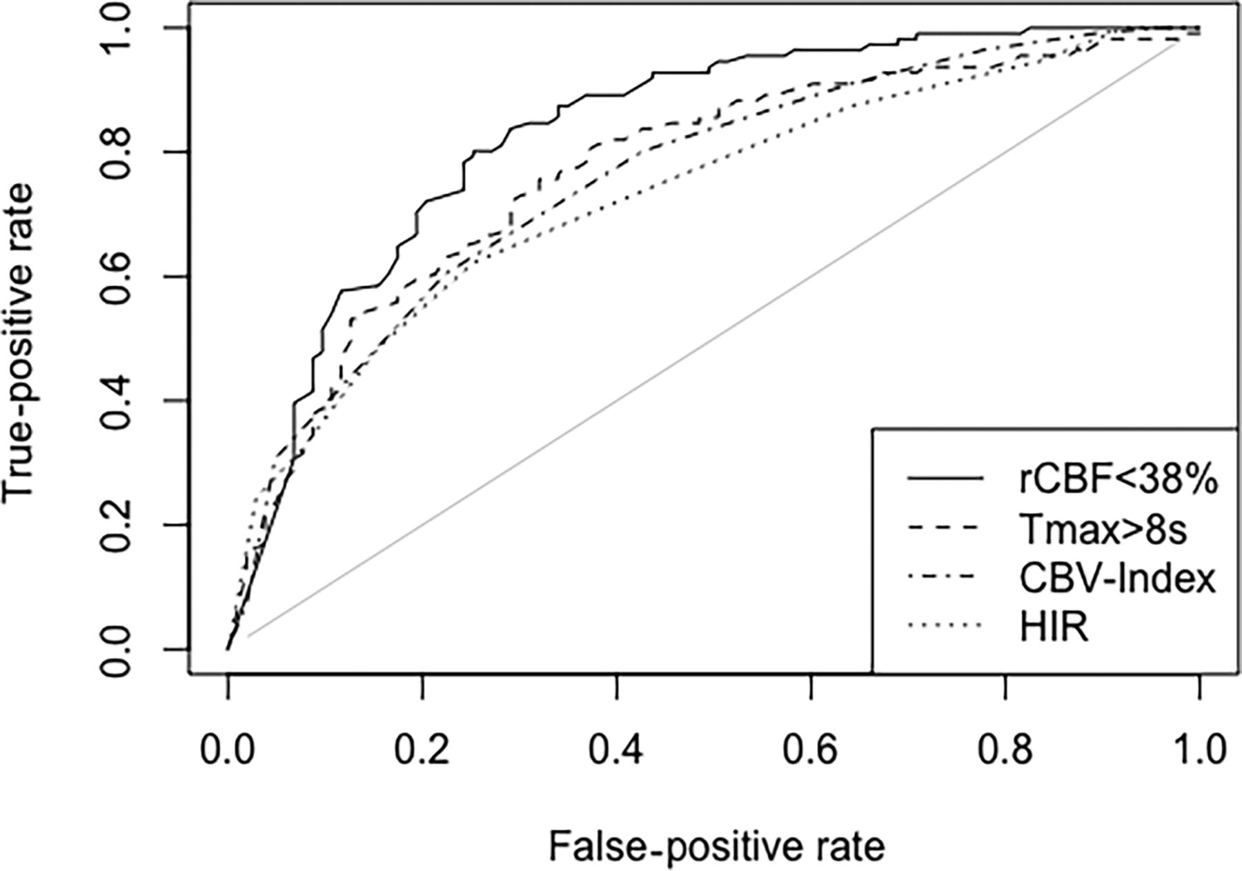
  • FIG 3.
    • Download figure
    • Open in new tab
    • Download powerpoint
    FIG 3.

    Receiver operating characteristics for rCBF < 38% (solid line), Tmax > 8 seconds (dashed line), the CBV-index (dot-dashed line), and HIR (dotted line) for the identification of good collateral status. The AUC was highest for rCBF < 38% with an AUC of 0.83.

Tables

  • Figures
    • View popup
    Table 1:

    CTP parameters in patients with good-versus-poor collateralsa

    Perfusion ParameterAll PatientsPatients with Poor Collaterals (Score, 0–3)Patients with Good Collaterals (Score, 4–5)P Value
    Tmax > 6 sec (mL)111 (102–119)135 (124–145)88 (77–99)<.001
    Tmax > 8 sec (mL)74 (67– 82)97 (88–107)53 (44–63)<.001
    Tmax > 10 sec (mL)48 (43–54)66 (58–74)32 (25–39)<.001
    rCBF < 30%, mL24 (19–28)41 (33–49)8 (5–11)<.001
    rCBF < 34%, mL30 (25–35)50 (41–58)11 (8–14)<.001
    rCBF < 38%, mL36 (31–42)59 (50–68)15 (12–19)<.001
    CBV-index0.68 (0.65–0.70)0.60 (0.56–0.63)0.76 (0.72–0.78)<.001
    HIR0.37 (0.34–0.40)0.46 (0.42–0.50)0.29 (0.24–0.33)<.001
    • ↵a Data are given as mean values and 95% confidence intervals.

    • View popup
    Table 2:

    Results from the Spearman rank correlation analysis for collateral score as a function of perfusion parameters (ρ [95% CI] and P value) and from ROC analysis for the identification of good collateral statusa

    Perfusion Parameterρ (95% CI)P ValueAUC (95% CI)Cutoff ValueSensitivitySpecificityAccuracy
    Tmax >6 sec (mL)−0.50 (−0.61 to −0.39)<.0010.75 (0.68–0.81)b124 mL56%82%69%
    Tmax >8 sec (mL)−0.54 (−0.64 to −0.43)<.0010.77 (0.71–0.83)b74 mL68%76%72%
    Tmax >10 sec (mL)−0.50 (−0.60 to−0.39)<.0010.77 (0.71–0.83)b53 mL64%80%72%
    rCBF < 30% (mL)−0.61 (−0.71 to −0.52)<.0010.81 (0.75–0.87)14 mL72%82%77%
    rCBF < 34% (mL)−0.64 (−0.73 to −0–55)<.0010.83 (0.77–0.88)25 mL67%87%77%
    rCBF < 38% (mL)−0.66 (−0.74 to −0.57)<.0010.83 (0.78–0.89)27 mL75%80%77%
    CBV-index+0.51 (0.40–0.63)<.0010.76 (0.69–0.81)b0.860%78%69%
    HIR−0.49 (−0.60−0.37)<.0010.73 (0.66–0.79)b0.475%62%65%
    • ↵a Overall, the volume with an rCBF < 38% performed best. Optimal cutoff values to identify good collateral supply were estimated according to the Youden index.

    • ↵b Significant differences in AUC compared with the AUC for the volume with rCBF < 38%.

    • View popup
    Table 3:

    Contingency table for collateral status compared with volume with rCBF < 38%a

    Volume with rCBF <38%:
    Collateral Status
    <27 mL≥27 mLTotal
    Good collateral status (score 4−5)8922111
    Poor collateral status (score 0–3)2677103
    Total11599214
    • ↵a Good collateral status was significantly associated with a smaller rCBF < 38% lesion size (Fisher exact test was significant with P < .001).

PreviousNext
Back to top

In this issue

American Journal of Neuroradiology: 43 (7)
American Journal of Neuroradiology
Vol. 43, Issue 7
1 Jul 2022
  • Table of Contents
  • Index by author
  • Complete Issue (PDF)
Advertisement
Print
Download PDF
Email Article

Thank you for your interest in spreading the word on American Journal of Neuroradiology.

NOTE: We only request your email address so that the person you are recommending the page to knows that you wanted them to see it, and that it is not junk mail. We do not capture any email address.

Enter multiple addresses on separate lines or separate them with commas.
RAPID CT Perfusion–Based Relative CBF Identifies Good Collateral Status Better Than Hypoperfusion Intensity Ratio, CBV-Index, and Time-to-Maximum in Anterior Circulation Stroke
(Your Name) has sent you a message from American Journal of Neuroradiology
(Your Name) thought you would like to see the American Journal of Neuroradiology web site.
CAPTCHA
This question is for testing whether or not you are a human visitor and to prevent automated spam submissions.
Cite this article
A. Potreck, E. Scheidecker, C.S. Weyland, U. Neuberger, C. Herweh, M.A. Möhlenbruch, M. Chen, S. Nagel, M. Bendszus, F. Seker
RAPID CT Perfusion–Based Relative CBF Identifies Good Collateral Status Better Than Hypoperfusion Intensity Ratio, CBV-Index, and Time-to-Maximum in Anterior Circulation Stroke
American Journal of Neuroradiology Jul 2022, 43 (7) 960-965; DOI: 10.3174/ajnr.A7542

Citation Manager Formats

  • BibTeX
  • Bookends
  • EasyBib
  • EndNote (tagged)
  • EndNote 8 (xml)
  • Medlars
  • Mendeley
  • Papers
  • RefWorks Tagged
  • Ref Manager
  • RIS
  • Zotero
0 Responses
Respond to this article
Share
Bookmark this article
RAPID CT Perfusion for Collateral Status in Stroke
A. Potreck, E. Scheidecker, C.S. Weyland, U. Neuberger, C. Herweh, M.A. Möhlenbruch, M. Chen, S. Nagel, M. Bendszus, F. Seker
American Journal of Neuroradiology Jul 2022, 43 (7) 960-965; DOI: 10.3174/ajnr.A7542
del.icio.us logo Twitter logo Facebook logo Mendeley logo
  • Tweet Widget
  • Facebook Like
  • Google Plus One
Purchase

Jump to section

  • Article
    • Abstract
    • ABBREVIATIONS:
    • MATERIALS AND METHODS
    • RESULTS
    • DISCUSSION
    • CONCLUSIONS
    • Acknowledgment
    • References
  • Figures & Data
  • Supplemental
  • Info & Metrics
  • Responses
  • References
  • PDF

Related Articles

  • PubMed
  • Google Scholar

Cited By...

  • Futile recanalization in patients with basilar artery occlusion: assessment of the underlying etiology and the role of perfusion imaging
  • Cerebral blood volume index in the era of thrombectomy-treated large and medium vessel ischemic strokes
  • Crossref (22)
  • Google Scholar

This article has been cited by the following articles in journals that are participating in Crossref Cited-by Linking.

  • Hypoperfusion Intensity Ratio Is Correlated With the Risk of Parenchymal Hematoma After Endovascular Stroke Treatment
    Laurens Winkelmeier, Jeremy J. Heit, Gautam Adusumilli, Vincent Geest, Soren Christensen, Helge Kniep, Noel van Horn, Paul Steffen, Matthias Bechstein, Peter Sporns, Maarten G. Lansberg, Gregory W. Albers, Max Wintermark, Jens Fiehler, Tobias D. Faizy
    Stroke 2023 54 1
  • Unfavorable cerebral venous outflow is associated with futile recanalization in acute ischemic stroke patients
    Christian Heitkamp, Laurens Winkelmeier, Jeremy J. Heit, Gregory W. Albers, Maarten G. Lansberg, Max Wintermark, Gabriel Broocks, Noel van Horn, Helge C. Kniep, Peter B. Sporns, Kamil Zeleňák, Jens Fiehler, Tobias D. Faizy
    European Journal of Neurology 2023 30 9
  • Relative Cerebral Blood Flow as an Indirect Imaging Surrogate in Patients With Anterior Circulation Large Vessel Occlusion and Association of Baseline Characteristics With Poor Collateral Status
    Hamza Adel Salim, Omar Hamam, Goksu Parilday, Rawan A. Moustafa, Samir Ghandour, Moustafa Rutgers, Muhanned Sharara, Andrew Cho, Ishan Mazumdar, Mahla Radmard, Christopher Shin, Daniel Montes, Ajay Malhotra, Javier M. Romero, Vivek Yedavalli
    Journal of the American Heart Association 2024 13 16
  • CT perfusion based rCBF &lt;38% volume is independently and negatively associated with digital subtraction angiography collateral score in anterior circulation large vessel occlusions
    Dhairya A Lakhani, Aneri B Balar, Manisha Koneru, Sijin Wen, Burak Berksu Ozkara, Richard Wang, Meisam Hoseinyazdi, Mehreen Nabi, Ishan Mazumdar, Andrew Cho, Kevin Chen, Sadra Sepehri, Nathan Hyson, Risheng Xu, Victor Urrutia, Licia Luna, Argye E Hillis, Jeremy J Heit, Greg W Albers, Ansaar T Rai, Vivek S Yedavalli
    The Neuroradiology Journal 2024 37 4
  • Pretreatment parameters associated with hemorrhagic transformation among successfully recanalized medium vessel occlusions
    Manisha Koneru, Meisam Hoseinyazdi, Richard Wang, Burak Berksu Ozkara, Nathan Z. Hyson, Elisabeth Breese Marsh, Rafael H. Llinas, Victor C. Urrutia, Richard Leigh, Luis Fernando Gonzalez, Risheng Xu, Justin M. Caplan, Judy Huang, Hanzhang Lu, Licia Luna, Max Wintermark, Adam A. Dmytriw, Adrien Guenego, Gregory W. Albers, Jeremy J. Heit, Kambiz Nael, Argye E. Hillis, Vivek S. Yedavalli
    Journal of Neurology 2024 271 4
  • Redefining CT perfusion‐based ischemic core estimates for the ghost core in early time window stroke
    Manisha Koneru, Meisam Hoseinyazdi, Dhairya A. Lakhani, Cynthia Greene, Karen Copeland, Richard Wang, Risheng Xu, Licia Luna, Justin M. Caplan, Adam A. Dmytriw, Adrien Guenego, Jeremy J. Heit, Gregory W. Albers, Max Wintermark, Luis F. Gonzalez, Victor C. Urrutia, Judy Huang, Kambiz Nael, Richard Leigh, Elisabeth B. Marsh, Argye E. Hillis, Rafael H. Llinas, Vivek S. Yedavalli
    Journal of Neuroimaging 2024 34 2
  • The negative effect of aging on cerebral venous outflow in acute ischemic stroke
    Christian Heitkamp, Laurens Winkelmeier, Jeremy J Heit, Fabian Flottmann, Christian Thaler, Helge Kniep, Gabriel Broocks, Lukas Meyer, Vincent Geest, Gregory W Albers, Maarten G Lansberg, Jens Fiehler, Tobias D Faizy
    Journal of Cerebral Blood Flow & Metabolism 2023 43 10
  • Using Deep-Learning-Based Artificial Intelligence Technique to Automatically Evaluate the Collateral Status of Multiphase CTA in Acute Ischemic Stroke
    Chun-Chao Huang, Hsin-Fan Chiang, Cheng-Chih Hsieh, Chao-Liang Chou, Zong-Yi Jhou, Ting-Yi Hou, Jin-Siang Shaw
    Tomography 2023 9 2
  • Uncertainty-aware deep learning for trustworthy prediction of long-term outcome after endovascular thrombectomy
    Celia Martín Vicario, Dalia Rodríguez Salas, Andreas Maier, Stefan Hock, Joji Kuramatsu, Bernd Kallmuenzer, Florian Thamm, Oliver Taubmann, Hendrik Ditt, Stefan Schwab, Arnd Dörfler, Iris Muehlen
    Scientific Reports 2024 14 1
  • The Relative Cerebral Blood Volume (rCBV) &lt; 42% Is Independently Associated with Collateral Status in Anterior Circulation Large Vessel Occlusion
    Dhairya A. Lakhani, Aneri B. Balar, Manisha Koneru, Sijin Wen, Burak Berksu Ozkara, Hanzhang Lu, Richard Wang, Meisam Hoseinyazdi, Janet Mei, Risheng Xu, Mehreen Nabi, Ishan Mazumdar, Andrew Cho, Kevin Chen, Sadra Sepehri, Nathan Hyson, Victor Urrutia, Licia Luna, Argye E. Hillis, Jeremy J. Heit, Greg W. Albers, Ansaar T. Rai, Adam A. Dmytriw, Tobias Faizy, Max Wintermark, Kambiz Nael, Vivek S. Yedavalli
    Journal of Clinical Medicine 2024 13 6

More in this TOC Section

  • The insula and malignant cerebral edema
  • ICH Shape & Density Predict Rebleeding after MICE
  • Circle of Willis Variants and Stroke Outcomes
Show more NEUROVASCULAR/STROKE IMAGING

Similar Articles

Advertisement

Indexed Content

  • Current Issue
  • Accepted Manuscripts
  • Article Preview
  • Past Issues
  • Editorials
  • Editor's Choice
  • Fellows' Journal Club
  • Letters to the Editor
  • Video Articles

Cases

  • Case Collection
  • Archive - Case of the Week
  • Archive - Case of the Month
  • Archive - Classic Case

More from AJNR

  • Trainee Corner
  • Imaging Protocols
  • MRI Safety Corner
  • Book Reviews

Multimedia

  • AJNR Podcasts
  • AJNR Scantastics

Resources

  • Turnaround Time
  • Submit a Manuscript
  • Submit a Video Article
  • Submit an eLetter to the Editor/Response
  • Manuscript Submission Guidelines
  • Statistical Tips
  • Fast Publishing of Accepted Manuscripts
  • Graphical Abstract Preparation
  • Imaging Protocol Submission
  • Evidence-Based Medicine Level Guide
  • Publishing Checklists
  • Author Policies
  • Become a Reviewer/Academy of Reviewers
  • News and Updates

About Us

  • About AJNR
  • Editorial Board
  • Editorial Board Alumni
  • Alerts
  • Permissions
  • Not an AJNR Subscriber? Join Now
  • Advertise with Us
  • Librarian Resources
  • Feedback
  • Terms and Conditions
  • AJNR Editorial Board Alumni

American Society of Neuroradiology

  • Not an ASNR Member? Join Now

© 2025 by the American Society of Neuroradiology All rights, including for text and data mining, AI training, and similar technologies, are reserved.
Print ISSN: 0195-6108 Online ISSN: 1936-959X

Powered by HighWire