
Article Figures & Data
Figures
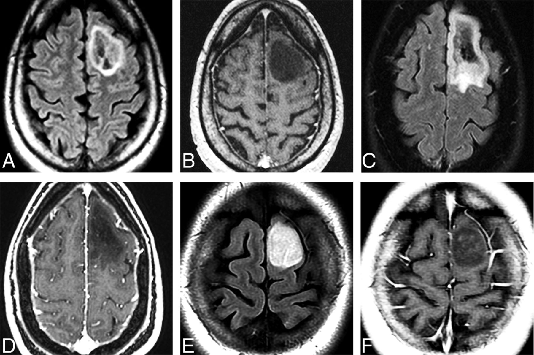
- FIG 1.
Adult-type diffuse gliomas. Series of 3 cases illustrates the importance of complete IDH mutation status determination and the investigation of other molecular markers in evaluation of adult-type diffuse astrocytomas. Axial FLAIR (A) and postcontrast T1WI (B) in a 54-year-old man with a first-time seizure shows a well-delineated left frontal lobe mass with a hyperintense rim surrounding a mixed signal mass. No enhancement is present. Pathology disclosed diffuse astrocytoma without necrosis or microvascular proliferation. Immunohistochemistry demonstrated that the tumor was IDH-mutant. Next generation sequencing disclosed CDKN2A/B homozygous loss, so the tumor was upgraded to WHO CNS grade 4. Axial FLAIR (C) and postcontrast T1WI (D) in a 44-year-old woman with a first-time seizure demonstrate a left frontal mass that was completely resected. Pathology findings were consistent with WHO CNS grade 3. Initial immunohistochemistry was negative for IDH1 mutation, but further investigation disclosed the presence of an IDH2 mutation. Final pathologic diagnosis is diffuse astrocytoma, IDH-mutant, grade 3. The patient is alive without evidence of disease 4 years after the initial diagnosis. Axial FLAIR (E) and postcontrast T1WI (F) in a 24-year-old woman with a first-time seizure show a well-delineated nonenhancing left frontal lobe mass that was surgically resected. Histologically, the tumor was WHO CNS grade 2 but IDH-wildtype on immunohistochemistry. No further investigation was conducted. One year later, the tumor recurred and re-resection demonstrated EGFR amplification and was, therefore, upgraded to glioblastoma (WHO CNS grade 4). The patient died of disseminated disease 18 months after the initial diagnosis.
- FIG 2.
Pediatric-type diffuse low-grade glioma. Axial T2 (A) MR image in a 7-year-old boy with a diffuse astrocytoma, MYB-altered, shows a hyperintense mass in the pons with no significant surrounding edema. There was no enhancement and no diffusion restriction of the mass (not shown). Axial FLAIR (B), postcontrast T1 (C), and arterial spin-labeling (ASL) (D) in a 1-year-old child with an angiocentric glioma show a FLAIR hyperintense mass involving the cortex and subcortical white matter of the left frontal lobe. There is no enhancement (C) and decreased perfusion (D) on ASL imaging.
- FIG 3.
Two patients with the WHO 2021 new-entity polymorphous low-grade neuroepithelial tumor of the young (PLNTY). Axial FLAIR (A) and postcontrast T1-weighted (B) MR images in a 19-year-old man with refractory epilepsy show a hyperintense, nonenhancing mass in the cortex and subcortical white matter of the left temporal lobe. Axial FLAIR (C) and susceptibility-weighted (D) MR images and a noncontrast CT image (E) in a 19-year-old woman with progressive seizure show a FLAIR-hyperintense, SWI-hypointense mass with characteristic calcification seen on CT in the right medial temporal lobe (Case courtesy of M. Castillo, MD).
- FIG 4.
Pediatric-type diffuse high-grade gliomas. Diffuse midline glioma, H3K27-altered in an 8-year-old girl with cranial neuropathies. Axial T2 (A) and FLAIR (B) MR images show an expansile, hyperintense pontine mass. Axial postcontrast T1 MR image (C) shows heterogeneous enhancement within the mass. Arterial spin-labeling (ASL) perfusion (D) shows increased perfusion. E, Axial FLAIR MR image shows a bithalamic hyperintense mass. Postcontrast T1WI showed no significant enhancement, and ASL perfusion showed increased perfusion within the bilateral thalami (not shown). These WHO grade 4 tumors have a poor prognosis.
- FIG 5.
Diffuse hemispheric glioma, H3 G34-mutant and IDH-wildtype tumor in an 8-year-old boy. A, Axial FLAIR shows a large, very heterogeneous right temporal lobe mass with minimal surrounding edema. B, An ADC map in the same case shows restricted diffusion consistent with a high-cellularity tumor. C, Arterial spin-labeling perfusion shows decreased perfusion in the tumor. In pediatric tumors, perfusion is often less helpful compared with diffusion-weighted imaging in discriminating tumor grade. Histology demonstrated necrosis, hemorrhage, and neovascularity in a glioblastoma-like pattern, consistent with grade 4 tumor.
- FIG 6.
Infant-type hemispheric glioma, NOS. A male neonate child with macrocephaly and bulging fontanelles had a large, heterogeneous-appearing mass on an emergent CT scan (not shown). Axial T2-weighted (A) and postcontrast T1-weighted (B) MR images show a very heterogeneous mass with enhancement involving almost the entirety of the left cerebral hemisphere.
- FIG 7.
Axial T2WI in a 19-month-old child with astroblastoma, MN1-altered. A, Axial T2WI shows a bubbly-appearing mixed-signal hemispheric mass with little surrounding edema. B, Postcontrast T1WI shows that the mass enhances strongly but heterogeneously.
- FIG 8.
Two cases of MGNT are illustrated. A, Axial T2WI in a 14-year-old boy shows an extremely hyperintense, slightly bubbly mass in the left juxtaventricular white matter. B, Axial FLAIR shows a hyperintense rim surrounding a largely isointense center of the mass. Smaller-but-similar-appearing lesions are adjacent to the mass. The mass did not enhance following contrast administration. C, Sagittal T1WI in a 39-year-old man shows a well-demarcated mass in the corpus callosum rostrum/septum pellucidum. D, The mass is extremely hyperintense on T2WI. E, FLAIR shows that the mass has a hyperintense rim with an isointense center. The mass is thought to represent an MGNT because of its classic location and signal characteristics but is not biopsy-proven.
- FIG 9.
ZFTA fusion–positive ependymoma in an 11-year-old girl. A, Axial T2WI shows a large, bulky, heterogeneous left frontal mass. B, Susceptibility-weighted scan shows intratumoral hemorrhage. C, Strong-but-very heterogeneous enhancement is seen on postcontrast T1WI.
- FIG 10.
Embryonal tumor with multilayered rosettes in a 1-year-old girl. A, An axial T2-weighted scan shows a large, left parieto-occipital mass with little surrounding edema. B, The mass exhibits hemorrhage on susceptibility-weighted imaging and no enhancement following contrast administration (C). D, Strikingly restricted diffusion is seen on the ADC map. E, Arterial spin-labeling perfusion shows decreased perfusion in the tumor.


Tables
Layered neuropathology diagnosisa
Brain (right frontal) Integrated diagnosis: glioblastoma, IDH-wildtypeb Histologic diagnosis: diffuse astrocytic tumor with mitotic figures WHO CNS grade: 4 Molecular information: IDH: wildtype (DNA sequencing) ATRX: retained nuclear expression, consistent with wild-type (immunohistochemistry) p53: rare positive cells, consistent with wild-type (immunohistochemistry) EGFR: amplified (FISH) ↵a Illustrative example of a layered neuropathology report beginning with site and identifier and with integrated diagnosis as the top line.
↵b Although this tumor lacks microvascular proliferation and necrosis, the presence of EGFR amplification necessitates a diagnosis of glioblastoma in an IDH-wildtype diffuse astrocytic glioma.


Jump to section
Related Articles
Cited By...
- Mesenchymal Nonmeningothelial Tumors of the CNS: Evolving Molecular Landscape and Implications for Neuroradiologists
- Ependymal Tumors: Overview of the Recent World Health Organization Histopathologic and Genetic Updates with an Imaging Characteristic
- High-Grade Astrocytoma with Piloid Features: A Dual Institutional Review of Imaging Findings of a Novel Entity
- Theranostics in Neurooncology: Heading Toward New Horizons
- Modeling Glioma Oncostreams In Vitro: Spatiotemporal Dynamics of their Formation, Stability, and Disassembly
- Comparison of the Diagnostic Performance from Patients Medical History and Imaging Findings between GPT-4 based ChatGPT and Radiologists in Challenging Neuroradiology Cases
- PiDeeL: Pathway-Informed Deep Learning Model for Survival Analysis and Pathological Classification of Gliomas
- Newly Recognized CNS Tumors in the 2021 World Health Organization Classification: Imaging Overview with Histopathologic and Genetic Correlation
- Amino Acid Tracer PET MRI in Glioma Management: What a Neuroradiologist Needs to Know
- Increasing Ciliary ARL13B Expression Drives Active and Inhibitor-Resistant SMO and GLI into Glioma Primary Cilia