
Article Figures & Data
Figures
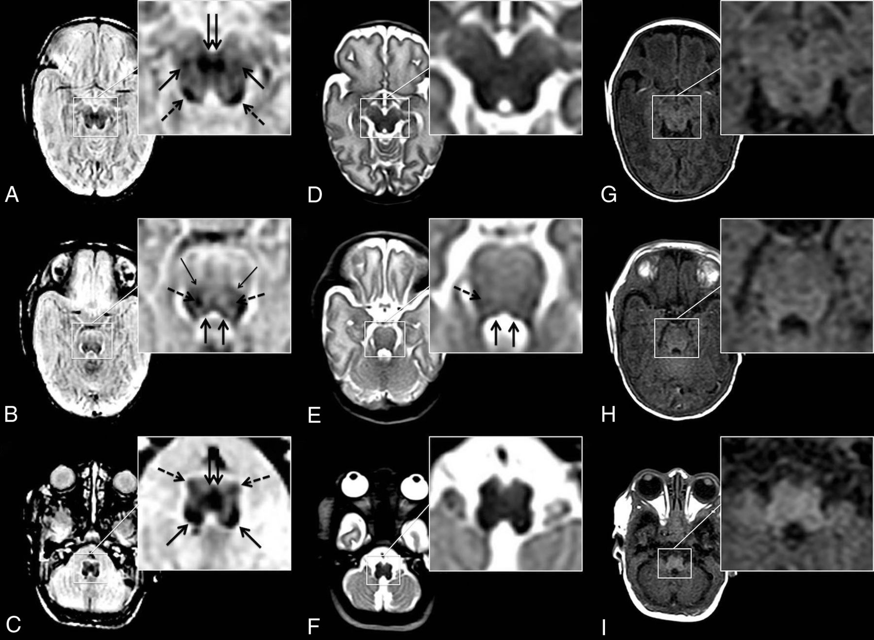
- FIG 1.
Neonatal brainstem anatomy is shown in an infant born at 24 + 3 weeks’ GA (MR imaging at 38 + 0 weeks postmenstrual age) at the level of the midbrain (A, D, and G), pons (B, E, and H), and the medulla oblongata (C, F, and I) on syWMSS data (A, B, and C), conventional T2-weighted contrasts (D, E, and F), and conventional T1-weighted contrasts (G, H, and I). syWMSS data (A, B, and C) depict early myelinating structures: DSCP (double arrow in A); ML (arrows in A; thin arrows in B) and its decussation (double arrow in C); inferior colliculus (dotted arrows in A); CTT (dotted arrows in B); MLF (arrows in B); amiculum of the inferior olivary nucleus (dotted arrows in C); and inferior cerebellar peduncle (arrows in C). Pontine bundles are depicted sufficiently on T2-weighted imaging data (D, E, and F) (CTT [dotted arrow in E] and MLF [arrows in E]). On the basis of T1-weighted contrasts, a reliable delineation of brainstem pathways is limited (G, H, and I). Center/width at the reader’s discretion.
- FIG 2.
Myelin-related signal intensity differences between preterm-born neonates and term-born infants are demonstrated. The midbrain is shown in a neonate born at 27 + 0 weeks’ GA (MR imaging at 37 + 0 weeks postmenstrual age) (A, B, and C) and an infant born at 40 + 5 weeks’ GA (MR imaging at 41 + 4 weeks postmenstrual age) (D, E, and F) on syWMSS data (A and D), conventional T2-weighted contrasts (B and E), and conventional T1-weighted contrasts (C and F). While proceeding myelination is perceivable on conventional T1-weighted MR contrasts (DSCP, double arrows in F), the information provided by conventional T2-weighted MR imaging contrasts regarding myelin-related signal alterations is limited. In contrast to standard-of-reference MR imaging acquisitions, WM signal suppression enables a reliable identification and assessment of progressively myelinating structures in the course of brain development (DSCP, double arrows in A and D and ML, dotted arrows in D). Center/width at the default setting.
- FIG 3.
Pearson correlations between GA at birth (x-axis) and myelin scorings (performed by rater 1) (y-axis) based on syWMSS data (r = 0.887, P < .001) (A), conventionally acquired T1WI contrasts (r = 0.546, P = .002) (B), and conventionally acquired T2WI contrasts (r = 0.117, P = .530) (C).


Tables
ICC syWMSS Moderate agreement: 0.535 (0.032–0.788) Conventional T1-weighted Poor agreement: 0.404 (–0.034–0.694) Conventional T2-weighted Poor agreement: −0.050 (−0.315–0.259) ↵a Numbers in parentheses are 95% confidence intervals.


Jump to section
Related Articles
Cited By...
- No citing articles found.