
Article Figures & Data
Figures
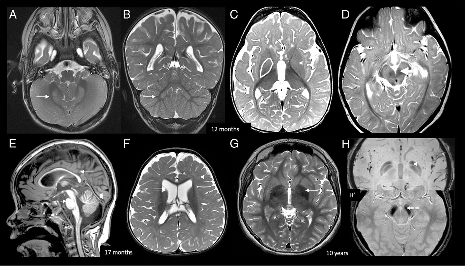
- FIG 1.
BPAN early radiologic signature. At 12 months of life, axial and coronal T2 (A and B) images show bilateral symmetric dentate nucleus hyperintensity and swelling (white arrows). Axial T2 (C) shows bilateral GP swelling and mild hyperintensity (white arrow). Bilateral SN swelling and T2 hyperintensity are demonstrated on D (white arrow). At 17 months, midsagittal T1 (E) shows diffuse callosal thinning (white arrow), a small pons (dotted arrow), and superior vermian volume loss (double arrow). Axial T2 (F) shows diffuse myelin reduction (white arrow) and prominent lateral ventricles (double arrow). Delayed myelination was infrequently seen in our cohort (1/15 patients). At 10 years of age, T2 axial image (G) in an older child shows GP iron deposition, which is confirmed on SWI (H). H, Increased iron deposition in the SN. The typically reported T1 halo sign was not seen in our pediatric cohort.
- FIG 2.
MR imaging findings in relation to age. GP iron deposition is not detectable early on but becomes more prominent after 4 years of age. Conversely, GP and SN swelling are findings present in young patients and persistent later on. GP swelling was absent in 1 patient (B14, 2 scans at 1.5 and 3 years of age) but was otherwise consistently present in all patients and all scans.


Jump to section
Related Articles
Cited By...
- Mutation in Wdr45 leads to early motor dysfunction and widespread aberrant axon terminals in a beta-propeller protein associated neurodegeneration (BPAN) patient-inspired mouse model
- Mutations in EPG5 are associated with a wide spectrum of neurodevelopmental and neurodegenerative disorders
- Medullary Tegmental Cap Dysplasia: Fetal and Postnatal Presentations of a Unique Brainstem Malformation
- Early Neuroimaging Markers in {beta} Propeller Protein-Associated Neurodegeneration