
Article Figures & Data
Figures
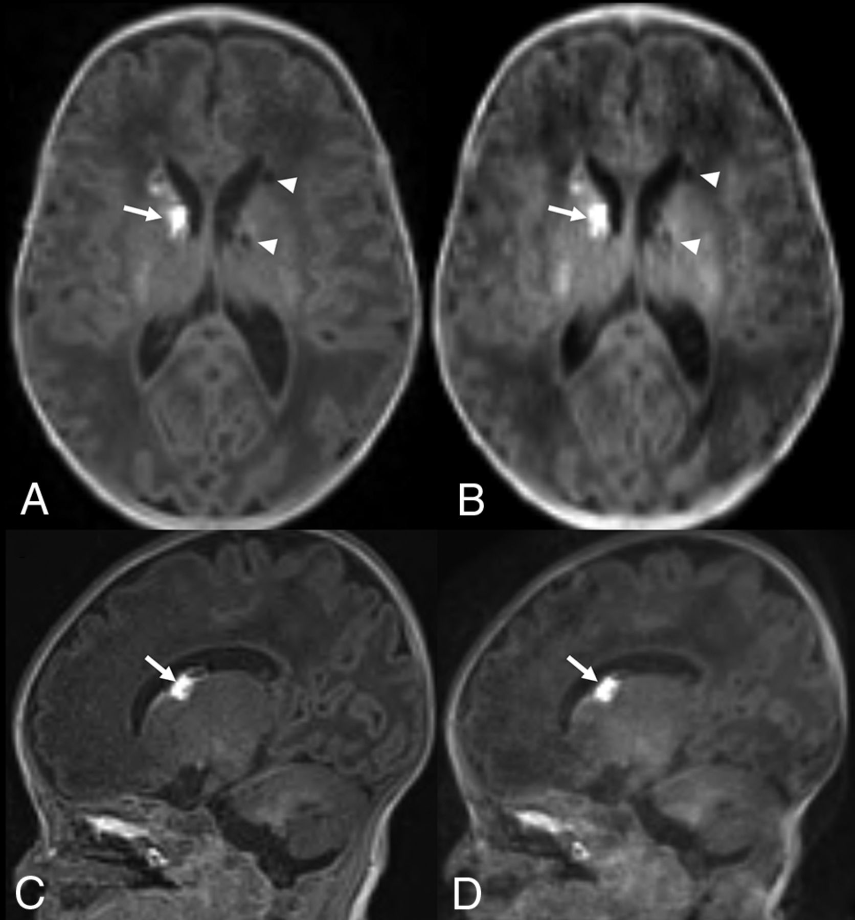
- FIG 1.
Term-equivalent-age MR imaging of a premature infant (29 weeks’ gestation). Nonenhanced standard MPRAGE images (A and C) show hyperintense germinal matrix hemorrhage (arrows) in the right caudothalamic groove and cystic changes of germinal matrix hemorrhage (arrowheads) in the left caudothalamic groove. Although wave-T1-MPRAGE images (B and D) demonstrate lower image quality than standard MPRAGE, both caudothalamic lesions are well-delineated in nonenhanced wave-T1-MPRAGE images.
- FIG 2.
Term-equivalent-age MR imaging of a premature infant (31 weeks’ gestation). Nonenhanced standard MPRAGE (A) shows decreased cerebral WM volume and a focal T1-hyperintense lesion (arrow) at the left corona radiata. The focal corona radiata lesion (arrows) demonstrates hypointensity on gradient recalled-echo (B) and T2-weighted (C) images. Although the wave-MPRAGE image (D) shows lower image quality than standard MPRAGE, the focal T1-hyperintense lesion (arrow) at the left corona radiata is also visible in the wave-MPRAGE image.
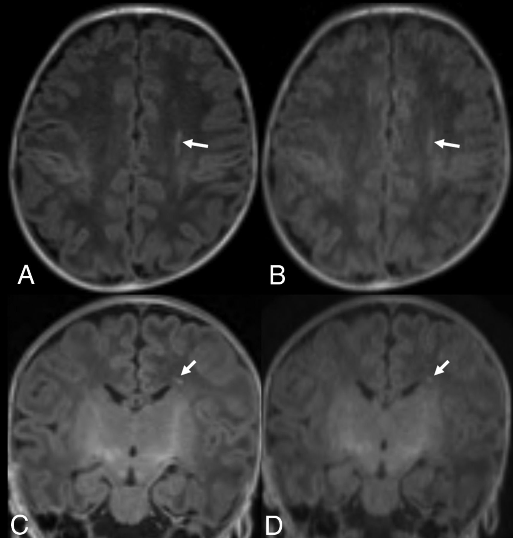
- FIG 3.
Term-equivalent-age MR imaging of a premature infant (33 weeks’ gestation). Nonenhanced standard MPRAGE images (A and C) show a focal hyperintense lesion (arrows) in the left periventricular WM. Although the wave-T1-MPRAGE images (B and D) also show the focal, hyperintense lesion (arrows) in the left periventricular WM, it is less prominently visualized than in the standard MPRAGE; thus, 1 observer missed the lesion.
- FIG 4.
Brain MR imaging of a premature infant (29 weeks’ gestation) 1 month after birth. Nonenhanced standard MPRAGE images (A–C) show severe motion artifacts, which are not acceptable for diagnostic use, while wave-T1-MPRAGE (D–F) shows mild motion artifacts, which are acceptable for diagnostic use.


Tables
Image parameters of standard MPRAGE and wave-T1-MPRAGE
Standard MPRAGE Wave-T1-MPRAGE FOV (mm2) 180 × 180 180 × 80 Voxel size (mm3) 0.8 × 0.8 × 0.9 0.8 × 0.8 × 0.9 TR (ms) 2400 2200 TE (ms) 2.96 3.05 Flip angle 9° 9° Bandwidth (Hz) 260 260 TI (ms) 1200 1090 Number of excitations 1 1 Parallel imaging method GRAPPAa CAIPIRINHAb Acceleration factor (phase) 2 2 Acceleration factor (section) – 2 Scan time 4 min 55 sec 2 min 14 sec


Jump to section
Related Articles
Cited By...
- No citing articles found.